Introduction
Following recent phylogenetic analyses of DNA sequence data, the order Candelariales was suggested for the genera Candelariella Müll. Arg. and Candelaria A. Massal., a group of lichen-forming ascomycetes belonging to the Candelariaceae (Hibbet et al. Reference Hibbett, Binder, Bischoff, Blackwell, Cannon, Eriksson, Huhndorf, James, Kirk and Lücking2007). This family (sometimes considered as a synonym of Lecanoraceae, (e.g., LaGreca & Lumbsch Reference LaGreca and Lumbsch2001; Eriksson et al. Reference Eriksson, Baral, Currah, Hansen, Kurtzman, Rambold and Laessøe2004), was previously placed in the Lecanorales, but Wedin et al. (Reference Wedin, Wiklund, Crewe, Döring, Ekman, Nyberg, Schmitt and Lumbsch2005) showed that the Candelariaceae was not closely related to the core group of Lecanorales, a conclusion which was later confirmed by Hofstetter et al. (Reference Hofstetter, Miądlikowska, Kauff and Lutzoni2007) and Miądlikowska et al. (Reference Hofstetter, Miądlikowska, Kauff and Lutzoni2007). The final classification at higher levels is somewhat uncertain but the group may be recognized as a subclass within the Lecanoromycetes in the future (Hofstetter et al. Reference Hofstetter, Miądlikowska, Kauff and Lutzoni2007; Miądlikowska et al. Reference Hofstetter, Miądlikowska, Kauff and Lutzoni2007).
Two further genera, Candelina Poelt and Placomaronea Räsänen are known to be closely related to Candelaria and Candelariella (e.g., Poelt Reference Poelt1974; Westberg et al. Reference Westberg, Arup and Kärnefelt2007). The morphological variation in the Candelariales ranges from minute lichenicolous species without a thallus to species with a well-developed foliose thallus, up to c. 2–3 cm wide, a range that is partly reflected in the generic classification (Candelariella – crustose; Candelina – placoid/subfoliose; Candelaria – foliose; Placomaronea – umbilicate, foliose). The generic division is, however, uncertain and an earlier analysis of nuclear ITS rDNA data did not resolve the relationship among different species groups (Westberg et al. Reference Westberg, Arup and Kärnefelt2007).
At species level the taxonomy in Candelariales must still be characterized as poorly known in many parts of the world. Major revisionary works include Hakulinen's (Reference Hakulinen1954) world monograph of Candelariella (although very little extra-European material was included), and a recent revision of Candelariella in western North America (Westberg Reference Westberg2005, Reference Westberg2007a, Reference Westbergb, Reference Westbergc). In other parts of the world the taxonomic studies on the group are few and not comprehensive. The Candelariales flora of South America especially should receive more attention, as there appears to be a great diversity on this continent, and all four genera occur here. A revision of South American material will be an important key to the understanding of the evolution and the phylogeny within this group. The only taxonomic works focusing on Candelariales in South America are Poelt's study of the three small genera Candelina, Candelaria and Placomaronea (Poelt Reference Poelt1974) and a short paper on Candelaria by Westberg & Frödén (Reference Westberg and Frödén2007). Although some species are common to both western North America and central South America, many specimens belonging to the Candelariales, collected by the second author in Argentina, Chile and Peru in 2001, could not be identified, and most likely represent undescribed species. Also, among the rich material from South America accumulated under Candelariella spp. at S, collected mainly by R. Santesson, there are obviously several undescribed and morphologically interesting taxa.
Placomaronea is a poorly known genus species of which grow in the montane partsof South America and SW North America (Poelt Reference Poelt1974; Santesson Reference Santesson1944; Westberg Reference Westberg, Nash, Ryan, Diederich, Gries and Bungartz2004). It was originally proposed for P. candelarioides Räsänen by Räsänen (Reference Räsänen1944) who suggested a position in the Acarosporaceae for the genus (Räsänen Reference Räsänen1943), but the close relationship to Candelariella was correctly recognized by Hakulinen (Reference Hakulinen1954), who transferred it to the Candelariaceae. Subsequently Candelariella lambii Hakul. was transferred to Placomaronea (Poelt Reference Poelt1974), and a third species was included in the genus by Westberg (Reference Westberg, Nash, Ryan, Diederich, Gries and Bungartz2004). In the present study, material tentatively identified as belonging to Placomaronea due to its thallus anatomy (thallus shiny, gelatinous epicortex present, epicortical pigments arranged as hoods on the outermost cell-layer, cortex well-developed and pseudoparenchymatous) and polysporous asci was analysed morphologically. The monophyly of the group was investigated with DNA sequence data.
We are delighted to dedicate this study to our good friend Professor Ingvar Kärnefelt; he has been a committed and patient supervisor for two of us (MW & PF) during our PhD studies.
Material and Methods
This study is based chiefly on material collected by Frödén during fieldwork in 2001 in South America (Peru, Chile and Argentina) deposited at LD, and on additional material, including types, from LD, S, H, HBG, ASU and MIN. Anatomical studies and measurements were carried out on hand-cut sections of thalli and apothecia mounted in water and in accordance with earlier studies in Candelariella by the first author (see e.g. Westberg Reference Westberg2007a). Spore measurements are given as (min. value observed –) range including 85% of the variation (– max. value observed). The hymenium height was measured from the base of the asci to the tip of the paraphyses.
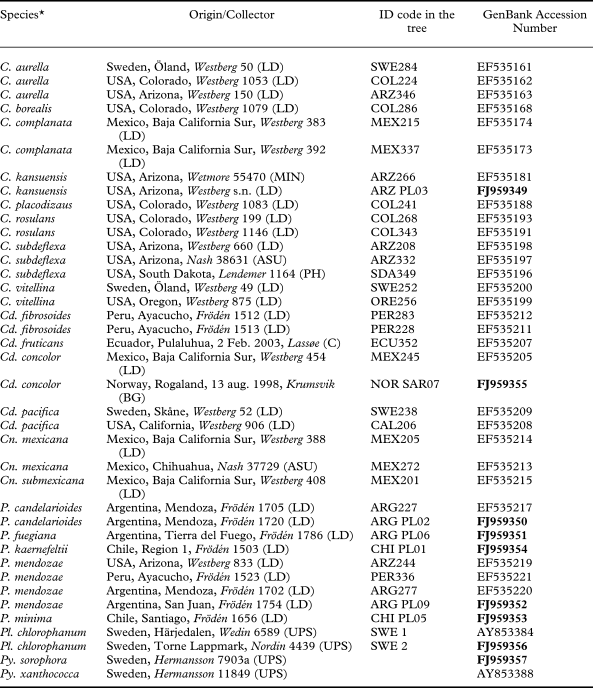
For the phylogenetic study we retrieved sequences already available in Genbank and produced new ones from relevant material (Table 1). Representatives of all genera in Candelariales, as well as most of the major groups retrieved in the analyses by Westberg et al. (Reference Westberg, Arup and Kärnefelt2007) are included. As outgroup, two representatives of the closely related Pycnora Hafellner were selected (Wedin et al. Reference Wedin, Wiklund, Crewe, Döring, Ekman, Nyberg, Schmitt and Lumbsch2005); in addition two representatives of Pleopsidium Körb. (Acarosporaceae) were included. For newly produced sequences we sampled thalli or apothecia of recently collected material of Placomaronea (suitable material of P. lambii was not available). Total DNA extraction, PCR amplification, PCR product purification and sequencing were performed as in Wedin et al. (Reference Wedin, Westberg, Crewe and Purvis2009). The sequences were aligned by hand using MacClade 4.08 for OS X. The phylogenetic maximum parsimony analyses were performed using PAUP* 4.0b10 (Swofford Reference Swofford2002) with the following settings: gaps treated as missing data, 1000 random additional sequence replicates, TBR branch swap, steepest descent off, collapse branches if minimum length is 0, and MulTrees ON. Uninformative characters were excluded from the analyses. Parsimony jackknifing for rapid identification of well-supported monophyletic groups (Farris et al. Reference Farris, Albert, Källersjö, Lipscomb and Kluge1996) were performed for the dataset, with the following settings: jackknife settings: 1000 jackknife replicates with ‘JAC’-emulation, nominal deletion of characters 37%, full heuristic search, retain groups with frequency >50%; heuristic search settings: 10 random additional replicates, otherwise as given above.
Specimen data and GenBank accession numbers of nuITS rDNA sequences newly produced (bold) or retrieved from Genbank and used in the phylogenetic analysis

* C=Candelariella, Cd=Candelaria, Cn=Candelina, P=Placomaronea, Pl=Pleopsidium, Py=Pycnora
Results
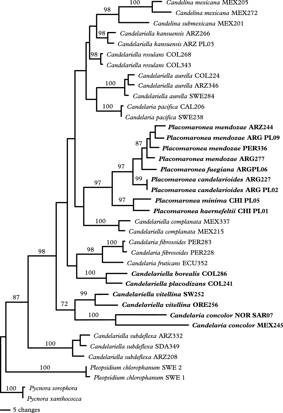
Six taxa were recognized based on the morphological studies. Five of these are represented in the phylogenetic analysis. Nine new nuITS rDNA sequences were obtained and aligned with 28 existing sequences. The data matrix produced contained 507 aligned sites; 332 uninformative characters were excluded and 175 informative sites were used in the analysis. The parsimony analysis resulted in 2 trees of 614 steps. The specimens identified as Placomaronea form a monophyletic clade with strong jackknife support in the resulting tree (Fig. 1), thus supporting the concept that the genus is a natural, monophyletic group. The five taxonomic species recognized by morphology that were included in the DNA analysis were all supported as distinct by the phylogeny.
Phylogeny of the Candelariaceae. One of two most parsimonious trees (length 614 steps, CI 46, RI 70), jack-knife support values (>70) above branches. Polysporous taxa are marked in bold. The back-bone topology is not supported, but Placomaronea constitutes a well-supported monophyletic group. All species represented by more than one sample likewise form well-supported monophyletic groups.

Taxonomy
Placomaronea Räsänen
Ann. Bot. Soc. Zool.-Bot. Fenn. ‘Vanamo’ 20(3): 29 (1944); type species P. candelarioides Räsänen.
Thallus areolate to peltate squamulose to placoid, subfoliose to umbilicate foliose; upper surface yellow to orange-yellow to brownish yellow; lower surface in foliose species whitish or brownish due to dust impregnation, without rhizines; cortex pseudoparenchymatous with gelatinized hyphae with rounded cells; epicortical pigments mostly conspicuously arranged as hoods on the outermost cells; epicortex present or not, thin, colourless and gelatinous; algae chlorococcoid; medulla white or not developed, in which case the algal cells are distributed throughout the thallus interior below the cortex; lower cortex present or not, similar to the upper cortex.
Apothecia lecanorine, rounded; thalline margin persistent; proper margin not visible from the outside; thalline margin with a cortex similar to that of the thallus; proper margin basally forming a stipe penetrating down to the thallus, below the hypothecium radiating towards the margin, cells elongated, becoming shorter and rounder towards the margin and finally similar to the cortex cells; subhymenial layers colourless, with oil-drops; epihymenium of small, granular, brownish yellow to golden red crystals, sometimes arranged as hoods on the tips of the paraphyses; hymenium colourless; paraphyses simple or furcate near the tips, tips cylindrical to clavate or almost capitate, sometimes almost submoniliform with the two-three uppermost cells widened; asci clavate, >30-spored; ascospores narrowly ellipsoid, colourless, simple or rarely with a thin septum.
Pycnidia small, one-chambered, immersed in the thallus, conidiophores of type II–III sensu Vobis (Reference Vobis1980), conidia colourless, narrowly ellipsoid.
Chemistry. Pigments consisting of pulvinic acid derivatives: calycin (major), pulvic acid lactone, vulpinic acid and pulvinic acid found by HPLC (Westberg Reference Westberg2005).
Habitat and distribution. All species grow on rocks, probably acidic in most or all cases. Most species grow at high altitudes, the only lowland species we are aware of is P. fuegiana. The genus has a South American – South African distribution, but is found mainly along the Andes in South America. One species extends as far north as Arizona in south-western North America (P. mendozae) and one species is known from southern Africa (P. minima). A similar distribution is known within Candelariales in Candelina (Poelt Reference Poelt1974) and in Candelaria fruticans (Kärnefelt & Westberg Reference Kärnefelt and Westberg2001).
Key to the genus Placomaronea
1 Thallus umbilicate, monophyllous, with a central holdfast ... P. candelarioides
Thallus composed of numerous areoles or lobes individually attached to the substratum or multi-lobed and with many holdfasts ... 2
2 (1) Thallus with distinct and elongated lobes, often rosette-like ... 3
Thallus areolate to peltate squamulose ... 5
3 (2) Marginal lobes up to twice as long as wide; hymenium 90–105 μm tall; thallus not stratified, i.e., algal layer filling the thallus interior below the cortex ... ... P. kaernefeltii
Marginal lobes 3–6 times as long as wide; hymenium to 90 μm tall; thallus stratified with a distinct medullary layer below the alga ... 4
4 (3) Lobes flattened with a smooth surface, attached individually to the substratum at the base ... P. candelarioides
Lobes rounded to flattened in section, uneven, forming large multi-lobed thalli attached to the substratum with many holdfasts ... P. lambii
5 (2) Paraphyses usually forked near the tips, submoniliform or with swollen end cells; hymenium up to 70-95 μm tall; apothecium disc epruinose, shiny ... ... P. mendozae
Paraphyses mostly simple, tips cylindrical to clavate; hymenium 90–120 μm tall; apothecium disc pruinose (in P. fuegiana only faintly) ... 6
6 (5) Thallus bullate areolate; apothecium disc often becoming convex; margin not distinctly raised above the disc ... P. fuegiana
Thallus areoles flattened, becoming lobate and somewhat raised from the substratum; apothecium disc remaining flat; margin often raised above the disc ... ... P. minima
Placomaronea candelarioides Räsänen
Ann. Bot. Soc. Zool.-Bot. Fenn. ‘Vanamo’ 20(3): 29 (1944); type: Argentina, Mendoza, Dep. Lujan, Estancia El Salto, Quebrada de los Mango, ad rupes. 27 August 1939, A. Ruiz Leal 6193 (H—holotypus (photo seen); S—isotypus!).
Placomaronea candelarioides var. lacinulata R. Sant. Ark. Bot. 31A: 9 (1944); type: Argentina, Jujuy, (Region de la Puna), Dept. de Santa Catalina, alt. 3650 m, 7 January 1901, F. Claren 11382 (S—holotypus!).
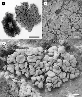
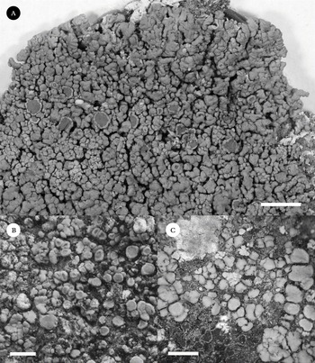
Placomoronea species. A, P. candelarioides (Santesson, Tehler & Thor, P82:1, S); B, P. fuegiana (Frödén 1786, LD—holotype); C, P. kaernefeltii (Frödén 1503, LD—holotype). Scales: A=5 mm; B & C=1 mm.

(Fig. 2A)
Thallus foliose, umbilicate, monophyllous, rounded in outline, to c. 20 mm wide, lobate from the centre (central parts often obscured by numerous apothecia), lobes 0·4–1·7 mm wide and up to 7 mm long, flattened but upper surface usually slightly convex, branched 2–4 times, sometimes with small lobules, lobes occasionally strongly convex, almost round in section and imbricate with numerous branchlets or lobules forming subfruticose cushions, occasionally the thallus consists of singular lobes attached at the base to the substratum; surface dark yellow to brownish yellow, smooth, shiny, epruinose; lower surface whitish or more often greyish to brownish due to dust impregnation; thallus in section up to 350 μm thick; epicortex 1–10 μm thick; cortex 15–35 μm thick, cells 4–7 μm wide; medulla distinct, white, composed of mesodermatous hyphae; lower cortex similar to the upper cortex, 25–45 μm thick and with somewhat larger cells, up to 10 μm wide.
Apothecia sessile, often numerous and covering the central parts of the thallus, 0·5–1·6 mm diam.; thalline margin persistent, entire, smooth or becoming slightly crenulate, to 0–15 mm thick, sometimes lobulate; disc darker yellow than the thallus, flat to becoming somewhat convex, smooth or dry-cracked, shiny, epruinose. Anatomy: thalline margin with a cortex similar to that of the thallus, 15-30 μm thick; epihymenium conspicuously arranged as hoods on the paraphyses tips; hymenium 65-85 μm tall; paraphyses simple or often furcate near the tips, 1·5–2·5 μm in mid-hymenium; tips clavate to more often capitate and sometimes almost submoniliform, to 6 μm wide; asci >30-spored, 50–70 × 18–26 μm (n=20); ascospores simple (but often with two large oil-drops and then appearing two-celled), narrowly ellipsoid, (8–)9–11·5(–12·5) × 3–4 μm (n=41).
Pycnidia common, visible as darker yellow pits on the thallus; conidia narrowly ellipsoid, 3–4 × 1·5 μm.
Spot tests. K+ weakly red, KC−, C−.
Habitat and distribution. Placomaronea candelarioides grows on acidic rocks in open montane habitats. It is known from many places along the Andes from Mendoza, Argentina in the south, to Ancash, Peru in the north.
Remarks. This species is mostly easily recognized by its umbilicate, monophyllous, growth form. A few thalli seen were uncharacteristically laciniate and almost subfruticose in their appearance. One specimen here identified as P. candelarioides was composed of numerous individual, little branched and ascending lobes attached to the substratum at the base. Such deviating specimens can only be confused with P. lambii, which normally has a darker yellow, somewhat brownish tinged colour. Furthermore, P. lambii has a multi-lobed thallus attached to the substratum with many holdfasts and has very uneven lobes with a swollen, rounded appearance.
Additional specimens examined: Argentina: Jujuy: 43 km E of La Quacia, lower part of Sierra de Santa Victoria, along route 5 (dirt road to Santa Victoria), 65°18′W, 22°08′S, alpine area, on acidic rock, c. 4000 m, 1989, T. H. Nash 27949 (ASU). Mendoza: by the road between Termas Villavicencio-Uspallata (NE of Mendoza), on rocks in an open landscape, eastern slope of the mountains, 32°32.39′S, 69°01.55′W, alt. c. 2200 m, 2001, P. Frödén 1667 (LD); by the road between Termas Villavicencio-Uspallata (NE of Mendoza), c. 34 km from Uspallata, on rocks near the top of a hill in an open landscape, eastern slope of the mountain, 32°31.09′S, 69°03.10′W, alt. c. 2550 m, 2001, P. Frödén 1705, 1706, 1707 (LD); NW of Cerro Cielo, c. 17 km SW of Santa Clara (between road 412 and 40), on rocks near the top of a hill in open dry area, 32°10.04′S, 69°04.85′W, alt. c. 2800 m, 2001, P. Frödén 1720 (LD). Rio Negro: Sierra Grande near Ruta 305, 63°59′W, 42°10′S, on acidic rock, c. 80 m, 1989, T. H. Nash 26484 (ASU). Salta: 35 km N of Molinos along hillside W of Rio Calchaqui and above route 40, 66°13′W, 25°13′S, on mudstone, c. 2000 m, 1989, T. H. Nash 27886 (ASU); 14 km W of pass through Cumbres de Obispo along route 33, 65°56′W, 25°14′S, c. 3000 m, 1989, T. H. Nash 27855 (ASU). San Juan: along road 436 between Puchuzan-Iglesia, just S of Tocota (NW of San Juan), by the statue of Jesus, open hill in semidesert, 30°41.32′S, 69°26.71′W, alt. c. 2600 m, 2001, P. Frödén 1758 (LD). Tucuman: 30 km NNW of Tafi del Valle along route 307 on W side of Cumbres Calchaquies, 65°49′W, 26°40′S, grassland, on acidic rock, c. 2800 m, 1989, T. H. Nash 28034 (ASU).—Bolivia: Potosi: 35 km from Potosi on road to Sucre, on rock, 1963, D. & V. Ugent (MIN). La Paz: Ruins of Tiwanaku, about 40 miles from La Paz on road to Desagradero, 1963, D. & V. Ugent (MIN).—Chile: Region 1. [Tarapacá]: Arica, close to the entrance of P.N Lauca, on a large boulder by the road, 18°09.7′S, 69°28.4′W, alt. 4400 m, 2001, P. Frödén 1504 (LD).—Peru: Ancash: Prov. Huaraz, road Huaraz-Casma, c. 26 km (road distance) WSW of Huaraz, alt. c. 3900 m, 9°33′S, 77°37′W, 1981, Santesson & Moberg P58:1 (S); road Huaraz-Casma, c. 10 km (road distance) SW of Huaraz, alt. c. 3500 m, 9°32′S, 77°35′W, 1981, Santesson & Moberg P59:1 (S). Cuzco: Prov. Urubamba, c. 4 km SE of Maras (WSW of Urubamba), alt. c. 3400 m, 13°18′S, 72°10′W, 1981, Santesson, Tehler & Thor P82:1 (S); Prov. Urubamba, 1–2 km E of Ollantaytambo, alt. c. 2800 m, 13°12′S, 72°18′W, 1981, Santesson, Tehler & Thor P83:6, P94:5 (S); Prov. Urubamba, Ollantaytambo, 2800 m, 13°15′S, 72°16′W, 2003, A. Thell (LD). Junin: Huancayo, Torre-Torre[?], 1960, G. Kunkel 3890 (HBG). Puno: 8 km from Puno on road to Juliaca, 1963, D. & V. Ugent (MIN). Lima: Huarochiri, valley of Rio Santa Eulalia, NE of Carampoma, alt. c. 3400 m, 11°39′S, 76°28′W, 1981, Santesson & Moberg P23:1 (S).
Placomaronea fuegiana M. Westb. & Frödén sp. nov
Thallus saxicola, areolatus, flavus. Areolae convexae, squamulascentes. Apothecia lecanorina, 0·4–1·5 mm lata, sessilia, disco plano vel convexescente. Hymenium 90–100 μm altum, in parte epihymeniale crystallis fulvis – rutilis inspersis. Paraphyses 1·5–2·0 crassae, simplices vel raro in parte epihymeniale furcatae, apicibus clavatis, ad 4 μm latis. Asci clavati, >30-spori. Ascosporae hyalinae, simplices, anguste ellipsoideae, (9–)9·5–13(–14) × (3·0–)3·5–4·0 μm.
Typus: Argentina, Tierra del Fuego, just after the border crossing from San Sebastian (Chile), on sandstone. 53°18.9′S, 68°34.09′W, 5 February 2001, P. Frödén 1786 (LD—holotypus).
(Fig. 2B)
Thallus crustose, composed of scattered to crowded areoles, areoles first beginning as small granules developing into rounded, bullate areoles, as they grow larger they become somewhat irregular in outline and slightly incised, up to 0·6 mm wide, the largest finally almost lobate but not raised from the substratum; surface, yellow, smooth and somewhat shiny, epruinose; epicortex 0–4 μm, cortex 15–25 μm thick, cells c. 4–8 μm wide; medulla indistinct, algae distributed throughout the interior of the thallus; lower cortex lacking.
Apothecia numerous, first immersed in the thallus, emerging through cracks in the thallus, becoming sessile, 0·4–1·5 mm diam., thalline margin entire, somewhat uneven, to 0·10 mm thick, in later stages often obscured by the disc; disc at first flat, becoming convex, slightly darker yellow than the thallus, smooth to somewhat coarse and faintly pruinose; thalline margin with a cortex similar to that of the thallus, 10–25 μm thick; epihymenium not distinctly arranged as hoods on the paraphyses tips; hymenium 90–100 μm tall; paraphyses simple or occasionally branched near the tips, 1·5–2·0 μm in midhymenium, tips clavate, to 4 μm wide; asci >30-spored, 58–70 × 22–25 μm (n=6); ascospores simple, narrowly ellipsoid, (9–)9·5–13(–14) × (3·0–)3·5–4·0 μm (n=37).
Pycnidia sparse, visible as orange, shallow pits on the thallus; conidia narrowly ellipsoid, 3–4 × 1·5 μm.
Spot tests. K− or faintly reddish, KC−, C−.
Etymology. This species is named fuegiana referring to its type locality in Tierra del Fuego, Argentina.
Habitat and distribution. Placomaronea fuegiana is known from two localities in south-eastern South America. In the type locality in north-east Tierra del Fuego, it grows on very soft sandstone in an open, lowland habitat. The second locality is on the mainland, in Parque Nacional Pali-Aike, about 150 km from the type locality. There it grows mainly in the small pits of volcanic rocks and on mosses and debris. It is likely to have a more extensive distribution in south-eastern South America.
Remarks. The numerous, convex apothecia, which often cover the mainly bullate, areolate thallus gives the species a characteristic appearance. Anatomically it is separated from P. mendozae by its less branched paraphyses with clavate tips. Anatomically it is not distinct from P. minima but its general appearance is different from this species. The ITS sequence data shows that it is more closely related to P. kaernefeltii than to P. minima.
Additional specimens examined. Argentina: Tierra del Fuego: just after the border crossing from San Sebastian (Chile), on sandstone, 53°18.9′S, 68°34.09′W, 2001, P. Frödén 1785, 1787, 1788 (filed under Caloplaca sp.) (LD).—Chile: Reg. XII [Magallanes]: P. N. Pali-Aike, on rocks, 52°06`85"S 69°42`78"W, 2001, P. Frödén 1875 (LD).
Placomaronea kaernefeltii M. Westb., Frödén & Wedin sp. nov
Thallus saxicola, lobatus, flavus. Lobae ad 1·5 mm longae, rosulas formantes. Apothecia lecanorina, 0·8–1·7 mm lata, sessilia, disco plano. Hymenium 90–105 μm altum, in parte epihymeniale crystallis fulvis – rutilis inspersis. Paraphyses 1·5–2·0 crassae, simplices vel raro in parte epihymeniale furcatae, apicibus cylindricis vel clavatis, ad 4·5 μm latis. Asci clavati, >30-spori. Ascosporae hyalinae, simplices, anguste ellipsoideae – fere oblongae, (10–)11–14 × 3·5–4·0(–4·5) μm.
Type: Chile, Region 1. [Tarapacá], Arica, between Socorome and Putre, on a large boulder, 18°35.98′ S, 69°32.70′ W, alt. 3750 m, 3 January 2001, P. Frödén 1503 (LD—holotypus).
(Fig. 2C)
Thallus crustose to squamulose to effigurate with raised marginal lobes, beginning as bullate areoles soon becoming lobate and raised from the substratum, attached to the substratum at the base, moderately elongated, up to 1·5 mm long and to 1·0 mm wide and, at most, twice as long as wide, growing scattered to crowded and forming rosette-like thalli; surface bright yellow, smooth, dull, epruinose; lower surface whitish or often brownish due to dust impregnation; epicortex lacking, cortical pigments forming hoods on the outermost cortex cells; cortex 15–45 μm thick, cells 3–8 μm wide; medulla not distinct, algae filling the interior of the thallus; lower cortex similar to the upper cortex, 20–35 μm thick.
Apothecia 0·8–1·7 mm diam., disc flat, concolorous with the thallus, smooth, faintly pruinose; margin entire, smooth to becoming somewhat uneven, to c. 0·2 mm thick, not distinctly raised above the disc. Anatomy: thalline margin with a cortex similar to that of the thallus, 45–50 μm thick; epihymenium not arranged as hoods on the paraphyses tips; hymenium 90–105 μm tall; paraphyses simple or sometimes branched near the tips, tips cylindrical to clavate, up to 4·5 μm wide; asci >30-spored, 63–70 × 20–22 μm wide (n=3); ascospores simple, narrowly ellipsoid to almost oblong, (10–)11–14 × 3·5–4·0(–4·5) μm (n=25).
Pycnidia not seen.
Spot tests. K+ orange, KC−, C−.
Etymology. This species is named after Professor Ingvar Kärnefelt in recognition of more than 30 years of his contributions to lichenology and in gratitude to all the inspiration and continuous support we have received from him through the years.
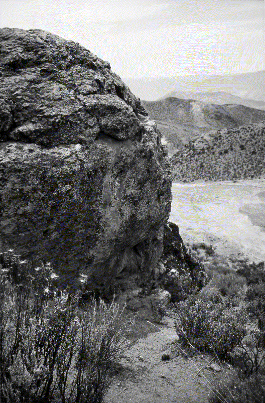
Habitat and distribution. The type collection from Chile is so far the only specimen known. However, the distribution is likely to be more extensive as the collection site was one of only three short stops at high altitudes in the area. Due to altitude sickness and a hail storm at even higher altitudes, this one-day visit to the mountains east of Arica had to be shortened considerably. It was found on a siliceous boulder in an open, dry mountain slope (Fig. 3), where it grew abundantly, mainly along small cracks and pits in the rock.
The type locality of P. kaernefeltii near Arica, Chile, where it grows abundantly along cracks and crevices on the large boulder in the photograph.

Remarks. A large species that when well developed approaches the larger species P. candelarioides and P. lambii in appearance but there are many differences. The lobes are shorter and not as elongated in P. kaernefeltii. Furthermore, the thallus in P. kaernefeltii has a brighter yellow colour, a dull surface and is not stratified, i.e., a distinct medulla is not developed. In addition, it has a taller hymenium and larger spores.
Placomaronea lambii (Hakul.) R. Sant
in Poelt, Phyton 16: 205 (1974).—Candelariella lambii Hakul. Ann. Bot. Soc. Zool.-Bot. Fenn. ‘Vanamo’ 27(3): 36 (1954); type: Argentina, Tucumén [Tucumán], Valle de Tafi, West slope of Cumbre Potrerillo, on the top of a block of schistose rock in alpine pasture, alt. 3100 m, 24 November 1947, I. M. Lamb 5413 (CANL—holotypus!, TUR-Hakulinen—isotypus!).
Placmoronea species. A, P. lambii (Lamb 5413, CANL—holotype); B, P. mendozae (Frödén 1702, LD); C, P. minima (Frödén 1656, LD—holotype). Scales: A=5 mm; B & C=1 mm.

(Fig. 4A)
Thallus rosette-like, composed of numerous lobes together forming a wide-spread multi-lobed thallus attached to the substratum with many holdfasts, lobes up to 4 mm long and to 0·75 mm wide, central lobes shorter, erect or ascending and somewhat imbricate, peripheral lobes raised from the substratum forming a rosette-like margin; surface brownish yellow, dull, epruinose; epicortex c. 1–4 μm thick, cortex 30–55 μm thick (7–12 μm in one specimen), cells 3–6 μm wide; medulla white, composed of mesodermatous hyphae; lower cortex 20–40 μm thick, similar to the upper cortex but with slightly larger cells, up to 8 μm wide and often somewhat elongated.
Apothecia 0·65 – 2·0 mm wide, disc darker yellow than the thallus, flat, smooth, epruinose, margin smooth, entire to crenulate, raised and often bent inwards over the disc, to 0·30 mm wid. Anatomy: thalline margin with a cortex similar to that of the thallus, 30–40 μm thick; epihymenium arranged as hoods on the paraphyses tips; hymenium 65–95 μm tall; paraphyses simple or branched near the tips, 1·5–2·0 μm in midhymenium, tips clavate to almost submoniliform, to 5·5 μm wide; asci >30-spored, 54–74(–80)× 18–23 μm (n=10), ascospores, narrowly ellipsoid to almost oblong, (8·5–)9·0–12·0 × 3·0–4·5 μm wide (n=37).
Pycnidia common, visible as darker yellow or somewhat brownish pits or shallow warts on the thallus; conidia narrowly ellipsoid, 3–4·5 × 1·0–1·5 μm.
Spot tests. K+ reddish, KC−, C−
Habitat and distribution. Placomaronea lambii grows at high altitude on acidic rocks in open habitats. It is known only from four localities in Peru and Ecuador. Hakulinen (Reference Hakulinen1958) reported this species from Uzbekistan from a sterile specimen but this is likely to be an erroneous record, the specimen probably belongs to Candelariella kansuensis H. Magn.
Remarks. Anatomically this species is close to P. candelarioides but the two species differ in their growth form; the thallus of P. lambii is composed of numerous lobes forming a large conglomerate thallus with many holdfasts to the substratum in contrast to the normally monophyllous, umbilicate thallus of P. candelarioides. Although occasional forms of P. candelarioides can cause some confusion as to their identity and, although we lack molecular data for P. lambii, we are convinced that it is a distinct species, which is generally easily separated from P. candelarioides. Placomoronea lambii differs from P. kaernefeltii in many characters. Placomaronea lambii has much longer than broad lobes, a lower hymenium, smaller spores and the thallus is also distinctly stratified with a well-developed, white medulla whereas P. kaernefeltii lacks a distinct medulla and the alga fills the thallus interior in the same way as in the smaller species of the genus. A deviating specimen from Ecuador with a very thin cortex (7–12 μm), but otherwise with similar anatomical characters, is here tentatively identified as P. lambii. This specimen is at a first glance more similar to Candelina submexicana, but this genus has 8-spored asci and a different cortex anatomy (Poelt Reference Poelt1974; Westberg & Nash 2000).
Additional specimens examined. Ecuador: Prov. Chimborazo, southern slope of Mt Chimborazo, on stone, 1939, Asplund 245 (S).—Peru: Ancash: Prov. Huaraz, road Huaraz-Casma, c. 31 km (road distance) WSW of Huaraz, alt. c. 4100 m, 9°33′S, 77°41′W, 1981, Santesson & Moberg P56:4, P64:31 (S). Lima: Prov. Huarochiri, valley of Rio Santa Eulalia, NE of Carampoma, alt. c. 3700 m, 11°38′S, 76°27′W, 1981, Santesson & Moberg P24:1 (S). Puno: on road from Puno to Arequipa, at km 131, 12 km SW Tincopalca (E of Paso de Condor), open grasslands with cliffs, 14500 ft, (c. 4400 m), 1963, H. H. & C. M. Iltis 2443 (MIN).
Placomaronea mendozae (Räsänen) M. Westb
Sonoran Lichen Flora Vol. 2: 419 (2004).—Candelariella vitellina var. mendozae Räsänen Anales Soc. Ci. Argent. 131: 100 (1941); type: Argentina, Mendoza, Las Heras, ad rupes multis locis sed non copiose, 18 July 1939, A. Ruiz Leal 5970 (H—lectotypus! here designated).
(Fig. 4B)
Thallus crustose, areolate to minutely peltate squamulose, areoles scattered to somewhat crowded, often spreading along small crevices in the substratum, soon raised from the substratum at the edge, forming small squamules, squamules flattened, somewhat incised, up to 1·2 mm long; surface yellow to brownish yellow, smooth, shiny, epruinose; epicortex 1–8 μm; cortex 10–25 μm thick, cells 3–9 μm wide; medulla not developed, algae filling the interior of the thallus; lower cortex only present as a continuation of the upper cortex near the margin of raised squamules.
Apothecia becoming sessile, 0·4 –0·8(–1·5) mm diam., thalline margin persistent, entire, smooth to uneven and weakly crenulate, to 0·15 mm thick, often rather uneven, level with the disc or somewhat raised, occasionally lobulate; disc flat, darker yellow than the thallus, smooth or dry-cracked, shiny, epruinose. Anatomy: thalline margin similar to that of the thallus, cortex to 40 μm thick; epihymenium not or sometimes indistinctly arranged as hoods on the paraphyses tips; hymenium 70–95 μm tall; paraphyses 1·5–2·5 μm in midhymenium, frequently furcate near the tips, tips clavate to almost capitate to 5 μm, often almost submoniliform; asci >30-spored, (50–)55–78 × (13–)17–22 μm (n=7);ascospores narrowly ellipsoid, simple, (8–)9·0–11·5(–12·5) × (2·5–)3·0–4·0(–4·5) μm (n=57).
Pycnidia common, immersed, visible as darker yellow pits on the thallus; conidia ellipsoid to narrowly ellipsoid, 2·5–4·0 × 1·5 μm.
Spot tests. K+ reddish, KC−, C−.
Habitat and distribution. Placomaronea mendozae grows on sun-exposed, siliceous rocks in open montane landscapes up to at least 3900 m. In addition to its localities along the Andes in Argentina and Peru, this species is also known from Arizona, USA and so far is the only species found in North America.
Remarks. This small species is perhaps morphologically more similar to a Candelariella, for example, C. vitellina (Hoffm.) Müll. Arg., than to the much larger P. candelarioides and P. lambii, the two species previously included in that genus. It was identified as a Placomaronea due to the unusual cortex anatomy (Westberg Reference Westberg, Nash, Ryan, Diederich, Gries and Bungartz2004). Its close relationship to P. candelarioides was confirmed by Westberg et al. (Reference Westberg, Arup and Kärnefelt2007) and it is reconfirmed to belong to the Placomaronea clade in this study (Fig. 1). The material described and informally named as P. lambii ‘f. microthallina’ by Poelt (Reference Poelt1974) belongs to this species. It is similar in appearance to P. minima but differs, for example, in the more branched paraphyses with more swollen tips. In addition it has a lower hymenium and can also be distinguished by the smooth apothecial disc which in P. minima shows a distinct pruina formed by the epihymenial pigments. There is a fairly large variation in the ITS sequences between the specimens in the phylogenetic analysis but with the current material at hand there is little data to support distinguishing more than one species. The specimens from Arizona however, are rather small with somewhat smaller asci and narrower spores, which could indicate that a future taxonomic revision may recognize more than one species.
Additional specimens. Argentina: Jujuy: Dept. Santa Catalina, Laguna Colorada, in rupibus ripae, alt. 3900 m, 1901, Fries 101 (S). Mendoza: near Mt Aconcagua, Puente del Incas, 16 ii 1903, Malme (S); by the road between Termas Villavicencio-Uspallata (NE of Mendoza), c. 34 km from Uspallata, on rocks in an open landscape, eastern slope of the mountain, 32°31.09′S, 69°03.10′W, alt. c. 2550 m, 2001, Frödén 1702 (LD). San Juan: along road 436 between Talacasto-Iglesia, c. 60 km NW of Talacasto, S of Huilian. 30°45.38′S, 68°58.06′W. alt. c. 1750 m, 2001, Frödén 1754 (LD); along road 436 between Puchuzan-Iglesia, just S of Tocota (NW of San Juan), by the statue of Jesus, open hill in semidesert, 30°41.32′S, 69°26.71′W, alt. c. 2600 m, 2001, Frödén 1757 (LD).—Peru: Ayacucho: Distr. Lencio-Prado, c. 32 km along the road between Nazca-Puquio, on exposed rocks in open pasture land, 2001, Frödén 1523 (LD).—USA: Arizona: Coconino Co., San Francisco Peaks. Talus slope (NE-facing) at timberline (Pinus aristata), SW above crossing of Abineau Trail and old secondary road, 35°21′N, 111°41′W, alt. 3300–3400 m, on volcanic rocks, 1998, M. Westberg 833 (LD); Coconino Co., above timberline on the N to NE slope on Mount Agassiz in the San Fransisco Peaks, 35°19′N, 111°40′W, on volcanic rock, 3540 m, 1988, T. H. Nash 25430 (ASU).
Placomaronea minima M. Westb. & Frödén sp. nov
Thallus saxicola, areolatus, flavus. Areolae minutae, planae, squamulascentes, ad 1 mm longae. Apothecia lecanorina, 0·2–0·6(–1·3) mm lata, sessilia, disco plano, pruinoso. Hymenium 90–120 μm altum, in parte epihymeniale crystallis fulvis – rutilis inspersis. Paraphyses 1·5–2·0 crassae, simplices vel raro in parte epihymeniale furcatae, apicibus cylindricis vel clavatis, ad 4 μm latis. Asci clavati, >30-spori. Ascosporae hyalinae, simplices, anguste ellipsoideae, (8–)8·5–11(–12) × 3·0–4·0(–5·0) μm.
Type: Chile, Region Metropolitana (Santiago), by the road from San José de Maipo to La Gunillas, on rocks in a hill just outside San José de Maipo, 33°39.64′S, 70°20.87′W, alt. c. 1200 m, 20 January 2001, P. Frödén 1656 (LD—holotypus).
(Fig. 4C)
Thallus crustose, composed of individual areoles forming a spreading crust, areoles along the margin subsquamulose, becoming minutely lobate and raised from the substratum, up to c. 1 mm long; surface yellow, smooth, shiny, epruinose; epicortex 2–5 μm; cortex 10–35 μm thick, cells 3–9 μm wide; medulla not developed, algae filling the interior of the thallus; lower cortex only present as a continuation of the upper cortex near the margin of raised squamules.
Apothecia 0·2–0·6(–1·3) mm diam., disc flat, darker yellow than the thallus, smooth, pruinose (pruina formed by the granular epihymenial pigments); thalline margin persistent, entire, somewhat uneven, distinctly raised above the disc, up to 0·15 mm thick. Anatomy: thalline margin with a cortex similar to that of the thallus, 15–25 μm thick; epihymenium not arranged as hoods on the paraphyses tips; hymenium 90–120 μm tall; paraphyses simple or sometimes branched near the tips, 1·5–2·0 μm in mid-hymenium, tips cylindrical to clavate, to 4 μm wide; asci >30 spored, 52–80 × 18–30 μm (n=9); ascospores colourless, simple (rarely 1-septate), narrowly ellipsoid, (8–)8·5–11(–12) × 3·0–4·0(–5·0) μm (n=26).
Pycnidia numerous, visible as small orange warts on the thallus; conidia narrowly ellipsoid 2·5–4·0 × 1·5 μm.
Spot tests. K− to K+ reddish, KC−, C−.
Etymology. Minima refers to the small size of this species.
Habitat and distribution. This species has a disjunct distribution, occurring in Chile and Argentina and in southern Africa. In the type locality in central Chile it grows on rocks in a fairly dry, semi-open hill slope.
Remarks. Placomaronea minima is similar in size and appearance to P. mendozae but can be distinguished by its simple paraphyses with cylindrical to clavate tips and usually the taller hymenium. In P. mendozae the paraphyses are frequently branched near the tips which are distinctly swollen and often submoniliform. Furthermore, the epihymenial pigments form a granular pruina on the apothecium disc in P. minima disc while in P. mendozae the disc is shiny and epruinose.
Additional specimens examined. Argentina: Mendoza: by the road between Termas Villavicencio-Uspallata (NE of Mendoza), c. 34 km from Uspallata, on rocks in an open landscape, eastern slope of the mountain, 32°31.09′S, 69°03.10′W, alt. c. 2550 m, 2001, Frödén 1702 (LD, filed under P. mendozae).—Chile. Region Metropolitana (Santiago): by the road from San José de Maipo to La Gunillas, on rocks on a hill just outside San José de Maipo, 33°39.64′S, 70°20.87′W, alt. c. 1200 m, 2001, Frödén 1657 (LD, filed under Caloplaca sp.).—Lesotho: summit of Masite Mountain, 6300 ft., 1929-30, Dr & Mrs Hewitt (LD, 2 specimens).
Discussion
There are some differences between the current analysis and the previous treatment in Westberg et al. (Reference Westberg, Arup and Kärnefelt2007), most notably the phylogenetic position of Candelariella vitellina, the type species of Candelariella. In this paper C. vitellina groups together with Candelaria concolor (Dicks.) Stein, although there is low support for this clade. This is interesting since both species have polysporous asci and the evolution of polyspory in Candelariales apparently occurred on a limited number of occasions (see also Westberg et al. Reference Westberg, Arup and Kärnefelt2007). We do not, however, wish to draw any conclusions from this result since the topology of the tree has proved to be highly sensitive to the choice of taxa included in the analysis. As previously stated (Westberg et al. Reference Westberg, Arup and Kärnefelt2007), ITS sequence data are clearly insufficient to resolve the phylogeny and to investigate the relationship between different species groups within Candelariales. Terminal groups including closely related species do, however, appear with strong support in the tree and Placomaronea is one such terminal group. This monophyly had previously been suggested by Westberg et al. (Reference Westberg, Arup and Kärnefelt2007), although only two of the species were included in that study.
Morphologically Placomaronea is characterized by its well-developed pseudoparenchymatous cortex and the pigments which form characteristic hoods on the outermost cortical cells and usually also by the presence of a thin, gelatinous epicortex. However, a similar cortex anatomy is also known in Candelariella kansuensis, a species that does not appear to be the closest relative to Placomaronea, and which only differs in having 8-spored asci (Westberg Reference Westberg2007c). Originally Placomaronea was characterized by its umbilicate, foliose growth form and separated from Candelariella by its ‘higher’ thallus organization, but only two species, P. candelarioides and P. lambii, have a stratified thallus with a distinctly developed medulla. The remaining species have a thallus similar to Candelariella, i.e., the alga is more or less evenly distributed throughout the interior of the thallus. Also in growth form they are similar to many Candelariella species in having an areolate to minutely peltate thallus. Nor have we found any significant differences in the anatomy of the apothecia and the pycnidia, and the conidia are similar to the rest of the species in Candelariales. The distinction between Placomaronea and Candelariella thus has to be based on the combination of morphological characters, i.e., cortex anatomy and polysporous asci. Nevertheless, without further data from other DNA markers a revision of the generic boundaries in the family is not possible and the new species proposed in this paper are with the current knowledge best placed in Placomaronea. Additional data from other DNA markers will be necessary together with a better sampling of the morphological variation, before a generic revision within Candelariales is possible. Further taxonomic revisions of South American material will without doubt contribute important information to understand the evolution and phylogeny within the Candelariales.
The study was supported by The Swedish Taxonomy Initiative (Svenska Artprojektet), administered by the Swedish Species Information Centre (ArtDatabanken). Additional support was granted from The SwedishResearch Council (VR 621-2006-3760). The field work by PF was financed by Lars Hiertas minnesfond, Per Westlings minnesfond, Stiftelsen Ekedahl-Lundbergska fonden, Stiftelsen Dagny, Eilert Ekvalls premie- och stipendiefond, and Ove Almborns fond. Gratitude is expressed to the curators of the different herbaria mentioned for loan of specimens and to Bodil Cronholm at MSL at the Natural History Museum, Stockholm, for valuable assistance during the laboratory work. We thank Mohammad Sohrabi who kindly provided photographs of type material at H and Torbjörn Tyler for checking the Latin diagnoses. Finally two anonymous referees are thanked for comments on the manuscript.