Background
The anomalies of the systemic venous connection are very rare cardiac anomalies. The most common is persistent left superior caval vein which is a normal variant in most situations.Reference Irwin, Greaves and Schmitt1 Biatrial drainage of the right superior caval vein (RSVC) with preferential streaming into the left atrium (LA) is an extremely rare variant, with approximately 10 cases found in our literature review.Reference Van Praagh, Geva, Lock, Del Nido, Vance and Van Praagh2,Reference Alghamdi, Elfaki, Al-Habshan and Aljarallah3 The most common presentation is cyanosis with normal clinical findings in the childhood.
Case report
A 5-month-old male presented for evaluation of cyanosis. He had been in his usual state of health until 1 month earlier when he presented to a local emergency room on two separate occasions due to symptoms of cough and rhinorrhoea. Additionally, he was noted to have an oxygen saturation (SpO2) of 87–89% on room air, which improved after oxygen administration. Finding of a murmur prompted an echocardiogram which demonstrated superior sinus venosus atrial septal defect (ASD) and biatrial drainage of the RSVC with preferential streaming into the LA. The pulmonary venous drainage was not completely delineated.
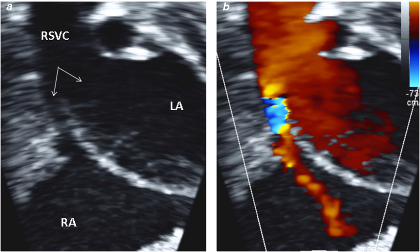
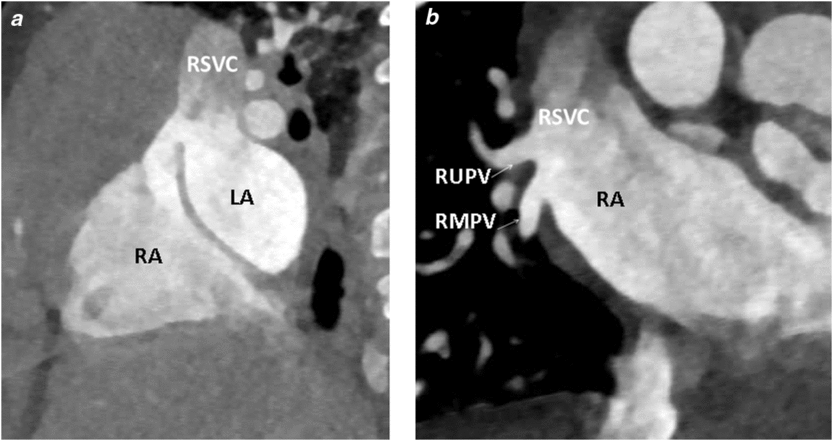
Physical examination was significant for a grade I/VI systolic ejection murmur at the left sternal border and mild peri-oral cyanosis (SpO2 = 79% by pulse oximetry). Electrocardiogram showed sinus rhythm with left axis deviation, prominent Q waves, and minimal ST elevation in lateral leads. Chest X-ray showed non-specific minor patchy atelectasis at the lung bases with mild cardiomegaly and normal pulmonary vascular markings. A transthoracic echocardiography showed normal segmental anatomy with a superior sinus venosus ASD. The RSVC was found straddling the atrial septum with flow predominantly draining into the LA (Fig 1). The LA was mildly enlarged. A gentle agitated saline injection confirmed the drainage pattern. To better assess the pulmonary veins and confirm the diagnosis, a contrast-enhanced CT was performed. This depicted the predominant drainage of the RSVC to the LA (Fig 2). The pulmonary venous drainage was anomalous with the right upper and middle pulmonary veins draining into the RSVC. The right lower and the left pulmonary veins drained into the LA. The patient underwent corrective cardiac surgery which consisted of closure of the ASD with rerouting of the anomalous pulmonary veins to the LA, transection of the RSVC and re-implantation into the right atrial appendage (Warden procedure). The post-operative course was uneventful. Post-operative echocardiography demonstrated no residual ASD, RSVC draining to the right atrium (RA) without obstruction and no pulmonary venous stenosis.
Transthoracic echocardiography showing the RSVC overriding the atrial septum with preferential flow into the LA. (a) 2D image without colour; (b) with colour, arrows denote the orifice of the RSVC into LA and RA. RSVC = right superior caval vein; LA = left atrium; RA = right atrium.

CT images. (a) showing RSVC draining predominantly into the LA with superior sinus venosus ASD. (b) Anomalous drainage of RUPV and RMPV to the RSVC. RSVC = right superior caval vein; LA = left atrium; RA = right atrium; ASD = atrial septal defect. RUPV = right upper pulmonary vein; RMPV = right middle pulmonary vein.

Discussion
Isolated drainage of the RSVC to the LA or biatrial drainage of the RSVC is a rare anomaly of the systemic venous connection. In review of the literature, we found 10 reported cases of biatrial drainage of RSVC published by Van Praagh and another case was reported by Alghamdi.Reference Van Praagh, Geva, Lock, Del Nido, Vance and Van Praagh2,Reference Alghamdi, Elfaki, Al-Habshan and Aljarallah3 Our case describes biatrial drainage of the RSVC with preferential flow into the LA in the presence of sinus venosus ASD and partial anomalous pulmonary venous return. Most of the reported cases in the literature were identified in association with persistent left superior caval vein, sinus venosus ASD, partial anomalous drainage of the right upper pulmonary veins, tetralogy of Fallot, pulmonary atresia, transposition of great vessels, and patent ductus arteriosus.Reference Van Praagh, Geva, Lock, Del Nido, Vance and Van Praagh2 The most common presentation was cyanosis with normal clinical findings in the childhood period. Symptoms of dyspnoea, fatigue, effort intolerance, cerebral emboli, or abscess were also described in adults.Reference Shapiro, Al-Sadir, Campbell, Thilenius, Anagnostopoulos and Hays4 The drainage of the RSVC to the LA leads to right-to-left shunting that can cause cyanosis and left ventricular overload. Our patient had preferential flow to the LA with smaller right atrial orifice causing significant right-to-left shunt.
Van Praagh proposed that sinus venosus defects appear to be secondary to a defect in the wall shared between the anterior wall of right upper pulmonary vein and the posterior wall of the superior caval vein.Reference Van Praagh, Geva, Lock, Del Nido, Vance and Van Praagh2 The absence of this wall unroofs the right pulmonary veins, resulting in overriding of the atrial septum by the superior caval vein and anomalous drainage of right upper pulmonary vein to the right side of the heart.Reference Van Praagh, Geva, Lock, Del Nido, Vance and Van Praagh2 Kirsch and associates postulated that drainage of the RSVC to the LA is likely caused by malposition of the right horn of the sinus venosus during fetal life.Reference Kirsch, Carlsson and Hartmann5 Another theory by Braudo et al suggested that the cephalic portion of the right valve of the sinus venosus fuses with the atrial septum superior to the coronary sinus inlet, forming a seal that prevents the superior caval vein from draining into the RA.Reference Braudo, Beanlands and Trusler6
The prevalence of sinus venosus ASD varies from 5 to 10% of patients with ASDs.Reference Webb and Gatzoulis7 Approximately 90% of patients with a superior sinus venosus defect have anomalous right pulmonary venous drainage, with right upper pulmonary vein being the most common anomalous connection.
Sinus venosus defect and anomalous venous connection are often difficult to diagnose with transthoracic echocardiography alone. Trans-oesophageal echocardiography is considered a sensitive and cost-effective imaging modality.Reference Pascoe, Oh, Warnes, Danielson, Tajik and Seward8,Reference Oliver, Gallego, Gonzalez, Dominguez, Aroca and Mesa9 Cardiac MRI and CT are very useful in confirming the diagnosis and assessment of anomalous pulmonary venous drainage and their precise point of drainage. Cardiac MRI can also help with quantifying Qp: Qs prior to surgical correction.
Conclusion
In summary, our case highlights the importance of recognising this rare anomaly of the systemic venous connection as a possible cause of cyanosis in children. Also, importantly, this was not detected by newborn screening for critical CHD. Persistent hypoxia which is out of proportion to the respiratory illness should raise the index of suspicion to evaluate for a right-to-left shunt. These lesions can be repaired completely successfully and safely once the diagnosis is made and the anatomy is clear.
Author ORCIDs
Omar Abu Anza, 0000-0003-2587-1139
Acknowledgements
None.
Financial Support
This research received no specific grant from any funding agency, or from commercial or not-for-profit sectors.
Conflict of Interest
None.