Introduction
Anisakid nematodes of the genera Anisakis Dujardin, 1845, Pseudoterranova Krabbe, 1878 and Contracaecum Railliet & Henry, 1913 (herein only considering species maturing in pinnipeds) are parasites of the alimentary tract of aquatic vertebrates, with indirect life cycles that involve various hosts at different levels across the food webs. In general, marine mammals (cetaceans and pinnipeds) serve as definitive hosts, fish and squid serve as intermediate or paratenic hosts, while planktonic or semi-planktonic crustaceans act as first intermediate hosts (fig. 1) (Mattiucci & Nascetti, Reference Mattiucci and Nascetti2008).
Schematic life cycle of species of the genera Anisakis, Pseudoterranova and Contracaecum occurring in NE Atlantic waters. Some of the main definitive (marine mammals) and intermediate/paratenic hosts (fish) of the actual anisakid species are shown, including the role of humans as accidental hosts.

Any biodiversity assessment of anisakid nematodes is incomplete if inferred only from morphology (Mattiucci & Nascetti, Reference Mattiucci and Nascetti2008), although morphological analyses and older (historical) considerations often share a considerable degree of congruence with modern molecular-based taxonomic assessments (Mattiucci et al., Reference Cipriani, Smaldone, Acerra, D'Angelo, Anastasio, Bellisario, Palma, Nascetti and Mattiucci2014). This highlights the importance of proper identification and delimitation of biological species within anisakid nematodes inferred from (a multilocus) molecular approach, as a prerequisite for precise assessment of biodiversity. This, in turn, provides the basis for studying patterns and processes in the evolution and ecology, including biogeography, host preference and co-evolution, of the parasites. Moreover, a more extensive investigation of the genetic diversity in anisakids facilitates our understanding of their temporal and spatial distribution, which is also related to demographic changes of hosts and food webs. It has recently been suggested that data on anisakid abundance and genetic variability could be used for monitoring the status of various marine trophic webs (Mattiucci & Nascetti, Reference Mattiucci and Nascetti2008; Zarlenga et al., Reference Zarlenga, Hoberg, Rosenthal, Mattiucci and Nascetti2014; Mattiucci et al., Reference Mattiucci, Cipriani, Paoletti, Nardi, Santoro, Bellisario and Nascetti2015). Thus, integrating molecular systematics of anisakid nematodes with ecological data allows us to assess properly their biodiversity on a global scale, and in space and time (Mattiucci et al., Reference Mattiucci, Cipriani, Paoletti, Nardi, Santoro, Bellisario and Nascetti2015). Recent findings suggest that environmental changes due to anthropogenic activities represent one of these influencing factors (Mattiucci & Nascetti, Reference Mattiucci and Nascetti2008; Zarlenga et al., Reference Zarlenga, Hoberg, Rosenthal, Mattiucci and Nascetti2014; Mattiucci et al., Reference Mattiucci, Cipriani, Paoletti, Nardi, Santoro, Bellisario and Nascetti2015).
The aim of this review was to provide an inventory of the biodiversity of anisakid species of the genera Anisakis, Pseudoterranova and Contracaecum identified so far in the NE Atlantic Ocean, with emphasis on: (1) taxa recognized as ‘biological species’ based on molecular/genetic markers; (2) current molecular/genetic approaches to identify the species at different developmental stages; (3) ecological data related to the actual geographical distribution and definitive host preferences of the species; (4) their distribution in various, commercially important fish species in northern European waters; (5) their possible occurrence in farmed fish; and finally, (6) an update of their zoonotic potential as causative agents of anisakidosis in humans.
Genetic/molecular markers to identify anisakid nematodes
Specific identification of anisakid nematodes based on molecular/genetic markers also represents a valuable tool for assessment of their biodiversity. Thus, unambiguous identification of potentially zoonotic anisakids is an essential requirement for properly designing and conducting epidemiological surveys of these parasites.
Valid nuclear and mitochondrial molecular markers, which have been developed over the past two decades, have allowed the disclosure of a number of cryptic or sibling species in different anisakid groups. The discovery and characterization of cryptic and sibling species represents one the most challenging aspects in marine parasitological studies. According to Nadler & Peréz-Ponce de León (Reference Nadler and Pérez-Ponce de León2011), detection of cryptic biodiversity should start with observations of: (1) variability in morphological characters in nominal species; and (2) host range, often including species of different ecology and belonging to different families. In further steps, the above aspects should be integrated with molecular/genetic analyses in order to evaluate if a given nominal species should be considered a complex of several ‘biological species’.
The biological species concept (BSC) (Mayr, Reference Mayr1963) has regained importance through the use of nuclear genetic markers which, in the case of anisakid nematodes, enabled us to demonstrate the existence of reproductive isolation between various sympatric and allopatric populations (Mattiucci & Nascetti, Reference Mattiucci and Nascetti2008). The concept was first supported by applying allozyme markers in certain anisakid morphospecies within the genera Anisakis, Pseudoterranova and Contracaecum (Mattiucci & Nascetti, Reference Mattiucci and Nascetti2008; Mattiucci et al., Reference Cipriani, Smaldone, Acerra, D'Angelo, Anastasio, Bellisario, Palma, Nascetti and Mattiucci2014). Reproductive isolation and absence of gene flow between sympatric and allopatric sibling species were demonstrated by allozyme loci, based on the Hardy–Weinberg equilibrium inference, which allowed the establishment of their specific status (Nascetti et al., Reference Nascetti, Cianchi, Mattiucci, D'Amelio, Orecchia, Paggi, Brattey, Berland, Smith and Bullini1993; Mattiucci et al., Reference Mattiucci, Paoletti and Webb2009, Reference Mattiucci, Cipriani, Webb, Paoletti, Marcer, Bellisario, Gibson and Nascetti2014). Thus, based on allozyme diagnostic loci for different anisakid taxa, easy and rapid identification of large numbers of individuals can be performed. The methodology is particularly useful for identifying larvae from multiple intermediate/paratenic hosts in which two or more sibling species may occur in mixed infections. Accordingly, genetic species identification methodologies have proven very informative for answering epidemiological questions involving geographical distribution, host preference/range and life cycle of these parasites. Numerous allozyme analyses have since been applied in thousands of individual larvae, thus contributing greatly to our knowledge on the occurrence and distribution of anisakids in fish and marine mammals from the NE Atlantic Ocean.
Anisakid individuals recognized at species level by allozymes have been used to develop DNA-based approaches for species identification, including direct sequencing of nuclear and mitochondrial DNA genes (Valentini et al., Reference Valentini, Mattiucci, Bondanelli, Webb, Mignucci-Giannone, Colom-Llavina and Nascetti2006; Mattiucci et al., Reference Cipriani, Smaldone, Acerra, D'Angelo, Anastasio, Bellisario, Palma, Nascetti and Mattiucci2014, Reference Cipriani, Acerra, Bellisario, Sbaraglia, Cheleschi, Nascetti and Mattiucci2016). This again enabled us to study phylogenetic relationships between closely related anisakids, based on the evolutionary lineage concept, PSC (phylogenetic species concept).
Polymerase chain reaction (PCR)-DNA molecularly derived methodologies to elucidate the systematics of anisakid nematodes have also gained a wider application. Thus, a multigene approach, including both nuclear and mitochondrial markers, together with allozymes, permits a robust identification of the considered taxa.
The following molecular/genetic techniques are available for the identification of species of Anisakis that occur in the NE Atlantic Ocean: (1) diagnostic allozyme markers (see Mattiucci & Nascetti, Reference Mattiucci and Nascetti2008; Mattiucci et al., Reference Mattiucci, Paoletti and Webb2009; Reference Mattiucci, Cipriani, Webb, Paoletti, Marcer, Bellisario, Gibson and Nascetti2014); (2) sequence analysis of the mitochondrial cytochrome oxidase 2 gene (mtDNA cox2) (Mattiucci & Nascetti, Reference Mattiucci and Nascetti2006; Valentini et al., Reference Valentini, Mattiucci, Bondanelli, Webb, Mignucci-Giannone, Colom-Llavina and Nascetti2006; Mattiucci et al., Reference Cipriani, Smaldone, Acerra, D'Angelo, Anastasio, Bellisario, Palma, Nascetti and Mattiucci2014) and the small subunit of rRNA (rrnS) gene (Nadler et al., Reference Nadler, D'Amelio, Dailey, Paggi, Siu and Sakanari2005; Mattiucci et al., Reference Cipriani, Smaldone, Acerra, D'Angelo, Anastasio, Bellisario, Palma, Nascetti and Mattiucci2014); (3) direct sequencing of nuclear DNA, such as the elongation factor-1 alpha 1 of nuclear DNA (EF1 α-1 nDNA region) (Mattiucci et al., Reference Mattiucci, Acerra, Paoletti, Cipriani, Levsen, Webb, Canestrelli and Nascetti2016); and (4) the internal transcribed spacer (ITS) region of rDNA (Nadler et al., Reference Nadler, D'Amelio, Dailey, Paggi, Siu and Sakanari2005). However it has been demonstrated that sequencing the ITS region of rDNA and restriction fragment length polymorphisms (RFLPs)-PCR of the same gene, should be combined with other nuclear and mitochondrial markers in order to separate A. simplex (s. s.) and A. pegreffii and their possible hybrids (Mattiucci et al., Reference Mattiucci, Acerra, Paoletti, Cipriani, Levsen, Webb, Canestrelli and Nascetti2016).
In the case of Pseudoterranova species, the following molecular markers allow us to identify the siblings occurring in seals and fish from the NE Atlantic Ocean: (1) diagnostic allozyme markers (see Mattiucci & Nascetti, Reference Mattiucci and Nascetti2008); (2) direct sequencing of the mitochondrial cytochrome oxidase 2 gene (mtDNA cox2) (Timi et al., Reference Timi, Paoletti, Cimmaruta, Lanfranchi, Alarcos, Garbin, George-Nascimento, Rodríguez, Giardino and Mattiucci2014); and (3) sequencing the ITS region of rDNA (Zhu et al., Reference Zhu, D'Amelio, Palm, Paggi, George-Nascimento and Gasse2002; Nadler et al., Reference Nadler, D'Amelio, Dailey, Paggi, Siu and Sakanari2005).
Finally, members of the Contracaecum osculatum (s. l.) complex infecting pinnipeds and fish in the NE Atlantic Ocean can be identified with the following molecular markers: (1) diagnostic allozyme markers (see Mattiucci & Nascetti, Reference Mattiucci and Nascetti2008); (2) direct sequencing of the mitochondrial cytochrome oxidase 2 gene (mtDNA cox2) (Mattiucci et al., Reference Mattiucci, Paoletti, Webb, Sardella, Timi, Berland and Nascetti2008); and (3) sequencing the ITS region of rDNA (Zhu et al., Reference Zhu, D'Amelio, Paggi and Gasser2000).
Anisakid species in fish and marine mammals
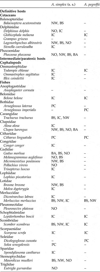
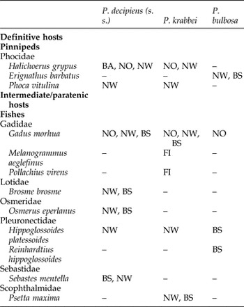
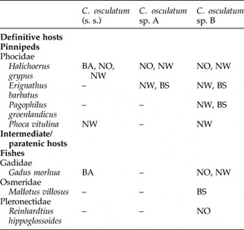
This section summarizes the occurrence of species belonging to the genera Anisakis, Pseudoterranova and Contracaecum (herein considering only the species maturing in pinnipeds) infecting definitive (marine mammals) and intermediate/paratenic fish hosts from the NE Atlantic Ocean, and which have been genetically characterized to date (see tables 1, 2, 3 and figs 4 and 6).
Definitive and intermediate/paratenic hosts detected so far by molecular/genetic markers, of the Anisakis spp. from the North-east Atlantic waters. Sampling locality codes: BA, Baltic Sea; NO, North Sea; NW, Norwegian Sea; BS, Barents Sea; IC, Iberian Atlantic Coast; PC, Portuguese coast. Additional species: in M. merluccius from the Iberian Atlantic Coast, A. physeteris, A. paggiae and A. ziphidarum were found; in Anoplogaster cornuta from the North Sea, A. paggiae was recorded. (Data from Mattiucci & Nascetti, Reference Mattiucci and Nascetti2008; Klimpel et al., Reference Klimpel, Kuhn, Busch, Karl and Palm2011; Kuhn et al., Reference Kuhn, Hailer, Palm and Klimpel2013; Levsen & Karl, Reference Levsen and Karl2014; Mattiucci et al., Reference Cipriani, Smaldone, Acerra, D'Angelo, Anastasio, Bellisario, Palma, Nascetti and Mattiucci2014; Mehrdana et al., Reference Mehrdana, Bahlool, Skov, Marana, Sindberg, Mundeling, Overgaard, Korbut, Strøm, Kania and Buchmann2014; Skrzypczak et al., Reference Skrzypczak, Rokicki, Pawliczka, Najda and Dzido2014; Bao et al., Reference Bao, Roura, Mota and Pascual2015; Münster et al., Reference Münster, Klimpel, Fock, MacKenzie and Kuhn2015.) Fish families are given in alphabetical order.

Definitive and intermediate/paratenic hosts detected so far, by molecular/genetic markers, of the Pseudoterranova spp. from the North-east Atlantic waters. Sampling locality codes: BA, Baltic Sea; FI, Faroe Islands; NO, North Sea; NW, Norwegian Sea; BS, Barents Sea. (Data from Mattiucci & Nascetti, Reference Mattiucci and Nascetti2008; Haarder et al., Reference Haarder, Kania, Galatius and Buchmann2014; Mehrdana et al., Reference Mehrdana, Bahlool, Skov, Marana, Sindberg, Mundeling, Overgaard, Korbut, Strøm, Kania and Buchmann2014; Skrzypczak et al., Reference Skrzypczak, Rokicki, Pawliczka, Najda and Dzido2014; Buchmann & Mehrdana, Reference Buchmann and Mehrdana2016). Fish families are given in alphabetical order.

Definitive and intermediate/paratenic hosts detected so far, by molecular genetic markers, for the Contracaecum spp. from the North-east Atlantic waters. Sampling locality codes: BA, Baltic Sea; NO, North Sea; NW, Norwegian Sea; BS, Barents Sea. (Data from Mattiucci & Nascetti, Reference Mattiucci and Nascetti2008; Haarder et al., Reference Haarder, Kania, Galatius and Buchmann2014; Mehrdana et al., Reference Mehrdana, Bahlool, Skov, Marana, Sindberg, Mundeling, Overgaard, Korbut, Strøm, Kania and Buchmann2014; Skrzypczak et al., Reference Skrzypczak, Rokicki, Pawliczka, Najda and Dzido2014; Klapper et al., Reference Klapper, Kuhn, Münster, Levsen, Karl and Klimpel2015; Münster et al., Reference Münster, Klimpel, Fock, MacKenzie and Kuhn2015; Buchmann & Mehrdana, Reference Buchmann and Mehrdana2016; Levsen et al, Reference Levsen, Paoletti, Cipriani, Nascetti and Mattiucci2016.) Fish families are given in alphabetical order.

Anisakis spp.
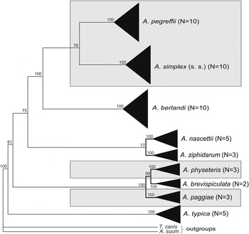
To date, nine species belonging to the genus Anisakis have been identified worldwide (Mattiucci et al., Reference Cipriani, Smaldone, Acerra, D'Angelo, Anastasio, Bellisario, Palma, Nascetti and Mattiucci2014). All species are characterized by distinct diagnostic genetic markers, possess distinct gene pools and are reproductively isolated. The existence of nine species as distinct phylogenetic units was also demonstrated by various concatenated phylogenetic analyses, inferred from nuclear and mitochondrial genes (Valentini et al., Reference Valentini, Mattiucci, Bondanelli, Webb, Mignucci-Giannone, Colom-Llavina and Nascetti2006; Mattiucci & Nascetti, Reference Mattiucci and Nascetti2008; Mattiucci et al., Reference Cipriani, Smaldone, Acerra, D'Angelo, Anastasio, Bellisario, Palma, Nascetti and Mattiucci2014). According to these analyses, four distinct clades within the genus Anisakis clearly exist. The first clade consists of the so-called A. simplex complex and includes the species A. simplex (s. s.), A. pegreffii and A. berlandi (= A. simplex sp. C) (see Mattiucci et al., Reference Cipriani, Smaldone, Acerra, D'Angelo, Anastasio, Bellisario, Palma, Nascetti and Mattiucci2014) (fig. 2). A second clade includes the two sibling species A. ziphidarum and A. nascettii; a third clade comprises A. physeteris, A. brevispiculata, and A. paggiae. Finally, concatenated phylogenetic trees obtained from the combined nuclear and mitochondrial sequences depicted A. typica as a separate lineage, thus comprising the forth clade within the group (Mattiucci et al., Reference Cipriani, Smaldone, Acerra, D'Angelo, Anastasio, Bellisario, Palma, Nascetti and Mattiucci2014) (fig. 2).
Maximum parsimony (MP) bootstrap consensus tree, inferred by PAUP*4.0 (bootstrap method with heuristic search) (Swofford, Reference Swofford2003), based on the combined mtDNA cox-2, rrnS rRNA and ITS rDNA from our sequence data for some specimens (previously sequenced) of all characterized species belonging to the genus Anisakis (based on sequence data from Mattiucci et al., Reference Cipriani, Smaldone, Acerra, D'Angelo, Anastasio, Bellisario, Palma, Nascetti and Mattiucci2014). Bootstrap values (≥70) were obtained over 1000 pseudoreplicates and are reported at the nodes. Black triangles are proportional to the number of specimens sequenced, reported in parentheses. Toxocara canis and Ascaris suum were used as outgroups; species reported to date from the NE Atlantic are shown within shaded boxes.

Below, we review the Anisakis species reported so far from the NE Atlantic Ocean. Sampling localities and intermediate/paratenic and definitive hosts for each sibling species are given in table 1. The A. simplex (s. s.) distribution ranges from 35°N, circumpolarly to the Arctic Seas (Mattiucci & Nascetti, Reference Mattiucci and Nascetti2008). Its southern limit of distribution in NE Atlantic waters seems to be the Spanish–Portuguese Atlantic coast around Gibraltar and the Alboran Sea; the latter area being oceanographically considered part of the Atlantic Ocean rather than the Mediterranean Sea (Mattiucci et al., Reference Cipriani, Smaldone, Acerra, D'Angelo, Anastasio, Bellisario, Palma, Nascetti and Mattiucci2014). Adults of A. simplex (s. s.) have so far been detected in several cetacean species (table 1) while the larvae occur in various demersal, pelagic or benthopelagic fish species, which act as intermediate or paratenic hosts (table 1). It has also been recorded in some squid species from NE Atlantic waters.
Anisakis pegreffii, on the other hand, seems to occur only occasionally in northern NE Atlantic waters. Indeed, the northerly limit of its geographical range is actually represented by the Spanish coast of the Bay of Biscay (Mattiucci et al., Reference Cipriani, Smaldone, Acerra, D'Angelo, Anastasio, Bellisario, Palma, Nascetti and Mattiucci2014). However, a small number of specimens of A. pegreffii have been recorded in migratory Atlantic mackerel, Scomber scombrus, from the southern Norwegian Sea (Levsen & Mattiucci, pers. obs.). Meanwhile, A. pegreffii is the species with the widest distribution in the Mediterranean Sea, but has also been reported from Austral regions between 30°S and 60°S (Mattiucci et al., Reference Cipriani, Smaldone, Acerra, D'Angelo, Anastasio, Bellisario, Palma, Nascetti and Mattiucci2014).
Anisakis simplex (s. s.) and A. pegreffii share the same definitive hosts and paratenic fish hosts in some geographical areas, except for some cetacean and fish species whose distribution is mostly Arctic (see table 1). Additionally, the species frequently co-infect the same individual definitive or intermediate/paratenic hosts in some sympatric areas, including the Spanish Atlantic coast and the Alboran Sea in the Mediterranean basin (table 1). In these areas, some F1 hybrids of the two Anisakis species have been recorded in commercially important fish hosts, by using a multilocus approach; however, no backcross individuals have been detected so far (Mattiucci et al., Reference Mattiucci, Acerra, Paoletti, Cipriani, Levsen, Webb, Canestrelli and Nascetti2016).
However, another valid species of the genus Anisakis, i.e. A. physeteris, has been also reported from the NE Atlantic Ocean. It occurs, at the adult stage, in Physeter macrocephalus from the Iberian Atlantic waters, while Type II larval stages belonging to this species have been indentified genetically from several fish hosts.
Finally, a parasitological study carried out by Klimpel et al. (Reference Klimpel, Kuhn, Busch, Karl and Palm2011) on the common fangtooth, Anoplogaster cornuta, a meso- and bathypelagic fish species from the North Atlantic, revealed the presence of Anisakis paggiae at high prevalence (P = 57.1%) and intensity (Im = 2.2), which was apparently not correlated with fish host size (standard length). Since Kogiid whales (i.e. Kogia breviceps, K. sima) are the final hosts of this nematode (Mattiucci et al., Reference Mattiucci, Nascetti, Dailey, Webb, Barros, Cianchi and Bullini2005; Mattiucci & Nascetti, Reference Mattiucci and Nascetti2008), Klimpel et al. (Reference Klimpel, Kuhn, Busch, Karl and Palm2011) suggested that A. paggiae larvae were introduced through the migratory kogiid final hosts. Moreover, the finding in this deep-sea fish seems to support an oceanic deep-water life cycle for A. paggiae in the North-east Atlantic Ocean (Klimpel et al., Reference Klimpel, Kuhn, Busch, Karl and Palm2011).
Pseudoterranova spp.
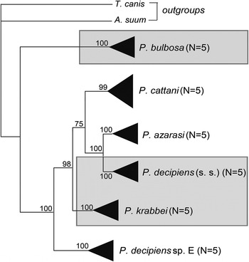
To date, six biological species are recognized in the Pseudoterranova decipiens complex (Mattiucci & Nascetti, Reference Mattiucci and Nascetti2008; Timi et al., Reference Timi, Paoletti, Cimmaruta, Lanfranchi, Alarcos, Garbin, George-Nascimento, Rodríguez, Giardino and Mattiucci2014), i.e. P. decipiens (s. s.) (see Paggi et al., Reference Paggi, Nascetti, Cianchi, Orecchia, Mattiucci, D'Amelio, Berland, Brattey, Smith and Bullini1991), P. krabbei (see Mattiucci et al., Reference Mattiucci, Paggi, Nascetti, Ishikura, Kikuchi, Sato, Cianchi and Bullini1998), P. bulbosa (see Mattiucci et al., Reference Mattiucci, Paggi, Nascetti, Ishikura, Kikuchi, Sato, Cianchi and Bullini1998), P. azarasi (see Mattiucci et al., Reference Mattiucci, Paggi, Nascetti, Ishikura, Kikuchi, Sato, Cianchi and Bullini1998), P. cattani (see George-Nascimento & Urrutia, Reference George-Nascimento and Urrutia2000; Timi et al., Reference Timi, Paoletti, Cimmaruta, Lanfranchi, Alarcos, Garbin, George-Nascimento, Rodríguez, Giardino and Mattiucci2014) and P. decipiens sp. E (Timi et al., Reference Timi, Paoletti, Cimmaruta, Lanfranchi, Alarcos, Garbin, George-Nascimento, Rodríguez, Giardino and Mattiucci2014). A synopsis of the geographical distribution of all the taxa belonging to the genus Pseudoterranova was provided by Mattiucci & Nascetti (Reference Mattiucci and Nascetti2008), while a phylogenetic analysis of all the members of the P. decipiens (s. l.) complex, which occur in both hemispheres, has recently been provided by Timi et al. (Reference Timi, Paoletti, Cimmaruta, Lanfranchi, Alarcos, Garbin, George-Nascimento, Rodríguez, Giardino and Mattiucci2014), based on sequence analysis of the mtDNA cox2 gene and direct sequencing of the ITS region of rDNA. A maximum parsimony (MP) inference obtained from a concatenated analysis of the mtDNA cox2 and ITS region of rDNA sequence datasets of all the Pseudoterranova taxa genetically characterized so far is given in fig. 3.
Maximum parsimony (MP) bootstrap consensus tree inferred by PAUP*4.0 (bootstrap method with heuristic search) (Swofford, Reference Swofford2003), based on the combined mtDNA cox2 and ITS rDNA sequences data for some specimens (previously sequenced) belonging to the species genetically charcterized so far of the genus Pseudoterranova (based on sequence data from Timi et al., Reference Timi, Paoletti, Cimmaruta, Lanfranchi, Alarcos, Garbin, George-Nascimento, Rodríguez, Giardino and Mattiucci2014). Bootstrap values (≥70) were obtained over 1000 pseudoreplicates and are reported at the nodes. Black triangles are proportional to the number of specimens sequenced, reported in parentheses. Toxocara canis and Ascaris suum were used as outgroups; species occurring in the NE Atlantic are shown within shaded boxes.

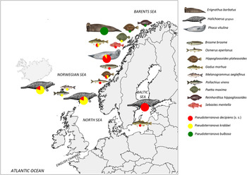
However, only three species are parasites of pinnipeds and fish in the NE Atlantic Ocean (fig. 4 and table 2). Indeed, population genetic analysis using allozyme markers on specimens of P. decipiens (s. l.) recovered and collected from various fish and seal species of the North Atlantic Ocean, demonstrated the existence of a remarkable genetic heterogeneity in allele frequencies among the samples (Paggi et al., Reference Paggi, Nascetti, Cianchi, Orecchia, Mattiucci, D'Amelio, Berland, Brattey, Smith and Bullini1991). The data clearly evidenced that three distinct cryptic species occur sympatrically in seal hosts from the actual sampling localities, with no gene flow between them. These three gene pools correspond to the three taxa which were provisionally designated as P. decipiens sp. A, P. decipiens sp. B and P. decipiens sp. C (Paggi et al., Reference Paggi, Nascetti, Cianchi, Orecchia, Mattiucci, D'Amelio, Berland, Brattey, Smith and Bullini1991). However, a deeper morphological analysis carried out on male specimens identified genetically as P. decipiens sp. A and sp. B, allowed the detection of significant differences in a number of characters between these two siblings. Based on the differences, the names Pseudoterranova krabbei for P. decipiens sp. A, and P. decipiens (s. s.) for P. decipiens sp. B were proposed, along with a formal description of the two taxa (see Paggi et al., Reference Paggi, Mattiucci, Gibson, Berland, Nascetti, Cianchi and Bullini2000). Later on, the name P. bulbosa (Cobb, 1888) was proposed for the former taxon P. decipiens sp. C (see Mattiucci et al., Reference Mattiucci, Paggi, Nascetti, Ishikura, Kikuchi, Sato, Cianchi and Bullini1998) which parasitizes the bearded seal, Erignathus barbatus, from the NE Atlantic Ocean. Indeed, it was demonstrated that P. decipiens sp. C agrees morphologically with Ascaris bulbosa, originally described by Cobb (1888) from the bearded seal at Spitzbergen (NE Atlantic Ocean). Therefore, some morphological features of diagnostic value were finally detected in the adults of the three sibling species of the P. decipiens complex that occur in NE Atlantic waters.
Relative proportions of the species of the genus Pseudoterranova, genetically characterized so far, in their main definitive host (seals) and in some intermediate/paratenic hosts (fish) from the north-eastern Atlantic Ocean.

The hitherto known geographical distribution of P. decipiens (s. s.) includes the Arctic Boreal region, covering both North Atlantic and North Pacific waters. In the NE Atlantic Ocean, its range comprises the following areas: the Norwegian Sea, Baltic Sea, Scottish waters, English Channel, southern Icelandic Sea and the waters around the Faroe Islands; thus closely reflecting the geographical distribution of its main definitive hosts, i.e. the common seal Phoca vitulina and the grey seal Halichoerus grypus (fig. 4, table 2). The species has so far been detected genetically in various fish hosts including Atlantic cod, Gadus morhua, in the Norwegian and Baltic Seas (Mehrdana et al., Reference Mehrdana, Bahlool, Skov, Marana, Sindberg, Mundeling, Overgaard, Korbut, Strøm, Kania and Buchmann2014).
Pseudoterranova krabbei has only been recorded from the NE Atlantic Ocean, including the Norwegian Sea, southern Icelandic waters and the English Channel. However, it seems to be absent in the NW Atlantic Ocean. It is a parasite, at the adult stage, of grey seals and common seals (table 2). In the latter host species, P. krabbei often appears in mixed infections with P. decipiens (s. s.). However, P. krabbei usually outnumbers P. decipiens (s. s.) when occurring syntopically in grey seals. Its larval stages have been identified genetically in various fish hosts, such as Atlantic cod Gadus morhua, haddock Melanogrammus aeglefinus and saithe Pollachius virens (table 2).
Pseudoterranova bulbosa has been recorded from both the NE and NW Atlantic Ocean, where the adults infect the bearded seal, E. barbatus. In the NE Atlantic, its range includes the Barents Sea and the Norwegian Sea. It has only rarely been recorded in Atlantic cod, while it seems to occur at higher prevalence and abundance in both long rough dab Hippoglossoides platessoides and Greenland halibut Reinhardtius hippoglossoides (table 2).
Contracaecum spp.
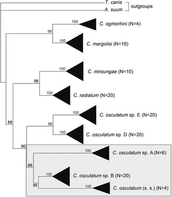
Members of the C. osculatum (s. l.) species complex, hosted at their adult stage by pinnipeds from Arctic Boreal regions (Mattiucci & Nascetti, Reference Mattiucci and Nascetti2008), are C. osculatum sp. A, C. osculatum sp. B and C. osculatum (s. s.) (Nascetti et al., Reference Nascetti, Cianchi, Mattiucci, D'Amelio, Orecchia, Paggi, Brattey, Berland, Smith and Bullini1993). These nematode species are morphologically very similar, both at their adult and third larval stages, but appear to be reproductively isolated (cryptic species). Indeed, no morphological characters have been found that may allow us to specifically separate the adults of the three members of the C. osculatum complex. Furthermore, these members represent distinct phylogenetic lineages compared to the Antarctic members of the C. osculatum (s. l.) species complex, i.e. C. osculatum sp. D and C. osculatum sp. E (Mattiucci et al., Reference Mattiucci, Cipriani, Paoletti, Nardi, Santoro, Bellisario and Nascetti2015) (fig. 5). Genetic studies of this species complex, based on allozyme markers, have demonstrated reproductive isolation and the absence of gene flow among sympatric and allopatric populations of C. osculatum (s. l.) collected from seals and fish in the NE Atlantic Ocean. Moreover, the identification of the species as distinct phylogenetic units was supported by sequence analysis of ITS rDNA (Nadler et al., Reference Nadler, D'Amelio, Dailey, Paggi, Siu and Sakanari2005) and the mitochondrial cox2 gene sequence analysis (Mattiucci et al., Reference Mattiucci, Paoletti, Webb, Sardella, Timi, Berland and Nascetti2008). Additionally, single-strand conformation polymorphism (SSCP) analysis of the ITS rDNA was carried out to screen for sequence variation within and among individuals of the C. osculatum (s. l.) species complex (Zhu et al., Reference Zhu, D'Amelio, Paggi and Gasser2000; Hu et al., Reference Hu, D'Amelio, Zhu, Paggi and Gasser2001).
Maximum parsimony (MP) bootstrap consensus tree inferred by PAUP*4.0 (bootstrap method with heuristic search) (Swofford, Reference Swofford2003), based on the combined mtDNA cox2 sequences data for some specimens of Contracaecum spp., having pinnipeds as final hosts. Bootstrap values (≥70) were obtained over 1000 pseudoreplicates and are reported at the nodes. Black triangles are proportional to the number of specimens sequenced, reported in parentheses (based on sequence data from Mattiucci et al., Reference Mattiucci, Cipriani, Paoletti, Nardi, Santoro, Bellisario and Nascetti2015). Toxocara canis and Ascaris suum were used as outgroups; species occurring in the NE Atlantic are shown within shaded boxes.

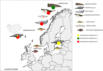
Allozyme markers and sequence analysis of the mtDNA cox2 gene strongly improved our knowledge about the geographical range and host distribution of the Arctic members of these taxa. Thus, C. osculatum (s. s.) is the only species of the genus to have been genetically characterized so far in the Baltic Sea. It is a parasite, at the adult stage, of the grey seal (Mattiucci & Nascetti, Reference Mattiucci and Nascetti2008; Mehrdana et al., Reference Mehrdana, Bahlool, Skov, Marana, Sindberg, Mundeling, Overgaard, Korbut, Strøm, Kania and Buchmann2014), while the larvae were genetically identified from Baltic cod. The reproductive isolation of C. osculatum sp. A from the other member C. osculatum sp. B was reported when occurring sympatrically in the same definitive seal species, i.e. grey seal H. grypus, from Icelandic waters (Nascetti et al., Reference Nascetti, Cianchi, Mattiucci, D'Amelio, Orecchia, Paggi, Brattey, Berland, Smith and Bullini1993). In contrast, C. osculatum sp. B was reported as adults in the common seal P. vitulina and harp seal Pagophilus groenlandicus from the Barents Sea, Norwegian Sea, Faroe Islands and Icelandic waters. Finally, the species assigned to C. osculatum sp. A seems to show a restricted geographical range, being a parasite, at the adult stage, of the bearded seal E. barbatus. However, only little information exists concerning the fish species that act as paratenic hosts for the two sibling species in NE Atlantic waters. However, C. osculatum sp. B larvae were recently found in capelin Mallotus villosus from the Barents Sea (Levsen et al., Reference Levsen, Paoletti, Cipriani, Nascetti and Mattiucci2016). The sibling species is also known to infect Atlantic cod Gadus morhua from the Norwegian Sea (Mattiucci & Nascetti, Reference Mattiucci and Nascetti2008) (table 3, fig. 6). Additionally, it has been recorded in beaked redfish Sebastes mentella (Klapper et al., Reference Klapper, Kuhn, Münster, Levsen, Karl and Klimpel2015) and Greenland halibut R. hippoglossoides (Karpiej et al., Reference Karpiej, Dzido, Rokicki and Kijewska2013), in basically the same area.
Relative proportions of the members of the Contracaecum osculatum (s. l.) species complex in the main definitive hosts (seals) and in some intermediate/paratenic hosts (fish) from the north-eastern Atlantic Ocean.

Ecological aspects of anisakid species
Genetic/molecular markers used to characterize anisakid species in NE Atlantic waters, have allowed us to screen and identify the fish species that act as intermediate/paratenic hosts, and the pinnipeds and cetaceans acting as definitive hosts. Genetic identification data of anisakids have also provided information on various ecological and evolutionary aspects, such as host preference and host–parasite co-evolutionary adaptations, including host–parasite co-phylogenetic processes (Mattiucci & Nascetti, Reference Mattiucci and Nascetti2008).
For example, differences in definitive host preferences have been suggested for the members of Pseudoterranova and Contracaecum maturing in pinnipeds, with differential distribution in various seal hosts. Indeed, P. bulbosa is the only Pseudoterranova species to infect the bearded seal E. barbatus in NE Atlantic waters (fig. 4). However, the species has also been recorded in bearded seals from NW Atlantic and North Pacific waters.
On the other hand, P. krabbei and P. decipiens (s. s.) show a different distribution from the other taxa of the genus, with a marked difference in their relative proportions when they occur sympatrically and syntopically in the same definitive hosts from the NE Atlantic Ocean, i.e. the common seal P. vitulina and the grey seal H. grypus, respectively. For example, in areas of the NW Atlantic where P. krabbei is absent, the species P. decipiens (s. s.) infects both common and grey seals in the same relative proportion. Differences in host preference could be the result of parasite–host adaptations, as well as interspecific competition. These mechanisms probably reduce the range of potential hosts, or promote differential use of host resources when infecting the same definitive host under syntopic conditions. In the case of P. decipiens (s. s.) and P. krabbei, different host adaptations could have resulted from interspecific competition, probably as a consequence of a secondary contact, i.e. when the two parasite species occurred in sympatry in NE Atlantic seals.
Interspecific competition between distinct species of Pseudoterranova and Contracaecum could also exist in seals, e.g. if they increase in numbers and are lodging in the stomach of the same individual seal host. For instance, McClelland et al. (Reference McClelland, Misra and Martell1985), McClelland (Reference McClelland2002) and Marcogliese (Reference Marcogliese1997) suggested that C. osculatum (s. l.) interfered with P. decipiens (s. l.) in grey seals from NW Atlantic waters by inducing a smaller female body size and lower egg counts in P. decipiens. It was also observed that differential use of resources could take place in cases of co-infection of the same individual host by members of the P. decipiens (s. l.) and C. osculatum (s. l.) complexes. For example, the former seems primarily to occupy the seal intestine if the stomach of the same host individual is heavily parasitized by C. osculatum (s. l.) (Mattiucci & Nascetti, Reference Mattiucci and Nascetti2008).
However, the geographical distribution of genetically recognized anisakid species could be the result of different feeding behaviour of their definitive seal hosts, as well, thus resulting in area-specific life cycles of the actual anisakid species. This seems to be the case in P. bulbosa from the NE Atlantic Ocean where it infects the bearded seal. This pinniped preys mostly upon benthic fish species, such as long rough dab H. platessoides, which is commonly infected with P. bulbosa larvae in this area. Since P. bulbosa also infects Atlantic cod G. morhua, another frequently occurring benthopelagic fish species in the area, it seems likely that P. bulbosa has primarily a benthic/benthopelagic life cycle in the NE Atlantic Ocean.
Differential spatial distribution patterns of anisakids infecting the same individual fish species have been found between A. simplex (s. s.) and A. pegreffii. Indeed, A. simplex (s. s.) outnumbers A. pegreffii in the musculature (flesh) of the same fish host, e.g. the European hake Merluccius merluccius. Despite marked differences in the relative proportions of the two species in the fish flesh, they infect the viscera of the fish in the same proportion (Cipriani et al., Reference Cipriani, Smaldone, Acerra, D'Angelo, Anastasio, Bellisario, Palma, Nascetti and Mattiucci2014). This phenomenon could be the result of differential resource utilization due to interspecific competition between the two siblings for the same trophic resource, such as the liver. Another explanatory model is linked to the different migrating ability of the species the tissues, which seems to be considerably higher in A. simplex (s. s.) and, again, could result from different ability of the two species to respond to the fish host's immune system.
Distribution of anisakid nematodes in wild fisheries
According to the Food and Agriculture Organization of the United Nations (FAO/WHO, 2014), the world fisheries and aquaculture production has greatly increased in the past decades. Two major changes in global seafood consumption have taken place. The per capita fish consumption has nearly doubled in the past four decades; at the same time, the global overall fish production, including aquaculture and wild-catch fisheries, has increased by many tons in order to meet the market demands. However, some of the most important fish species captured in European fishing grounds (including the Mediterranean Sea) and distributed in European markets, are at risk of carrying zoonotic parasites. Among the latter, anisakid nematodes have been recognized by the European Food Safety Authority (EFSA, 2010) as representing the most important ‘biological hazard’ in seafood products. Consequently, EFSA (2010) recommended the collection of systematic data on the complete life cycle, geographical and seasonal distribution, prevalence, intensity and anatomical location of parasites of public health importance in wild-caught fishery products.
However, and despite the fact that the occurrence of genetically recognized larval anisakids from many fish species from the NE Atlantic Ocean has been reported, comprehensive data on the distribution, infection levels and preferred infection sites in different fish hosts throughout their geographical range, are still scarce. Indeed, data on anisakids infecting wild fish stocks, are largely based on randomly and locally collected fish samples. Additionally, there is still a lack of harmonization of the methodological procedures applied during parasitological fish inspections, along with ubiquitous and commonly applied genetic/molecular markers for specific parasite identification. This has partly resulted in some puzzling data on the occurrence and distribution of anisakids in some of the commercially most important fish species. Recently, a well-designed systematic epidemiological survey of some of the economically most valuable fish species from European fishing grounds has been carried out within the framework of a European Union 7th Framework Program project (EU FP7 PARASITE Grant agreement #312068). Large samples of fish (up to several thousands of individual fish) belonging to 16 teleost species from some of the richest European fishing grounds, were recently examined for the presence of anisakid nematodes. The uniform application of the so-called UV-press method (Karl & Leinemann, Reference Karl and Leinemann1993, Levsen & Karl, Reference Levsen and Karl2014) by all surveyors permitted investigation of the presence and the infection site of anisakid larvae with high accuracy, not only in the fish viscera but also in fish muscle. All subsamples of anisakid larvae were identified to species level, using the same set of congruent genetic/molecular markers throughout the survey, thus providing the basis for analysing and monitoring anisakid prevalence and abundance in each of the surveyed fish host species. Indeed, following EFSA's recommendations, the survey emphasized the acquisition of data on the occurrence and distribution of potentially zoonotic anisakid species in the edible parts (mostly flesh) of fish, which represent the main exposure risk factor for the consumer. In this respect, some aspects related to the presence and infection level of anisakids in the actual fish host species were more thoroughly explored: (1) anisakid spatial distribution in the fish; (2) differences in larval infection level with respect to fishing ground/fish host sampling locality; and (3) fish-host body size (length/weight).
In a fish host, the majority of anisakid larvae typically infect the visceral organs, often, as in Anisakis spp. larvae, encapsulated and coiled up in tight spirals on the surface of the organs. Moreover, some larvae may migrate from the visceral cavity into the musculature of the fish host, sometimes deeply into the fillets. However, the reason for and the molecular basis of this behaviour are not yet fully understood, despite the fact that the larvae secrete enzymes from glandular cells in the oesophageal lining which probably facilitate larval penetration of the stomach wall and peritoneum, to eventually reach the fish muscle. However, it was demonstrated that this phenomenon occurs both intra vitam (Roepstorff et al., Reference Roepstorff, Karl, Bloemsma and Huss1993; Karl et al., Reference Karl, Baumann, Ostermeyer, Kuhn and Klimpel2011; Cipriani et al., Reference Cipriani, Acerra, Bellisario, Sbaraglia, Cheleschi, Nascetti and Mattiucci2016) and post mortem of the fish host (Šimat et al., Reference Šimat, Miletić, Bogdanović, Poljak and Mladineo2015; Cipriani et al., Reference Cipriani, Acerra, Bellisario, Sbaraglia, Cheleschi, Nascetti and Mattiucci2016). It was further demonstrated by the latter authors that there is a significantly positive correlation between the post-mortem migration of A. pegreffii in anchovy Engraulis encrasicolus and increasing storage temperature and time (Cipriani et al., Reference Cipriani, Acerra, Bellisario, Sbaraglia, Cheleschi, Nascetti and Mattiucci2016). Additionally, Levsen et al. (Reference Levsen, Paoletti, Cipriani, Nascetti and Mattiucci2016) observed that some C. osculatum sp. B specimens infecting capelin M. villosus from the Barents Sea were about to penetrate the peritoneum which separates the visceral cavity from the fish flesh, indicating that the larvae had started the migration process post mortem, i.e. after death of the fish host.
The presence of anisakid larvae in fish fillets is of high concern to food safety. Generally, most larvae parasitize the ventral portion of the fish flesh surrounding the visceral cavity, even though some may migrate deeply into the dorsal parts of the fillets. The larval muscle-migrating behaviour represents a major consumer health risk because Anisakis larvae, especially Anisakis spp., appear almost transparent in the fish flesh and are thus very hard to detect during HACCP (hazard analysis and critical control points) procedures or other routine quality inspections.
A number of different anisakid species of the genera Anisakis, Pseudoterranova and Contracaecum occurring in the NE Atlantic waters have been reported to infect the musculature of fish. For example, Levsen et al. (Reference Levsen, Paoletti, Cipriani, Nascetti and Mattiucci2016) found high prevalence of C. osculatum sp. B larvae in the flesh of capelin, although most of the larvae seemed to occur in the fatty tissue layer between the peritoneum and the actual muscle tissue of the belly flaps, i.e. the hypaxial part of the body musculature surrounding the abdominal cavity (Levsen et al., Reference Levsen, Paoletti, Cipriani, Nascetti and Mattiucci2016).
However, the spatial distribution and infection level of larval anisakid species belonging to the above genera may vary greatly, depending on parasite species, fish host species and sampling locality. Indeed, the relative distribution of A. simplex (s. s.) in both ventral and dorsal portions of the flesh of European hake M. merluccius was significantly higher compared to that of A. pegreffii, even if their relative occurrence in the visceral organs remained unchanged (Cipriani et al., Reference Cipriani, Smaldone, Acerra, D'Angelo, Anastasio, Bellisario, Palma, Nascetti and Mattiucci2014). This trend was seen in both single infections with the two species in allopatry, and when occurring in mixed infections in sympatric areas (Cipriani et al., Reference Cipriani, Smaldone, Acerra, D'Angelo, Anastasio, Bellisario, Palma, Nascetti and Mattiucci2014). Significantly higher abundances of A. simplex (s. s.) in the muscle of the fish host compared to A. pegreffii in the same individual fish host have been reported by Suzuki et al. (Reference Suzuki, Murata, Hosaka and Araki2010) and Cipriani et al. (Reference Cipriani, Smaldone, Acerra, D'Angelo, Anastasio, Bellisario, Palma, Nascetti and Mattiucci2014). However, the factors and the molecular mechanisms that are responsible for the clearer tendency of A. simplex (s. s.) to penetrate the fish muscle still remain to be understood.
In general, A. simplex (s. s.) may reach comparatively high prevalence in the flesh of several commercially important fish species from the NE Atlantic Ocean, including herring Clupea harengus (Levsen & Lunestad, Reference Levsen and Lunestad2010), grey gurnard Eutrigla gunardus (Levsen & Karl, Reference Levsen and Karl2014), European hake M. merluccius (Cipriani et al., Reference Cipriani, Smaldone, Acerra, D'Angelo, Anastasio, Bellisario, Palma, Nascetti and Mattiucci2014) and Atlantic cod (Mouritsen et al., Reference Mouritsen, Hedeholm, Schack, Møller, Storr-Paulsen, Dzido and Rokicki2010; Münster et al., Reference Münster, Klimpel, Fock, MacKenzie and Kuhn2015). High prevalence and abundance of A. simplex (s. s.) in some fish host species were apparently also related to sampling locality, i.e. the actual fishing ground. Thus, Klimpel (Reference Klimpel2005) found differences in the prevalence of anisakid nematodes in haddock M. aeglefinus, withing Merlangius merlangius and saithe P. virens, when comparing different sampling localities in the North Sea, e.g. higher prevalence was found in haddock from the central North Sea compared to other areas. Similarly, Levsen & Karl (Reference Levsen and Karl2014) found statistically significant differences in infection with A. simplex (s. s.) larvae in grey gurnard E. gurnardus, fished in different areas of the North Sea. Thus, gurnard from two localities off the north-eastern coast of the British mainland showed significantly higher infection levels than the fish caught in northern and eastern areas of the North Sea. The same authors (Levsen & Karl, Reference Levsen and Karl2014) suggested that the differences may be related to the differential distribution of preferred prey items of the fish host, which may act as an intermediate or paratenic host for the parasite in the different fishing areas. On the other hand, it is well known that the repeated transmission of larvae between fish species, through a prey–predator relationship, may result in the accumulation of infections with anisakid larvae, which is largest in piscivorous fish at the top of a food chain. Thus, the parasite load in larger and older piscivorous fish could be characterized by hundreds of larvae in the same individual host. Indeed, a significantly positive correlation between the fish host length (or weight), i.e. fish size, and the prevalence and abundance values of the infections by anisakids has been reported in several fish species. For instance, Münster et al. (Reference Münster, Klimpel, Fock, MacKenzie and Kuhn2015) found that small Atlantic cod G. morhua sampled in West Greenland waters, which probably preyed almost entirely on small crustaceans, showed significantly lower infections with anisakids than larger cod, which likely accumulated the larvae by preying upon other infected fish. A similar trend was observed in Atlantic cod sampled in the Baltic Sea by Mehrdana et al. (Reference Mehrdana, Bahlool, Skov, Marana, Sindberg, Mundeling, Overgaard, Korbut, Strøm, Kania and Buchmann2014). The study showed that cod ≥50 cm were more strongly infected with P. decipiens (s. s.) than cod measuring ≤50 cm in length. The authors found high prevalence and abundance of P. decipiens (s. s.) (P = 55% and maximum abundance value, A = 56 parasites/fish), especially in the musculature of cod, also showing a significantly negative correlation between worm intensity and fish host condition factor. The same study, looking at infection in the cod by the sibling species C. osculatum (s. s.) (mainly found in the cod liver), showed a slight negative correlation between body condition index and high intensity value, in spite of the higher values of prevalence and abundance of the infection (P = 100% and A = 320 worms/fish) (Mehrdana et al., Reference Mehrdana, Bahlool, Skov, Marana, Sindberg, Mundeling, Overgaard, Korbut, Strøm, Kania and Buchmann2014). In contrast, only a very weak relationship between fish length (weight) and parasite abundance was found in capelin M. villosus in the Barents Sea, infected with C. osculatum sp. B and A. simplex (s. s.) (Levsen et al., Reference Levsen, Paoletti, Cipriani, Nascetti and Mattiucci2016). The latter finding is likely associated with the fish host's short lifespan and the fact that capelin start feeding on macroplankton, such as krill and amphipods (which represent the first intermediate hosts of the actual anisakids), at a rather late stage during growth and maturation (Levsen et al., Reference Levsen, Paoletti, Cipriani, Nascetti and Mattiucci2016). Thus, Barents Sea capelin may acquire the C. osculatum sp. B larvae over the entire post-larval lifespan, including the juvenile stages when copepods appear to be the main prey (Gjøsæter, Reference Gjøsæter1998).
However, despite the fact that anisakid infection levels generally increase with size and age of the fish host, likely due to ontogenetic changes related to larger prey items and feeding volume, the spatial distribution of anisakid larvae in a given fish species may remain largely unchanged and independent of fishing ground and fish size. This was shown by Levsen & Karl (Reference Levsen and Karl2014) and Levsen & Lunestad (Reference Levsen and Lunestad2010), who found that the relative occurrence of A. simplex (s. s.) larvae in the flesh of grey gurnard E. gurnardus and herring C. harengus remained the same, independent of both fish host size and sampling locality.
Since the life cycle of anisakid nematodes involves various marine organisms, each being at a different position in the food web of the actual ecosystem, the transmission routes of parasites are closely linked to the trophic relationships between their successive hosts. Therefore, any successful completion of the anisakid life cycle requires stable trophic webs (Mattiucci & Nascetti, Reference Mattiucci and Nascetti2008; Mattiucci et al., Reference Mattiucci, Cipriani, Paoletti, Nardi, Santoro, Bellisario and Nascetti2015). Consequently, the infection levels of anisakid species in a particular geographic area may be strongly affected by the population size of any of the host organisms that are involved in the parasite's life cycle.
Interestingly, in central and eastern parts of the Baltic Sea where only two anisakid species, C. osculatum (s. s.) and P. decipiens (s. s.), seem to occur, Mehrdana et al. (Reference Mehrdana, Bahlool, Skov, Marana, Sindberg, Mundeling, Overgaard, Korbut, Strøm, Kania and Buchmann2014) reported high prevalence and abundance of the two species in Baltic cod G. morhua, which seemed to have increased in the past decades. The authors suggested that the increase in anisakid prevalence and abundance in cod, compared to the lower infection levels reported in the past, was likely associated with increasing populations, in the Baltic Sea, of grey seal H. grypus, which acts as main definitive host of both P. decipiens (s. s.) and C. osculatum (s. s.) in the area.
However, the demography of the obligate intermediate crustacean host(s) in the life cycle of anisakids may also be responsible for the infection levels found in a given fish host. For example, Levsen et al. (Reference Levsen, Paoletti, Cipriani, Nascetti and Mattiucci2016) observed that a marked decline in overall prevalence and abundance of C. osculatum sp. B in Barents Sea capelin M. villosus correlated with a general tendency of decreasing body size and condition factor over the entire sampling period. The authors suggested that there appears to be an interrelationship between nematode abundance in capelin and the general condition of the spawning fraction of the stock over time. Indeed, Gjøsæter (Reference Gjøsæter2002) suggested that the mean annual growth of capelin is positively correlated with the average zooplankton density. Thus, lower overall nematode infections may indicate less favourable feeding conditions in terms of zooplankton availability during the feeding season that preceded the actual sampling year. Moreover, Levsen et al. (Reference Levsen, Paoletti, Cipriani, Nascetti and Mattiucci2016) further observed a pronounced effect of gender on the abundance of anisakid larvae in Barents Sea capelin, i.e. male capelin showed generally higher overall nematode abundance than the females. Since male capelin are significantly larger than females, they probably ingest comparatively larger volumes of zooplankton during their lifespan, which again would increase the probability of the males to acquire anisakid nematodes belonging to the species C. osculatum sp. B.
Anisakids in farmed fish
Fish cultured in floating sea-cages in coastal waters have generally not been regarded at risk of acquiring anisakid nematodes. This view was based largely on the results of several studies from a number of different countries or regions showing that sea-caged salmonid fish species, including Atlantic salmon Salmo salar, coho salmon Oncorhynchus kisutch and rainbow trout Oncorhynchus mykiss, apparently do not carry anisakid nematodes (Pacific North America: Deardorff & Kent, Reference Deardorff and Kent1989; Japan: Inoue et al., Reference Inoue, Oshima, Hirata and Kimura2000; Norway: Lunestad, Reference Lunestad2003; Denmark: Skov et al., Reference Skov, Kania, Olsen, Lauridsen and Buchmann2009; Scotland: Wootten et al., Reference Wootten, Yoon and Bron2010). Marty (Reference Marty2008) recorded a single anisakid larva during routine histopathological inspection of the visceral organs of farmed Atlantic salmon from British Columbia, Canada, but its further identification was not performed. Thus, the apparent absence of anisakids in sea-caged salmonids was explained by the fact that the fish are exclusively fed a diet consisting of extruded pelleted dry-feed, which cannot contain viable parasites.
However, two recent investigations in Norway revealed the presence of both A. simplex (s. s.) and the non-zoonotic ascaridoid species Hysterothylacium aduncum in runts (loser fish) of cultured Atlantic salmon in southern and western Norway (Mo et al., Reference Mo, Gahr, Hansen, Hoel, Oaland and Poppe2014; Levsen & Maage, Reference Levsen and Maage2016). In the latter study, 4184 farmed Atlantic salmon originating from 37 salmon farms from the Skagerrak coast in the south to Finnmark in the north-east of Norway, were examined for anisakid nematodes between January 2014 and August 2015, using the UV-press method (Karl & Leinemann, Reference Karl and Leinemann1993; Levsen & Karl, Reference Levsen and Karl2014). The samples consisted of 3527 salmon processed for human consumption and 657 discarded fish, including 395 runts. Although only very few nematodes were found in the viscera of three runts (two A. simplex (s. s.) larvae and three adult H. aduncum), i.e. none of the harvest-quality salmon were infected, the findings demonstrated that both nematode species are evidently able to enter the sea-cages in a way that renders them infective to the fish. Since both species exhibit broadly similar life-cycle features at the intermediate and paratenic host level, the vectors of transfer of the parasites from the surroundings into the cages may be sought among copepods, euphausiids or smaller wild fish which may occasionally cross the side nettings. While the healthy and normally developing salmon seem to feed exclusively on the pelleted dry-feed, runts may also have to rely on feeding opportunistically on whatever prey is available, in order to survive. In this respect, keeping wild-caught wrasse or lumpfish (e.g. Ctenolabrus rupestris, Labrus bergylta, Cyclopterus lumpus) in the cages for delousing purposes, may represent a potential source of nematode infection for the salmon (Hansen & Solgaard, Reference Hansen and Solgaard2011).
Another farming method that may increase the risk of anisakid infection in farmed fish is the capture of juvenile wild fish for subsequent on-growing in captivity. This practice occurs in both cod G. morhua and tuna cultures in Europe. Although currently not practised on an industrial scale, cod for on-growing has traditionally been captured at sizes of 3–5 g juveniles and 1–2 kg adults. When comparing the parasite fauna of wild and farmed cod from four localities in western and northern Norway, Heuch et al. (Reference Heuch, Gettinby and Revie2011) recorded 100% prevalence of A. simplex larvae in the viscera of 35 wild-caught and subsequently sea-caged cod in northern Norway. No additional information on infection descriptors, such as abundance or intensity, was reported in the study. However, at the time of capture, the body weight of the actual cod was around 400 g, indicating that the fish acquired the infection while still free-living.
Anisakid species in fish with a zoonotic importance
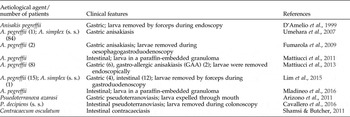
Live anisakid larvae infecting the fillets of fish and the edible part of squids to be consumed raw or undercooked can, if accidentally ingested by humans, cause the zoonotic disease known as anisakidosis. In this respect, the identification of anisakids becomes relevant not only for veterinarians and public or industrial food inspectors, but also for human medicine and pathology. References to the literature reporting human cases of anisakidosis and the aetiological agents involved, as well as the main pathological effects, are shown in table 4.
Cases of anisakidosis so far identified in humans, by means of molecular/genetic markers. The aetiological agent (belonging to the genera Anisakis, Pseudoterranova and Contracaecum), clinical features (number of cases in parentheses) and number of patients found infected (in parentheses) are reported.

Anisakiasis, so-called due to A. simplex (s. l.) larvae as the causative agent, has gained increasing health relevance, in particular in countries or regions where the consumption of raw or only lightly processed fish and squid is frequent or has become increasingly popular (e.g. sushi, sashimi, etc.). Consequently, human cases are reported increasingly from European countries, especially Spain, Italy and France. A number of seafood dishes are considered to be of high risk for the contraction of human anisakiasis in these countries, including boquerones in Spain, and marinated anchovies in Spain and Italy. However, among the nine nominal species of Anisakis, only A. simplex (s. s.) and A. pegreffii are hitherto reported as causative agents of human anisakiasis (D'Amelio et al., Reference D'Amelio, Mathiopoulos, Brandonisio, Lucarelli, Doronzo and Paggi1999; Moschella et al., Reference Moschella, Mattiucci, Minigazzini, De Angelis, Assenza, Lombardo, Monaco, Paggi and Modini2004; Umehara et al., Reference Umehara, Kawakami, Araki and Uchida2007; Fumarola et al., Reference Fumarola, Monno, Ierardi, Rizzo, Giannelli, Lalle and Pozio2009; Mattiucci et al., Reference Mattiucci, Paoletti, Borrini, Palumbo, Macarone Palmieri, Gomes, Casati and Nascetti2011, Reference Mattiucci, Fazii, De Rosa, Paoletti, Salomone Megna, Glielmo, De Angelis, Costa, Meucci, Calvaruso, Sorrentini, Palma, Bruschi and Nascetti2013; Arai et al., Reference Arai, Akao, Seki, Kumagai, Ishikawa, Ohta, Hirata, Nakaji, Yamauchi, Hirai, Shiartori, Koabayashi, Fujii, Ishii, Naito, Saitoh, Yamaguchi, Shibata, Shimo and Tokiwa2014; Lim et al., Reference Lim, Jung, Cho, Yooyen, Shin and Chai2015; Mladineo et al., Reference Mladineo, Popović, Drmić-Hofman and Poljak2016). Anisakis simplex (s. s.) commonly occurs in both the viscera and flesh of many different fish hosts from the NE Atlantic (table 1), while mixed infections of A. simplex (s. s.) and A. pegreffii are common in fish along the Atlantic Iberian coast (table 1). Anisakis pegreffii may cause gastric, intestinal and gastro-allergic anisakiasis (GAA). Indeed, the parasite has been identified in invasive cases of anisakiasis, after observing viable larvae during endoscopy and/or colonoscopy (Mattiucci et al., Reference Mattiucci, Fazii, De Rosa, Paoletti, Salomone Megna, Glielmo, De Angelis, Costa, Meucci, Calvaruso, Sorrentini, Palma, Bruschi and Nascetti2013; Lim et al., Reference Lim, Jung, Cho, Yooyen, Shin and Chai2015), or in surgically removed eosinophilic granulomas (Mattiucci et al., Reference Mattiucci, Paoletti, Borrini, Palumbo, Macarone Palmieri, Gomes, Casati and Nascetti2011; Mladineo et al., Reference Mladineo, Popović, Drmić-Hofman and Poljak2016). Similarly, A. simplex (s. s.) has mostly been associated with invasive cases of gastric and intestinal anisakiasis in Japan (Umehara et al., Reference Umehara, Kawakami, Araki and Uchida2007; Arai et al., Reference Arai, Akao, Seki, Kumagai, Ishikawa, Ohta, Hirata, Nakaji, Yamauchi, Hirai, Shiartori, Koabayashi, Fujii, Ishii, Naito, Saitoh, Yamaguchi, Shibata, Shimo and Tokiwa2014; Lim et al., Reference Lim, Jung, Cho, Yooyen, Shin and Chai2015); however, no cases of GAA due to A. simplex (s. s.) have been reported so far.
Experimental studies with rats, rabbits and pigs have demonstrated that not only A. simplex (s. s.) and A. pegreffii, but also the larvae of Pseudoterranova spp. and Contracaecum spp., are able to penetrate the mucosa and the gastrointestinal tract of mammals (Fagerholm, Reference Fagerholm1988; Strøm et al., Reference Strøm, Haarder, Korbut, Mejer, Thamsborg, Kania and Buchmann2015). However, only very few cases of human anisakidosis caused by the latter two anisakids have been reported to date. The first reports of the disease in humans known as ‘pseudoterranoviasis’ or ‘pseudoterranovosis’ was from North America (Margolis, Reference Margolis1977; Lee et al., Reference Lee, Kim and Choi1985), followed by cases described as transient infections in California. In Korea, human pseudoterranovosis was first described by Lee et al. (Reference Lee, Kim and Choi1985). In those cases, however, the aetiological agent was not identified to species level. More recently, a human case of pseudoterranoviasis caused by P. azarasi was reported from Japan (Arizono et al., Reference Arizono, Miura, Yamada, Tegoshi and Onishi2011); while another one caused by P. decipiens (s. s.) was identified in Italy, likely related to imported infected fish consumed raw or only lightly processed (Cavallero et al., Reference Cavallero, Scribano and D'Amelio2016). Due to the scattered distribution of Pseudoterranova species, it was suggested that its pathological effects in humans could be different from those of the other zoonotic anisakids (Arizono et al., Reference Arizono, Miura, Yamada, Tegoshi and Onishi2011; Mattiucci et al., Reference Mattiucci, Fazii, De Rosa, Paoletti, Salomone Megna, Glielmo, De Angelis, Costa, Meucci, Calvaruso, Sorrentini, Palma, Bruschi and Nascetti2013). Additionally, regional differences likely related to the main anisakid present in the area, seem to exist in the degree of pathology caused by larval Pseudoterranova. For example, in Japan, most patients showed severe pathology caused by larval penetration of the alimentary tract, whereas most cases diagnosed in Europe and Chile have been classified as ‘transient luminal infection’, with worms being expelled by coughing, vomiting or defecation (Smith, Reference Smith1999; McClelland, Reference McClelland2002). Thus, the clinical symptoms caused by Pseudoterranova sp. larvae seem to be weaker than those observed in A. simplex (s. s.) or A. pegreffii infections, or may even be asymptomatic. In some cases, infections were accidentally discovered in patients showing symptoms due to other causes. Clinical symptoms of Pseudoterranova may appear from 2 to 3 h, up to some days, after consumption of the infected fish (Nagano, Reference Nagano, Ishikura and Namiki1989; Cavallero et al., Reference Cavallero, Scribano and D'Amelio2016). In addition, Pseudoterranova spp. have not been reported to induce eosinophilic granulomas, even though light infections of non-penetrating larvae may be accompanied by vomiting and gastric or intestinal pain. No allergic hypersensitivity reactions have been reported so far in these cases, although experimental studies (Asaishi, Reference Asaishi1974; Mattiucci, pers. comm.) have shown that A. simplex (s. l.) and P. decipiens (s. l.) share some common antigens. Taking the latter findings into account, it is possible that the symptoms associated with Pseudoterranova infections may have arisen from cross-reactions to secondary infections, after previous exposure to A. simplex (s. l.) larvae.
Experimental infections of pigs with the larvae of C. osculatum (s. s.) from Baltic cod, have demonstrated that the worm is able to elicit immune responses in homeothermic hosts (Strøm et al., Reference Strøm, Haarder, Korbut, Mejer, Thamsborg, Kania and Buchmann2015), implying that also C. osculatum (s. s.) has to be considered a zoonotic worm. However, scanty data are available so far on human contracaeciasis or contracaecosis. In the past, a single case was reported from the Baltic region (Schaum & Müller, Reference Schaum and Müller1967); however, no species identification was carried out. More recently, two cases were reported from Japan (Nagasawa et al., Reference Nagasawa2012), but again, no molecular species identification was performed. Additionally, a human case of Contracaecum sp. infection was described in Australia (Shamsi & Butcher, Reference Shamsi and Butcher2011); however, in both the latter cases, the symptoms appeared to be milder and were restricted to vomiting, gastric or intestinal pain, and, in some cases, the worm was even expelled spontaneously with the stool.
Studies on the zoonotic potential of these nematodes should be extended, since human cases of anisakidosis are most likely underreported, probably due to unspecific symptoms related to acute and chronic infections. Thus, more accurate diagnostics of human anisakidosis by applying sensitive, rapid and specific molecular methodologies, such as real-time (RT)-PCR-based probe systems (Paoletti et al., Reference Paoletti, Nascetti and Mattiucci2015), or combined with serodiagnostics (immunoblotting) would greatly improve our knowledge on the epidemiology of this fish-borne disease.
Acknowledgements
We wish to thank Alessandra Colantoni and Marialetizia Palomba, for their help in preparing figures and tables. We are very grateful to anonymous referees for their comments on the paper.
Financial support
Part of these studies was supported by Grant no. C26H15SBJ4 of ‘Sapienza – University of Rome’ (year 2015).
Conflict of interest
None.