Introduction
Obesity is a serious public health problem for children and adolescents. According to two National Health and Nutrition Examination Surveys (1976–1980 and 2003–2006), the prevalence of obesity among children increases with age. This prevalence has also increased markedly over the past 25–30 years: for children aged 2–5 years, the prevalence of obesity increased from 5.0% to 12.4%; for those aged 6–11 years, from 6.5% to 17.0%; and for those aged 12–19 years, from 5.0% to 17.6%.Reference Ogden, Carroll and Flegal1 Obese children have higher risk for several health problems, such as cardiovascular disease (including high blood pressure and high cholesterol) and type 2 diabetes mellitus, during childhood and adulthood.Reference Freedman, Kahn, Mei, Grummer-Strawn, Dietz and Srinivasan2, Reference Freedman, Khan, Dietz, Srinivasan and Berenson3 Importantly, obese children and adolescents are more likely than are lean children to remain obese as adults. About 80% of children who were overweight at 10–15 years of age remained overweight or became obese by age 25, according to two studies.Reference Serdula, Ivery, Coates, Freedman, Williamson and Byers4, Reference Whitaker, Wright, Pepe, Seidel and Dietz5 Another study found that 25% of obese adults were overweight as children.Reference Freedman, Khan, Dietz, Srinivasan and Berenson3 The prevalence of overweight or obese children ages 5–19 in the Mexican-American population is even higher, as we have shown in our recent study in which 47% and 44% of the boys and girls, respectively, were either overweight or obese,Reference Hernández-Valero, Bustamante-Montes, Hernandez, Halley-Castillo, Wilkinson and Bondy6 as compared to about 17% in the same age group in the overall US population.Reference Ogden, Carroll and Flegal1
Many imprinted genes are involved in regulating embryonic and early postnatal growth.Reference Bartolomei and Tilghman7 This suggests that the effect of imprinted genes involved in growth may be particularly important in determining body size in children. Indeed, our previous study found multiple linkage peaks with a significant parent-of-origin effect that was associated with body mass index (BMI) in Caucasian children 5–11 years old.Reference Gorlova, Amos, Wang, Shete, Turner and Boerwinkle8 Several genetic syndromes that have a pleiotropic effect on obesity also involve imprinted genes.Reference Cassidy and Driscoll9–Reference Liu, Litman, Rosenberg, Yu, Biesecker and Weinstein12 Linkage studies allowing for the imprinting effect have identified several genetic variants and regions associated with BMI and obesity, and several chromosomal regions with significant parent-of-origin effects were identified in both adultsReference Gorlova, Lei, Zhu, Weng, Shete and Zhang13, Reference Guo, Shen, Liu, Wang, Xiong and Xiao14 and children.Reference Gorlova, Amos, Wang, Shete, Turner and Boerwinkle8
Insulin-like growth factor II (IGF2) plays a key role in mammalian growth, influencing fetal cell division, differentiation and possibly metabolic regulation. Roth et al.Reference Roth, Schrager, Metter, Riechman, Fleg and Hurley15 reported that Caucasian individuals with the AA genotype at the IGF2 ApaI site rs680 (n = 427) had significantly greater fat mass than did Caucasians with the GG genotype (P < 0.05). Martin et al.,Reference Martin, Holly, Davey Smith and Gunnell16 in a 65-year follow-up study of 728 Boyd Orr cohort participants, noted a positive association of childhood BMI and waist-to-hip ratio with circulating levels of IGF2 later in life.
There are several differentially methylated regions (DMRs) in the IGF2/H19 locus, and IGF2 is maternally imprinted (silenced). Heijmans et al.Reference Heijmans, Kremer, Tobi, Boomsma and Slagboom17 investigated the variation in DNA methylation in the IGF2/H19 locus owing to single-nucleotide polymorphisms (SNPs) in 196 adolescent and 176 middle-aged twins. In general, substantial variation in DNA methylation of the H19 and IGF2 DMRs across individuals was observed, underscoring that DNA methylation is a quantitative trait. The heritability of methylation of individual CpG sites varied between 20% and 74% for the H19 DMR and was even higher (between 57% and 97%) for the IGF2 DMR0. SNPs in the IGF2/H19 locus were significantly associated with DNA methylation of the IGF2 DMR0 (P = 0.004). The combined influence of environmental and stochastic factors on DNA methylation was not greater in middle age than in adolescence, suggesting a limited role for age-related degeneration of methylation patterns at this locus. Intriguingly, a negative association was observed between H19 DMR methylation and body size (P < 0.05).Reference Heijmans, Kremer, Tobi, Boomsma and Slagboom17 Another study on the association of methylation in the IGF2/H19 region with body weightReference Perkins, Murphy, Murtha, Schildkraut, Jirtle and Demark-Wahnefried18 found that European and African-American children overweight or obese at 1 year of age had higher levels of methylation in the H19 DMR at birth.
In the current pilot study, we assessed methylation patterns of the H19 DMR in 75 Mexican-American children. The studied H19 DMR has a CpG island that contains five CpG sites, one of which, CpG4, is polymorphic for the rs10732516, an SNP that destroys the CpG site. We hypothesized that interindividual variation in H19 DMR methylation is associated with obesity in children. We chose a Mexican-American population because of the high prevalence of obesity, which is unlikely to be solely caused by environmental factors.
Methods
Study population
The participants in this study were a subsample of the 384 Mexican-American children aged 5–18 years enrolled with their mothers in the Biomarkers of Genetic Susceptibility in Environmentally Exposed Migrant Farmworker (MF) Women and Children Study (From Mother to Child Project), a molecular epidemiological to investigate the health risks associated with chronic pesticide exposure among children of Mexican origin whose mothers worked in agriculture as MFs. The methodology for this study has been previously described elsewhere.Reference Hernández-Valero, Bustamante-Montes, Hernandez, Halley-Castillo, Wilkinson and Bondy6, Reference Hernández-Valero, Herrera, Zahm and Jones19 Briefly, From Mother to Child was a cross-sectional epidemiological study conducted from August 2004 to August 2007 in Baytown, Harris County, and La Joya, Hidalgo County, Texas. The inclusion criteria for the MF children in the study included (a) being of Mexican origin, (b) ages 5–18 years, (c) residents of Baytown or La Joya, Texas and (d) having a mother who had worked as an MF during the 3 years prior to enrollment in the study. A comparison group of non-MF children were matched to the MF children by ethnicity, age and residential status. For the non-MF children to be eligible neither parent must have ever worked in agriculture.
For this study, we included 90 children who had participated in the From Mother to Child study (54 boys and 36 girls), aged 5–11 years at the study entry (median age, 8.28); all of the children were prepubertal. The purpose of our study was to compare the methylation status of the H19 gene between lean and overweight or obese Mexican-American children; we only included children with an age- and sex-specific BMI below the 50th percentile (whom we categorized as lean) and children with a BMI at or above the 85th percentile (whom we categorized as overweight/obese), in accordance with the US Center for Disease Control definitions.Reference Hammer, Kraemer, Wilson, Ritter and Dornbusch20 We excluded children with BMIs in the normal range between the 50th and 85th percentiles in order to enhance the contrast between the two groups.
DNA methylation assay
DNA methylation frequency was determined from genomic DNA from whole blood that was extracted as part the From Mother to Child project in the Biospecimen Extraction Facility at The University of Texas MD Anderson Cancer Center (Houston, TX, USA). We converted 600 ng of DNA with sodium bisulfite treatment using the EZ Methylation kit (Zymo Research Corporation, Irvine, CA, USA), which specifies an input of 2 pg–2 μg of DNA; we found that the input of 600 ng worked well with nested primers. This process transforms unmethylated cytosines into uracils, leaving behind methylated cytosines. The template was then amplified using post-bisulfite treatment-specific primers. Methylation status was determined by Sanger sequencing on the 3730xl DNA Analyzer (Applied Biosystems, Carlsbad, CA, USA; described below). Methylated and unmethylated controls were used during bisulfite treatment. Sanger sequencing showed C peaks in the methylated controls, indicating the presence of methylation. Unmethylated controls showed T peaks, indicating mostly unmethylated DNA.
DNA methylation was assessed in a 5′ region of the H19 transcript encompassing a CpG island corresponding to the sixth CTCF binding site, which is part of the H19 DMR. The H19 amplicon was amplified using nested primers that spanned a 637-bp region [National Center for Biotechnology Information (NCBI) Homo sapiens chromosome 11 genomic contig, GRCh37.p10 Primary Assembly: 1960756–1961393]. These constructs were designed on the reverse complement strand. The first primer sequences were (forward) 5′-GGTGTTTTAGTTTTATGGATGATGG-3′ and (reverse) 5′-TCCTAAATTAACACCTCAAACCTACA-3′. The second primer sequences were previously reported:Reference Takai, Gonzales, Tsai, Thayer and Jones21 (forward) 5′-GTAGGGTTTTTGGTAGGTATAGAGT-3′ and (reverse) 5′-CACTAAAAAAACAATTATCAATTC-3′. The sequencing primer was 5′-GGAGGTTTTTTTTTYGGTTTTATYG-3′. This amplicon contained five CpG sites of interest, the fourth of which is also an SNP (rs10732516). These CpGs are located at positions 1961220/210/208/206 (also a SNP)/203 (NCBI Homo sapiens chromosome 11 genomic contig, GRCh37.p10 Primary Assembly, Ref seq NT009237.18). Sequencing was performed by the Sequencing and Microarray Facility at MD Anderson.
SNP detection
We amplified rs10732516 in samples of genomic DNA not treated with bisulfite. We used the primers (forward) 5′-CGGATGGTGCAGAATTGGTTGTAGC-3′ and (reverse) 5′-CCGCCTCTCGGCCATTTCCG-3′, which encompass a 1787-bp region (NCBI Homo sapiens chromosome 11 genomic contig, GRCh37.p10 Primary Assembly: 1960278–1962065). The sequencing primer 5′-CGTGGAAACATCCCAGGTCATCCAA-3′ was used to determine the frequency of rs10732516 using Sanger sequencing (Supplementary Fig. S1). We successfully genotyped 80 children (48 boys and 32 girls).
Sanger sequencing
We analyzed polymerase chain reaction products using a sequencing primer that binds to the reverse complement strand. Competitive binding of nucleotides (dNTPs) and fluorescently labeled dideoxynucleotides (ddNTPs) occurred in the presence of a DNA polymerase. Whereas incorporating dNTPs allowed strand elongation, the addition of ddNTPs, which lack a 3′-OH group, resulted in extension termination. The template comprised four separate reactions, each of which contained normal dNTPs (dATP, dCTP, dGTP and dTTP) along with only one ddNTP (ddATP, ddCTP, ddGTP or ddTTP). This resulted in the formation of extension products of differing lengths. Capillary electrophoresis was carried out using POP (performance optimized polymer)-7 matrix (Life technologies, Carlsbad, CA, USA) to separate the amplified DNA products. Once separation was achieved, we used a laser beam to excite the dye molecules, causing them to fluoresce. Fluorescence emissions were captured simultaneously, separated by a spectrograph, and then recorded. 3730xl instrument and data collection software (Life technologies, Carlsbad, CA, USA) was used to display the information as an electropherogram. Of the 80 genotyped children, we successfully generated methylation data for 75 children (44 boys and 31 girls).
Sanger sequencing showed that methylation frequency at CpG4 fell into one of two categories. All samples either showed partial methylation (both a C and a T peak were present at that site) or no methylation (only a T peak was present at that site). The other four non-polymorphic CpG sites all showed both C and T peaks, which we interpreted as partial methylation. Because these CpG sites belong to an imprinted region, we expected partial methylation, with methylated paternal DNA and unmethylated maternal DNA.
Statistical analysis
We used the χ 2-test to compare the binary/categorical variables between lean and overweight children and to evaluate either the association between the methylation of the CpG4 site and body size-related phenotypes or the association of methylation status with maternal factors. When the number of observations in any cell was below 5, we used the Fisher's exact test. We used the t-test to compare quantitative traits. We also performed binary logistic regression analysis, treating overweight status as the outcome and methylation status (unmethylated v. one allele methylated) or rs10732516 genotype as a predictor; we adjusted for maternal BMI, residential status (urban or rural) and sex where appropriate.
Results
Methylation patterns in the H19 DMR
Of the five CpG sites in the studied DMR, only one of them, CpG4, is polymorphic – the SNP rs10732516 destroys the CpG site in the carriers of the minor allele. The four non-polymorphic CpG sites all showed partial methylation (both C and T peaks) as expected for this imprinted region. Unfortunately, Sanger sequencing is not quantitative and therefore did not allow us to evaluate the degree of a loss of imprinting (resulting in biallelic methylation), unless the loss of imprinting was complete (i.e. producing only C peaks), which was not observed in this study. Likewise, we did not observe any case of complete loss of methylation for any of these CpG sites (manifested by T peaks only).
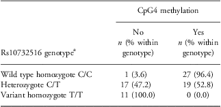
The genotype frequencies of the SNP rs10732516 did not deviate from the Hardy–Weinberg equilibrium. Owing to the presence of the SNP, CpG4 demonstrates a distinct methylation pattern that can be largely derived from the SNP genotype (Table 1). Among wild-type homozygotes, only one child, a girl with a BMI in the 10th percentile, had no detectible methylation at this site. All others had partial methylation at this site. Among heterozygotes, about half of the children had partial methylation at this site (indicating that the SNP was located on the maternal allele, normally unmethylated), and the rest had no methylation (indicating that the SNP was located on the paternal allele, which is normally methylated). Finally, in the variant homozygotes without a CpG4 site, methylation was absent.
Rs10732516 genotype and methylation at the CpG4 site in Mexican-American children
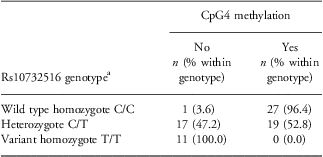
aThe data on both genotyping and methylation were successfully generated on 75 children.
Association between childhood obesity and CpG4 methylation status in a Mexican-American population
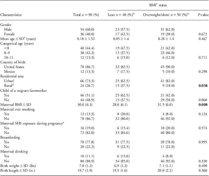
As shown in Table 2, lean and overweight children did not significantly differ by age, sex, country of birth (United States or Mexico), or birth weight and length. The urban group had a higher proportion of overweight children than did the rural group. The lean group had an insignificantly higher proportion of mothers who were MFs than did the overweight group. A higher maternal BMI was strongly associated with the child's overweight status. No other maternal factor was associated with overweight status.
Characteristics of 75 Mexican American children, including weight status

aBMI, body mass index; SD, standard deviation; SHS, secondhand smoke.
bLean: BMI < 50th percentile; overweight and obese, BMI ⩾ 85th percentile (according to the CDC's definition for age and sex).
cUrban: Baytown, Harris County, Houston Metropolitan Area.
dRural: La Joya, Lower Río Grande Valley, Texas-Mexico Border Area.
P-values below 0.05 are shown in bold.
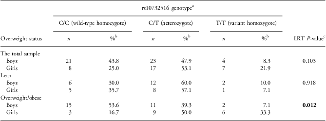
Girls had a higher frequency of the variant T allele at rs10732516 (minor allele frequency (MAF) = 0.484) than did boys (MAF = 0.323; P = 0.043), perhaps because of the limited number of study participants. Interestingly, the genotype distribution did not differ between girls and boys among the lean children or the overall sample; however, boys were significantly more likely to carry a wild-type C allele than were girls among the overweight and obese children (P = 0.012; Table 3). Accordingly, overweight or obese girls less often showed partial methylation of the CpG4 site and more often showed no methylation at this site as compared with overweight or obese boys (P = 0.002). In the logistic regression model for the overweight v. lean status, which included sex, rs10732516 genotype and their interaction term, the effects of genotype and of the interaction term itself were significant (P = 0.033 and 0.023, respectively), whereas the effect of sex did not reach statistical significance (P = 0.055; Supplementary Table S1).
Sex difference in the rs10732516 genotype by overweight status in Mexican-American children
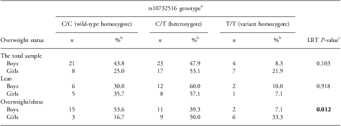
aThe genotyping data were successfully obtained from 80 children.
bWithin sex.
cLRT, likelihood ratio test.
P-values below 0.05 are shown in bold.
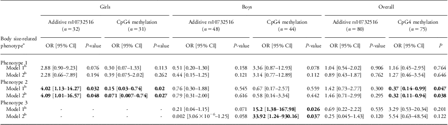
In accordance with the genotype difference by sex among overweight and obese children, both CpG4 methylation and the genotype of rs10732516 had opposite effects on the overweight/obese status in boys and girls, although in neither sex did the association reach statistical significance (Table 4). CpG4 methylation and the wild-type genotype had a borderline positive association with an overweight/obese status (85th percentile and above v. 50th and below) in boys, whereas in girls, both of these variables were associated with a lean status.
Association of rs10732516 genotype and CpG4 methylation status with body size-related phenotypes in Mexican-American children
OR, odds ratio; CI, confidence interval; BMI, body mass index.
aPhenotype 1 = Overweight (1) v. lean (0).
Phenotype 2 = birth weight above (1) v. below (0) the median.
Phenotype 3 = low birth weight (<2500 g) and currently lean (<50th percentile); only one girl had this phenotype.
bModel 1, no adjustment; model 2, adjusted for maternal BMI, residence (urban v. rural), and sex where appropriate.
P-values below 0.05 and corresponding adds ratios and confidence intervals are shown in bold.
Because methylation of growth-associated genes has been shown to play an important role in prenatal development,Reference Seki, Williams, Vuguin and Charron22, Reference Simmons23 we performed a secondary analysis of the association between birth weight on the one hand and CpG4 methylation and rs10732516 genotype on the other. We observed that girls with the variant genotype or lower methylation were significantly more likely to have a birth weight above the median (which was equal to 7.1 lbs in this sample). There was no association in boys, while for both sexes, lower CpG4 methylation was associated with a higher birth weight (P = 0.047). Boys with CpG4 methylation had a more than 15 times greater risk of being overweight/obese and of having a normal or high birth weight than did boys without CpG4 methylation (P = 0.026; Table 4). Since only one girl had a low birth weight (<2500 g), this analysis was not done for the girls alone, although they were included in the overall analysis.
To disentangle the effects of CpG4 methylation and SNP rs10732516 on obesity, we performed a stratified analysis that included only heterozygotes, who did not differ in genotype but did differ in methylation status. We were not able to detect any significant association of the methylation status, either with birth weight or with overweight/obese status, either overall or by sex. The lack of significant difference may have resulted from the smaller sample, owing to stratification.
Maternal factors such as BMI, smoking, drinking or breast feeding and its duration were not associated with the children's methylation status of any of the studied CpG sites. Only high maternal BMI was associated with an overweight/obese status of the children, but adjusting for BMI (along with adjusting for type of residence and sex in the overall analyses) did not change the significance of the observed associations (Table 4).
Discussion
Several studies have suggested that imprinting and methylation can play a role in weight control and childhood obesity.Reference Campion, Milagro and Martinez24–Reference Pinnick and Karpe27 To our knowledge, ours is the first study to pinpoint a discrete methylation event associated with weight at birth and obesity.
The results from our current pilot study demonstrate a complex pattern of methylation in the H19 DMR and suggest that CpG4 methylation status may be associated with childhood obesity among Mexican-Americans in a sex-specific manner. This study demonstrates the importance of taking into account the polymorphic status of CpGs when interpreting the results of methylation analysis. This is especially important considering that CpG sites in the human genome are often polymorphic.Reference Hodgkinson and Eyre-Walker28 Our results also suggest that methylation of a DMR on the polymorphic background of the methylated site may contribute to childhood obesity.
We noted a higher frequency of a wild-type rs10732516 allele in overweight/obese boys compared with overweight/obese girls. This results in a higher frequency of CpG4 methylation among boys than girls in overweight and obese children. Interestingly, boys of Mexican origin have higher obesity rates than girls.Reference Hernández-Valero, Bustamante-Montes, Hernandez, Halley-Castillo, Wilkinson and Bondy6 It has been also shown that male offspring of smoking pregnant women have higher methylation at the IGF2 DMR than male offspring of non-smoking women, while maternal smoking status had no impact on the methylation in that region among female offspring.Reference Murphy, Huang and Hoyo29 Taken together, those results suggest that IGF2 DMR methylation can be gender sensitive and may contribute to a higher obesity incidence in boys.
Not all studies demonstrate association of the IGF2/H19 methylation with birth weight. For example, Huang et al.Reference Huang, Galati, Burrows, Beilin, Li and Pennell30 found a negative association of IGF2/H19 methylation with head circumference but not birth weight or birth length. A study by Burris et al.Reference Burris, Braun, Byun, Tarantini, Mercado and Wright31 did not detect an association between birth weight and methylation in the imprinted IGF/H19 region (among other loci). However, in an analysis stratified by the methylation status of the CpG dinucleotide in the IGF2 imprinting control region (ICR)1 (the same site as the one denoted in our study as CpG4, which harbors the SNP rs10732516) infants with this site unmethylated showed an association of birth weight with mean methylation status of both IGF2 ICR1 and ICR2. Specifically, among infants with the absence of methylation at this position, increased mean methylation of ICR1 was associated with a lower birth weight. Similarly, there was a trend between increased methylation of ICR2 and lower birth weight among the infants in whom this site was unmethylated. This indicates importance of taking into account the polymorphic status of CpG sites. We could not perform the same analysis because our study was lacking the quantitative data on ICR1 methylation and did not consider methylation in ICR2.
The differences between the studies may be related to both the methods used to assess methylation (pyrosequencing in Burris et al.,Reference Burris, Braun, Byun, Tarantini, Mercado and Wright31 Sequenom MassARRAY in Huang et al.,Reference Huang, Galati, Burrows, Beilin, Li and Pennell30 and Sanger sequencing in our study) and to the differences in the population characteristics – methylation being assessed at age 17 in Huang et al.,Reference Huang, Galati, Burrows, Beilin, Li and Pennell30 in newborns in Burris et al.,Reference Burris, Braun, Byun, Tarantini, Mercado and Wright31 and in prepubertal children in our study. Although the age-dependent variation in methylation is well documentedReference Hackett and Surani32–Reference Teschendorff, Menon, Gentry-Maharaj, Ramus, Weisenberger and Shen34 much less is known about the specific DMRs regulating imprinted domains.Reference Murphy, Huang and Hoyo35 The studies point out that methylation of these regions can serve as a biosensor of exposure,Reference Murphy, Huang and Hoyo35 and there may have been differences between these study populations in terms of various unmeasured exposures.
Because this was a pilot study, we had a small sample population. Thus, the strong effects we observed might have been caused by random fluctuations owing to the limited sample. Because of the sample size and the multiple comparisons performed, we cannot rule out a chance association, especially with birth weight. Our study was limited in the phenotypic measures we used (BMI and birth weight), and since IFG2 might be more relevant to muscle mass than the overall body size,Reference Heijmans, Kremer, Tobi, Boomsma and Slagboom17 the measures of body composition would be important to assess in future studies.
Another limitation of our study was that we assessed CpG4 methylation not at birth, but 6 to 11 years later. Although a recent study demonstrated remarkable stability in methylation profiles of several imprinted gene DMRs, including the H19 DMR, between birth and 1 year of age,Reference Murphy, Huang and Hoyo35 it remains unknown whether this stability can be extrapolated further in time. Factors other than imprinting are shown to affect methylation status; in particular, it has also been related to stress exposure in children. Two studies have demonstrated differences in methylation between children exposed to early adversity and untraumatized children.Reference Bick, Naumova, Hunter, Barbot, Lee and Luthar36, Reference Yang, Zhang, Ge, Weder, Douglas-Palumberi and Perepletchikova37 Our study did not take stress exposure into consideration. Larger studies may be helpful to further explore this association.
Acknowledgments
The authors are grateful to Diane Hackett and Jill Delsigne for their help with the editing of this manuscript.
Financial Support
The study was supported by the pilot project award to M.A.H-V. and O.Y.G. from the National Institute of Environmental Health Sciences grant 5 P30 ES00778413, and by the Project EXPORT awarded to M.A.H-V. from the National Center for Minority Health and Health Disparities grant P60 MD000503-04.
Conflicts of Interest
None
Ethical Standards
The authors assert that all procedures contributing to this work comply with the ethical standards of the relevant national guidelines on human experimentation and with the Helsinki Declaration of 1975, as revised in 2008, and have been approved by the Institution Review Board of The University of Texas MD Anderson Cancer Center. The study included a bilingual (Spanish and English) written informed consent. All study participants signed the informed consent (informed assent for children younger than 7) at the time of enroll.