Staphylococcus aureus is considered the most frequently isolated pathogen from bovine IMI worldwide, primarily causing chronic subclinical mastitis (Calvinho and Dallard, Reference Calvinho and Dallard2023). Chronically infected lactating cows are probably the main reservoir of the organism, and the primary transmission mode is through contact of non-infected susceptible mammary quarters with fomites contaminated with infected milk during milking (Zecconi et al., Reference Zecconi, Calvinho and Fox2006). However, S. aureus IMI have been detected in heifer calves and preparturient heifers (Oliver et al., Reference Oliver, Gillespie, Headrick, Lewis and Dowlen2005), leading to the identification of extramammary sources of infection such as body sites of dairy animals, flies and dairy farm environments (Haveri et al., Reference Haveri, Hovinen, Roslöf and Pyörälä2008; Capurro et al., Reference Capurro, Aspán, Ericsson Unnerstad, Persson Waller and Artursson2010; Anderson et al., Reference Anderson, Lyman, Moury, Ray, Watson and Correa2012). Heifers can acquire IMI at a very early age or during pregnancy, impairing mammary growth and development, compromising future milk production and becoming a potential source of infection as they calve and enter the milking herd (Oliver et al., Reference Oliver, Gillespie, Headrick, Lewis and Dowlen2005). Although several studies have detected the presence of S. aureus IMI in heifers before parturition (antepartum: Oliver et al., Reference Oliver, Gillespie, Headrick, Lewis and Dowlen2005), only a few have evaluated clonal relationships between S. aureus isolated from heifer mammary gland secretion shortly before or after calving and cows in the milking herd, noting that within a given herd, isolates belonging to these two origins were either related (Haveri et al., Reference Haveri, Hovinen, Roslöf and Pyörälä2008; Anderson et al., Reference Anderson, Lyman, Moury, Ray, Watson and Correa2012) or not (Castelani et al., Reference Castelani, Silva Santos, dos Santos Miranda, Zafalon, Rodrigues Pozzi, Rodrigues Pozzi and Arcaro2013; Camussone et al., Reference Camussone, Molineri, Signorini, Neder, Vitulich and Calvinho2020). In addition, the duration of IMI naturally acquired by heifers antepartum, their potential persistence through their first lactation, and the ability of these isolates to infect herd mates during lactation have not been explored. Therefore, the present study aimed to determine the persistence and spread of IMI during the first lactation of heifers that were naturally infected with S. aureus antepartum in a herd with a high prevalence of this organism in lactating dairy cows, characterizing the isolates at the molecular level.
Materials and methods
Study description
Quarter milk samples were obtained from all primigravid heifers due to calve during the period from June 2018 to May 2019, 15 d before their expected calving date. Those heifers from which S. aureus was isolated antepartum (naturally infected) were followed up during their first lactation to determine potential persistence of IMI and characteristics of the isolates causing IMI. Milk samples were obtained from all the cows in the lactating herd at the beginning of the study and one year later. S. aureus isolated at both sampling times were compared with those obtained from heifers to establish their clonal relationships (online Supplementary Fig. S1). All procedures adhered to the Guide for the Care and Use of Agricultural Animals in Agricultural Research and Teaching (FASS, 2010) and were approved by the Ethics and Security Committee of the Centro Regional Santa Fe, Instituto Nacional de Tecnología Agropecuaria (CICUAE, No 18-006).
Herd description and animals
The study was carried out in a dairy herd located in the Santa Fe province of Argentina. Dairy heifers included in the present study were selected based on the presence of naturally acquired S. aureus IMI antepartum. A Holstein × Jersey dairy herd (≈ 170 milking cows) that reported mastitis problems due to S. aureus during the four years prior to the initiation of the experiment was used, with an S. aureus IMI prevalence at cow level higher than 15%. Mastitis control practices consisted of administration of dry cow antibiotic therapy with rifaximin in all quarters of all cows at drying off and treatment of clinical mastitis with a macrolide antibiotic (spiramycin) as first choice protocol, and a beta-lactam antibiotic (cloxacillin) in case of clinical failure of first choice protocol. All S. aureus isolated during this study and previously were susceptible, in vitro, to both antibiotics. Pre milking routine consisted of forestripping and teat washing with water before attaching the milking units. Milkers used latex gloves inconsistently. Post milking teat disinfection was carried out with a 0.5% iodophor formulation. The average milk yield per cow was 22.4 l and the average somatic cell count (SCC) was 790 × 103 cells/ml in the year prior to the start of this study. Cows were kept under grazing conditions on alfalfa-based pastures, milked twice daily in a single 10 herringbone parlor and received concentrates in the milking parlor. Heifer calves were raised in a collective facility near to the dairy farm and fed pasteurized milk from a different dairy herd. Fly control on heifer calves was carried out by applying pour-on insecticide (cypermethrin) from 60 d and insecticide-impregnated ear tags (organophosphate) in adult cows. Heifers were artificially inseminated at approximately 13 months and 15 d before the expected calving date returned to the dairy farm and were located in a group maternity area, fed a balanced TMR, alfalfa hay and had free access to water.
A vaccination study with an experimental S. aureus immunogen was carried out concomitantly in the herd, including primigravid heifers and second-lactation cows free from S. aureus IMI before parturition (Camussone et al., Reference Camussone, Reidel, Molineri, Cicotello, Miotti, Suarez Archilla, Curti, Veaute and Calvinho2022). The heifers included in the present study were not enrolled in the vaccination trial.
Sampling of lactating dairy cows
At the beginning of the study and one year later all of the lactating cows (n = 163 and n = 178, respectively) were sampled. Composite milk samples were taken and cultured for bacteriological detection of S. aureus according to standard procedures (Oliver et al., Reference Oliver, Gonzalez, Hogan, Jayarao and Owens2004). At the time of first sampling 24.7% were first-lactation cows, 26% second-lactation and 49.3% third-lactation or higher. At the time of second sampling, 37.3% were first-lactation cows, 18.2% were second-lactation, and 44.5% were third-lactation or higher. Eighty-two cows were present both at the first and second sampling times.
Sampling of heifers before parturition
Seventy-seven heifers that calved between June 2018 and April 2019 were included in the study. First quarter samples were taken 15 d before the expected calving date (before heifers returned to the original dairy herd) and cultured for bacteriological detection of S. aureus, following standard procedures (Oliver et al., Reference Oliver, Gonzalez, Hogan, Jayarao and Owens2004). Heifers included in this study showed no teat lesions and/or macroscopic signs of inflammation at the time of sampling. Heifers with IMI due to S. aureus antepartum confirmed by bacteriological culture entered the dairy herd after calving and milk samples were collected from individual quarters of these heifers at monthly intervals throughout lactation (for nine months or at least for three months for the last ones that were included in the study) to determine the presence and duration of the IMI (i.e. elimination of the infection or progression towards chronicity: online Supplementary Materials and Methods). These animals did not receive antimicrobial therapy during the evaluation period. In a separate experiment, heifers that were uninfected before parturition, as well as 37 uninfected second-lactation cows were enrolled in an experimental vaccination trial and entered the lactating herd either as vaccinated or controls (Camussone et al., Reference Camussone, Reidel, Molineri, Cicotello, Miotti, Suarez Archilla, Curti, Veaute and Calvinho2022).
Bacteriological examination
Milk and mammary secretion samples were cultured to detect mastitis pathogens according to standard procedures, seeding 10 μl of sample in a quarter of a blood agar plate (Columbia agar supplemented with 5% bovine blood: Oliver et al., Reference Oliver, Gonzalez, Hogan, Jayarao and Owens2004). Presumptive phenotypical identification was performed based on colony morphology, Gram staining, catalase test and tube coagulase test using rabbit plasma. The presence of one colony of coagulase-positive Staphylococcus on blood agar was considered a positive identification, therefore, the detection limit was 100 CFU/ml. Milk samples with 3 or more different bacterial species (based on colony morphology) were considered contaminated. Isolates were kept at −80°C in BHI-glycerol 20% until further use (online Supplementary Materials and Methods).
Molecular characterization of isolates
Genomic DNA was extracted with a commercial kit (Pure PCR Template Preparation Kit, Roche, Germany) and quantified using a spectrophotometer (NanoDrop 2000, Thermo Fisher Scientific, USA). Coagulase-positive Staphylococci were confirmed as S. aureus by PCR amplification of the nucA gene (Brakstad et al., Reference Brakstad, Aasbakk and Maeland1992). The presence of cap5 and cap8 loci, clfA, clfB, fnbA, fnbB and the accessory gene regulator (agr) were determined by PCR (Bardiau et al., Reference Bardiau, Caplin, Detilleux, Graber, Moroni, Taminiau and Mainil2016; Camussone et al., Reference Camussone, Molineri, Signorini, Neder, Vitulich and Calvinho2020). Clonality of S. aureus isolates was assessed by PFGE of SmaI-digested chromosomal DNA fragments using a CHEF-DR II apparatus (BioRad Laboratories, CA, USA: Camussone et al., Reference Camussone, Molineri, Signorini, Neder, Vitulich and Calvinho2020). A dendrogram was constructed based on the UPGMA algorithm with TREECON 1.3b. Cluster similarity was set at 80%, and clusters were identified with upper cases. Within a cluster, isolates with identical PFGE band patterns were considered indistinguishable genotypes (clonal). Clonal isolates comprised a pulsotype (PT) and were identified with upper cases plus a numeric suffix, if needed. One of the clonal isolates per PT was subjected to multi-locus sequence typing (MLST) based on DNA sequencing of the 7 housekeeping genes arcC, aroE, glpF, gmk, pta, tpi, and yqiL (Enright et al., Reference Enright, Day, Davies, Peacock and Spratt2000) and sequence types were defined using PubMLST (Jolley et al., Reference Jolley, Bray and Maiden2018). Fourier Transform Infrared (FTIR) spectroscopy was employed to subtype isolates according to their molecular fingerprint, primarily based on their glycostructural composition, including capsular polysaccharide (CP) expression (Johler et al., Reference Johler, Stephan, Althaus, Ehling-Schulz and Grunert2016). Capsular serotyping was examined based on an artificial-neural net using the raw, unprocessed FTIR spectra (Grunert et al., Reference Grunert, Wenning, Barbagelata, Fricker, Sordelli, Buzzola and Ehling-Schulz2013). Isolates were discriminated into either encapsulated strains (CP5 or CP8) or non-encapsulated strains (NT). Three independent experiments with minimum technical duplicates were run per sample.
Definition of intramammary infection
Staphylococcus aureus IMI was defined as two consecutive or interspersed samples with S. aureus positive identification (Sears et al., Reference Sears, Smith, English, Herer and Gonzalez1990), and identical PT. The isolation of S. aureus from one mammary quarter milk sample of a heifer at only one sampling time was considered a non-persistent (transient) IMI, while persistent IMI were those in which the same organism that established an IMI was isolated at least once from the same mammary quarter during the remainder of the lactation. Since lactating cows were sampled only in two fixed periods, the presence of transient or persistent IMI was not defined for this category of animals.
Statistical analysis
The associations between PT/cluster and origin of isolate (cow vs. heifer), number of lactations and presence of a given virulence factor were tested by a generalized linear mixed model with multinomial distribution, animal ID as random factor, and identity link function. The distribution of PTs between first and second sampling times was compared using the Chi-square test. (InfoStat software,Universidad Nacional de Córdoba, Argentina).
Results
Prevalence of S. aureus in heifers antepartum and lactating cows
At the time of the first and second sampling of lactating cows, 30 (18.4%) and 28 cows (15.7%), respectively, were positive for S. aureus. Two cows showed signs of clinical mastitis (CM) at the first sampling time. Six cows were positive for S. aureus in both the first and second sampling (online Supplementary Table S1). Seven heifers (7.8%) were positive for S. aureus in at least 1 of the quarters at antepartum sampling. From these animals, 13 isolates were obtained since one heifer presented infection in all 4 quarters, one in 3 quarters, one in 2 quarters and four in only 1 quarter (4.2% quarter level prevalence: online Supplementary Table S2).
Clonality of S. aureus isolates
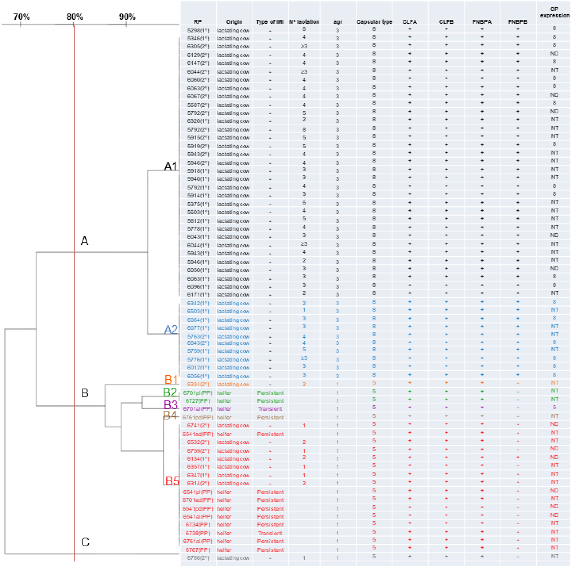
PFGE typing produced three clusters: A (43 isolates; A1 = 33 isolates, A2 = 10 isolates), B (21 isolates; B1 = 1 isolate, B2 = 2 isolates, B3 = 1 isolate, B4 = 1 isolate, B5 = 16 isolates) and C (1 isolate: Fig. 1). A significant association between cluster and origin of isolates (cow vs. heifer) was observed (P < 0.01: online Supplementary Table S3). Cluster A was composed entirely of isolates obtained from lactating cows (A1 n = 33; A2 n = 10; total n = 43, 82.7% of lactating cows), while all isolates obtained from heifers antepartum were grouped in cluster B (B2 n = 2; B3 n = 1; B4 n = 1; B5 n = 9; total n = 13). Only 15.4% of lactating cow isolates were clustered in B (B1 n = 1; B5 n = 7; total n = 8) and 1.9% in C (n = 1). Both isolates obtained from cows with CM belonged to PT A1.
Dendrogram of S. aureus SmaI PFGE profiles isolated from milk of 52 lactating cows and mammary secretions of 13 quarters from heifers antepartum. Type of IMI, Isolate origin (cow/heifer), agr type (1 to 4), Capsular type (5 or 8), presence (+) or absence (−) of selected S. aureus virulence factor genes, and Capsule (CP) expression, are shown.
References: RP: isolate identification; (1°): isolate obtained at first sampling; (2°) isolate obtained at second sampling. NT, non-encapsulated; ND, not determinable.

Regarding S. aureus isolated from lactating cows, a significant association was observed between cluster and number of lactations (P < 0.001: online Supplementary Table S3). All isolates obtained from lactating cows grouped in cluster B corresponded to isolates obtained from first- and second-lactation cows (n = 8), while all isolates obtained from cows with more than 3 lactations were grouped in cluster A (n = 38). Considering isolates of the same PT to be clonal, MLST for one isolate per PT was performed. The two isolates from PT A1 and A2 were assigned to CC1/ST1, while the five isolates from PT B1 to 5 were assigned to CC97/ST2187, and the single isolate of PT C to ST350 (CC350).
Molecular characterization and FTIR-spectroscopic fingerprinting of S. aureus isolates
All S. aureus tested were positive for fnbA, clfA and clfB, and 67.7% were positive for fnbB (Fig. 1). Origin of isolates (cow vs. heifer) was associated with the presence of fnbB, being more frequent in isolates from cows (84.6%) than from heifers (0%: P < 0.001). A significant association was also observed between PT and the presence of fnbB (P < 0.001). All isolates from cluster A were positive for fnbB (n = 43), while 95.2% of isolates from cluster B and the only isolate in cluster C were negative for fnbB.
Only agrI and agrIII were found among the isolates evaluated, being 33.8% positive for agrI and 66.2% for agrIII. Origin of isolates (cow vs. heifer) was associated with agr type; agrIII was more frequent in isolates from cows (n = 43, 82.7%), while agrI was more frequent in isolates from heifers (n = 13; 100%: P < 0.001).
All S. aureus from PTs in cluster A were genotyped as agrIII, cap8, fnbA, fnbB, clfA, and clfB, while 95.2% and 100% of isolates in clusters B and C, respectively, were genotyped as agrI, cap5, fnbA, fnbB(−), clfA, clfB (Table 1). FTIR-based capsule serotyping showed that 19 isolates were found to be encapsulated (CP5, n = 1; CP8 n = 18), 35 were NT, and 8 were not determinable (Fig. 1). Isolates of PFGE cluster A had a similar proportion of encapsulated (CP8, 47.4%) and NT (52.6%) isolates, while isolates of PFGE cluster B were nearly all NT (93.3%). Hierarchical cluster analysis of the FTIR data discriminated eleven distinct clusters divided into two major clusters, A and B (online Supplementary Table S4). FTIR cluster A was the most prominent cluster (58 isolates, 95%), which was further divided into nine distinct subclusters. One of the two predominant subclusters (A1.1.1, n = 16) comprised CP8 isolates, while mainly NT isolates were assigned to subcluster A1.2.1 (n = 25). FTIR cluster B comprised three isolates of PT A1 and the single isolate of PT C, all isolates lacking capsule expression. Within PFGE cluster A, nine biotypes could be distinguished using FTIR spectroscopy, and the PFGE cluster B lineage revealed four different biotypes.
Molecular characterization of Staphylococcus aureus isolated from mammary secretions of different origins (lactating cows vs. heifers before parturition)

MLST, muti-locus sequence typing; ND, undetermined.
Evolution and spread of S. aureus IMI in heifers infected before parturition
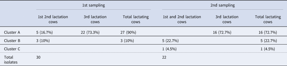
Eleven isolates (84.6%, from 7 animals) obtained from heifers antepartum belonged to PT B5, B2 and B4, and led to persistent IMI that lasted, in some cases, until the end of lactation (confirmed by same PT in PFGE analysis). The other 2 isolates (from 2 different animals), belonged to PT B5 and B3, and yielded non persistent (transient) infections (online Supplementary Table S2). At the beginning of the study (1st sampling of lactating cows), 90% of the isolates circulating in the lactating herd belonged to PTs in cluster A (A1 n = 19; A2 n = 8; total n = 27) and 10% to PT B5 (n = 3). At the end of the study (2nd sampling of lactating cows), after introduction of infected heifers to the herd, 72.7% of the circulating isolates belonged to PTs in cluster A (A1 n = 14; A2 n = 2; total n = 16), 22.7% to cluster B (B1, n = 1; B5 n = 4; total n = 5) and 4.5% to cluster C (n = 1: Table 2). The distribution of PTs between 1st and 2nd sampling of lactating cows was not statistically different (P > 0.05). Pulsotypes B2, B3 and B4 were restricted to heifers infected antepartum since they were not found in cows of first, second or more lactations.
Distribution of Staphylococcus aureus clones at first and second sampling among lactating cows according to parity

First sampling was May 2018 and second was 2019. Heifers infected before parturition entered the milking herd from June 2018 to April 2019.
Discussion
The prevalence of S. aureus and coagulase-positive Staphylococcus (CPS) IMI in primigravid heifers before parturition varies widely between studies, ranging from 0.4 to 14% at quarter level (reviewed by Oliver et al., Reference Oliver, Gillespie, Headrick, Lewis and Dowlen2005 and De Vliegher et al., Reference De Vliegher, Fox, Piepers, McDougall and Barkema2012). In the present study, the heifer and quarter S. aureus IMI prevalence before parturition was 7.8 and 4.2%, respectively. Several potential risk factors associated with heifer IMI before parturition and after calving have been identified, and among them lack of fly control, seasonal effects and increased susceptibility to mastitis in the last trimester of pregnancy have been associated with a higher prevalence of S. aureus IMI (Fox et al., Reference Fox, Chester, Hallberg, Nickerson, Pankey and Weaver1995). Exploring these potential factors was beyond the objectives of this study. Our research was conducted in a herd with a high prevalence of S. aureus IMI in lactating cows. However, in previous studies, this variable was not considered to be significantly associated with the prevalence of CPS IMI in heifers shortly after parturition (Roberson et al., Reference Roberson, Fox, Hancock, Gay and Besser1994) or of S. aureus before parturition (Camussone et al., Reference Camussone, Molineri, Signorini, Neder, Vitulich and Calvinho2020).
Several studies detected the presence of S. aureus or CPS IMI in heifers antepartum (Haveri et al., Reference Haveri, Hovinen, Roslöf and Pyörälä2008; Castelani et al., Reference Castelani, Silva Santos, dos Santos Miranda, Zafalon, Rodrigues Pozzi, Rodrigues Pozzi and Arcaro2013) or shortly after parturition (Roberson et al., Reference Roberson, Fox, Hancock, Gay and Besser1994; Anderson et al., Reference Anderson, Lyman, Moury, Ray, Watson and Correa2012). However, in these studies, only phenotypic characterization of the isolates was performed to confirm IMI, and the persistence of S. aureus IMI during the first lactation of heifers naturally infected antepartum was not explored. Here, the establishment of an IMI was confirmed based on molecular methods. Following this criterion, more than 80% (11/13) of S. aureus isolated from heifers antepartum yielded an IMI that persisted during lactation, underscoring the ability of these isolates to establish in the mammary gland and potentially be transmitted during milking time.
Molecular epidemiology studies of S. aureus isolated from bovine IMI are crucial to assess the dynamics and diffusion of circulating clones and potential sources of infection within a herd or between herds (Zadoks et al., Reference Zadoks, Middleton, McDougall, Katholm and Schukken2011). In previous studies by several research groups (Haveri et al., Reference Haveri, Hovinen, Roslöf and Pyörälä2008; Capurro et al., Reference Capurro, Aspán, Ericsson Unnerstad, Persson Waller and Artursson2010; Anderson et al., Reference Anderson, Lyman, Moury, Ray, Watson and Correa2012; Castelani et al., Reference Castelani, Silva Santos, dos Santos Miranda, Zafalon, Rodrigues Pozzi, Rodrigues Pozzi and Arcaro2013; Camussone et al., Reference Camussone, Molineri, Signorini, Neder, Vitulich and Calvinho2020; Vaughn et al., Reference Vaughn, Abdi, Gillespie and Kerro Dego2020), PFGE typing showed the presence of a few S. aureus genotypes circulating in a herd, with one or two genotypes predominating in each herd. Similarly, our isolates were grouped in seven different PTs within two main clusters, A and B, with only one exception that belonged to a third minor cluster, C. FTIR spectroscopy showed 11 different biotypes, with different FTIR biotypes for one and the same PT, and inversely, one biotype grouping several different PTs.
Regarding origin of isolates, there is only a limited number of reports that addressed clonal relationships between S. aureus isolated from lactating cows or heifers antepartum. Haveri et al. (Reference Haveri, Hovinen, Roslöf and Pyörälä2008) found that S. aureus isolated from milk from lactating cows were indistinguishable from the isolates obtained from two heifer colostrum samples obtained antepartum in a dairy herd with a high prevalence of S. aureus IMI. Likewise, Anderson et al. (Reference Anderson, Lyman, Moury, Ray, Watson and Correa2012) found identical or very similar genotypes between S. aureus isolated from cow milk samples, heifer colostrum samples and extra-mammary sites. On the contrary, but similar to previous findings by our group and others (Castelani et al., Reference Castelani, Silva Santos, dos Santos Miranda, Zafalon, Rodrigues Pozzi, Rodrigues Pozzi and Arcaro2013; Camussone et al., Reference Camussone, Molineri, Signorini, Neder, Vitulich and Calvinho2020), the S. aureus isolates we obtained here from heifers antepartum and lactating cows clustered into differing PT. Cluster A was associated with lactating cows and cluster B with IMI of heifers antepartum. Determining the origin and time of establishment of heifer IMI was beyond the scope of the present study. These heifers were raised in a nearby collective facility and offered milk from a different herd of unknown IMI prevalence and only returned to the original dairy herd before parturition where they shared the facility with older cows. Since heifers were sampled before coming into contact with lactating cows in the original dairy herd, and different PT were found in heifers antepartum and lactating cows, heifer IMI cases were most likely acquired before returning to the original dairy herd.
A concomitant experimental vaccination study was being carried out in the herd, implying that: (i) a small and selected proportion (half of the heifers and second lactation cows) of a herd population were vaccinated and could have been protected against S. aureus IMI (Camussone et al., Reference Camussone, Reidel, Molineri, Cicotello, Miotti, Suarez Archilla, Curti, Veaute and Calvinho2022), (ii) vaccination can indirectly affect the incidence of IMI in the herd (Schukken et al., Reference Schukken, Bronzo, Locatelli, Pollera, Rota, Casula, Testa, Scaccabarozzi, March, Zalduendo, Guix and Moroni2014) and (iii) vaccination could have potential effects on the diffusion of particular genotypes. However, the main objective of this study was to follow up non-vaccinated heifers infected antepartum that developed persistent IMI before the vaccination study started and were unlikely to have been affected by indirect effects of vaccination. In addition, indirect effects of vaccination against S. aureus mastitis have only been evaluated in a large trial carried out in two dairy herds with a novel experimental design that included vaccination of 50% of the cows before starting a randomization process (Schukken et al., Reference Schukken, Bronzo, Locatelli, Pollera, Rota, Casula, Testa, Scaccabarozzi, March, Zalduendo, Guix and Moroni2014), which differs substantially from vaccinating only a small and selected proportion (only heifers and second lactation cows) of a herd population (Camussone et al., Reference Camussone, Reidel, Molineri, Cicotello, Miotti, Suarez Archilla, Curti, Veaute and Calvinho2022), precluding a potential estimation of an indirect vaccine effect. Finally, potential effects of vaccination with selected antigens on the diffusion of particular genotypes were previously suggested in the vaccination study (Camussone et al., Reference Camussone, Reidel, Molineri, Cicotello, Miotti, Suarez Archilla, Curti, Veaute and Calvinho2022). Since variations in the virulence gene profiles among PT have been reported (Haveri et al., Reference Haveri, Roslöf, Rantala and Pyörälä2007, Reference Haveri, Hovinen, Roslöf and Pyörälä2008), these effects, although unknown, cannot be ruled out. Further studies of the distribution of the antigens included in the vaccine in different PT will be needed to confirm this hypothesis.
Only 15% of S. aureus isolated from lactating cows were clustered in B. B5 isolates 6314(2°) and 6532(2°) and B1 isolate 6334(2°) belonged to second lactation cows that were negative for S. aureus at the first sampling. In addition, two 1st lactation cows that had uninfected quarters when sampled antepartum showed IMI of cluster B5 origin at the 2nd sampling (6579(2°) and 6741(2°: online Supplementary. Fig S2). Therefore, these 5 cases appeared to be IMI acquired during lactation. It has to be pointed out that new cases by PT B were not observed in cows of 3 or more lactations. The nature of this latter phenomenon cannot be explained within the context and limitations of this study. In addition, although the proportion of isolates from cluster A with respect to cluster B in the first sampling (90%/10%) was not statistically different to the same proportion in the second sampling (72.7%/22.7%), a trend towards an increase in the number of isolates from cluster B was observed in the second sampling. Taken together, these observations indicate a potential transmission of these clones within the herd during the experimental period. Longer follow-up would have been necessary to confirm or refute this trend, as well as to assess the ability of these strains to generate new IMI in cows with more than 3 lactations and/or to become the predominant clone within the lactating herd (Grunert et al., Reference Grunert, Stessl, Wolf, Sordelli, Buzzola and Ehling-Schulz2018).
Three major clusters discovered by PFGE seem to be linked to three CCs (CC97, CC1, and CC350). PFGE has a higher discriminatory power than MLST. Thus, considering isolates of the same pulsotype to be clonal, it is expected that they may belong to the same ST type (McDougal et al., Reference McDougal, Steward, Killgore, Chaitram, McAllister and Tenover2003). Selected isolates of PFGE-cluster A were assigned to CC1/ST1, whereas selected isolates from PFGE-cluster B to CC97/ST2187. CC1 and CC97 are among the most disseminated lineages of S. aureus isolated from bovine IMI worldwide (Campos et al., Reference Campos, Pickering, Rocha, Aguilar, Fabres-Klein, de Oliveira Mendes, Fitzgerald and de Oliveira Barros Ribon2022). CC1/ST1 is associated with human and livestock infections, which has been shown to carry genes mostly related to subclinical IMI (Hoekstra et al., Reference Hoekstra, Zomer, Rutten, Benedictus, Stegeman, Spaninks, Bennedsgaard, Biggs, De Vliegher, Herrera, Reglindis, Katholm, Kovács, Krömker, Lequeux, Moroni, Pinho, Smulski, Supré, Swinkel, Holmes, Lam and Koop2020). CC97, initially originating in humans, is strongly linked to bovine mastitis, can potentially switch back to infecting humans (Yebra et al., Reference Yebra, Harling-Lee, Lycett, Aarestrup, Larsen, Cavaco, Seo, Abraham, Norris, Schmidt, Ehlers, Sordelli, Buzzola, Gebreyes, Gonçalves, Dos Santos, Zakaria, Rall, Keane, Niedziela, Paterson, Holmes, Freeman and Fitzgerald2022) and is notably associated with subclinical mastitis (Smith et al., Reference Smith, Green, Medley, Bird, Fox, Schukken, Kruze, Bradley, Zadoks and Dowson2005; Niedziela et al., Reference Niedziela, Murphy, Grant, Keane and Leonard2020). Cluster C (PT C) contains a single strain belonging to CC350/ST350, a clonal complex occasionally detected in cows with mastitis in Argentina, Germany, and the USA (Smith et al., Reference Smith, Green, Medley, Bird, Fox, Schukken, Kruze, Bradley, Zadoks and Dowson2005; Thomas et al., Reference Thomas, Chothe, Byukusenge, Mathews, Pierre and Kariyawasam2021; Engler et al., Reference Engler, Renna, Beccaria, Silvestrini, Pirola, Pereyra, Baravalle, Camussone, Monecke, Calvinho and Dallard2022).
S. aureus has a variety of virulence factors and characteristics that have been associated with the establishment, severity and persistence of bovine IMI (Calvinho and Dallard, Reference Calvinho and Dallard2023). However, the factors and mechanisms associated with these different traits and their link to the stages of IMI development are not yet fully understood. In the present study isolates from cluster A, related with lactating cows, were associated with agrIII, cap8, presence of fnbB, and approximately half of them expressed CP8. On the other hand, most of the isolates in cluster B, isolated from heifers antepartum and shown to cause IMI that persisted through lactation, were associated with agrI, cap5, absence of fnbB and were mostly NT. Similarly, in a study by Buzzola et al. (Reference Buzzola, Alvarez, Tuchscherr, Barbagelata, Lattar, Calvinho and Sordelli2007), it was found that all bovine S. aureus carrying agrIII expressed CP8, whereas isolates carrying agrI were either CP5 or NT. In addition, Bardiau et al. (Reference Bardiau, Caplin, Detilleux, Graber, Moroni, Taminiau and Mainil2016) also reported the existence of a group of strains cap5-agrI, but no correlation between the origin of the isolate regarding chronicity and genotype was found. There is growing evidence about the existence of different clusters of isolates with different adaptations to the host and/or the surrounding environment. The strains cap5-agrI are better adapted to establish and persist in the mammary gland environment for long periods of time without apparent signs of inflammation, whereas the agrII and agrIII groups have been associated with clinical mastitis isolates (Buzzola et al., Reference Buzzola, Alvarez, Tuchscherr, Barbagelata, Lattar, Calvinho and Sordelli2007; Melchior et al., Reference Melchior, van Osch, Graat, van Duijkeren, Mevius, Nielen, Gaastra and Fink-Gremmels2009; Bardiau et al., Reference Bardiau, Caplin, Detilleux, Graber, Moroni, Taminiau and Mainil2016; Rossi et al., Reference Rossi, Bonsaglia, Pantoja, Santos, Gonçalves, Fernandes and Rall2021). In the present study, the only two cases of clinical mastitis observed were due to cap8-agrIII isolates, expressing or not CP8. Also, isolates from mammary secretions of heifers antepartum, cap5-agrI-fnbp(−), mostly NT, could generate chronic IMI, that persisted during first lactation. These results agree with previous studies in which agrI CP5 or NT isolates showed greater internalization capacity in MACT cells and greater persistence in mice mammary glands than isolates belonging to other agr groups (II, III, IV) (Buzzola et al., Reference Buzzola, Alvarez, Tuchscherr, Barbagelata, Lattar, Calvinho and Sordelli2007).
In conclusion, our data demonstrate that S. aureus isolated from mammary secretions of heifers antepartum established in the mammary gland and evolved towards chronicity at least during the first lactation. Consequently, these infected heifers could serve as a potential source of infection for their herd mates. Differences both in the presence and expression of genes associated with virulence determinants among the major pulsotypes infecting lactating cows and those from heifers that developed persistent IMI were detected, which are indicative of distinct adaptive capacities to generate IMI.
Acknowledgements
The authors express their appreciation to O. Warnke and G. Lobo for field technical assistance. This work was supported by the Instituto Nacional de Tecnología Agropecuaria (INTA; 2019-PD-E5-I105-001), Agencia Nacional de Promoción Científica y Tecnológica (ANPCyT; PICT-2013-01175), Agencia Santafesina de Ciencia Tecnología e Innovación (ASACTEI; IO-2016-06616) and Asociación Cooperadora de INTA Rafaela.
Supplementary material
The supplementary material for this article can be found at https://doi.org/10.1017/S0022029925000202