




No CrossRef data available.
Article contents
Effect of phenotypic variation in Xenorhabdus nematophila on its mutualistic relationship with the entomopathogenic nematode Steinernema carpocapsae
Published online by Cambridge University Press: 28 June 2005
Abstract
The entomopathogenic nematode Steinernema carpocapsae is mutualistically associated with the bacterium Xenorhabdus nematophila. Infective Juveniles (IJs) transport X. nematophila cells that provide them with good conditions to reproduce within the insect. In the laboratory, long term stationary-phase culture conditions sometimes lead X. nematophila's variant 1 cells, which were previously isolated from the worms, to spontaneously and irreversibly change into a new phenotypic variant (variant 2). In this paper, we tested the ability of each phenotypic variant to (i) be transmitted by IJs, (ii) to optimize the worm's fitness within the insect, and (iii) to counteract the effect of closely related antagonistic bacteria previously shown as being able to totally prevent S. carpocapsae's reproduction within the insect. We found that IJs did associate with cells of both phenotypes but that the variant 2 cells were preferentially retained by the nematodes when both variants were present in the insect. Both phenotypic variants led to the same fitness of S. carpocapsae in insects not infected by antagonistic bacteria. In insects infected by antagonistic bacteria, both variants were able to provide protection to S. carpocapsae. Nevertheless, this protection depended on the phenotypic variant and the antagonistic bacteria that were co-injected into the insect. Further analysis conduced in vitro showed that this variability could be partly linked to the sensitivity of each antagonistic bacterium to xenorhabdicin, produced by X. nematophila.
Keywords
Information
- Type
- Research Article
- Information
- Copyright
- © 2005 Cambridge University Press
INTRODUCTION
The mutualism between the entomopathogenic nematode Steinernema carpocapsae and the bacterium Xenorhabdus nematophila plays a role in several important life-history traits of each partner. It is known that the nematodes (the Infective Juveniles IJs) act as vectors dispersing the bacteria, by carrying them from host to host inside a structure called a ‘vesicle’ and protecting them in the soil where they cannot survive a long time (Bird and Akhurst, 1983; Morgan et al. 1997; Sicard et al. 2004a). In turn, bacteria clearly increase the nematode's access to insect resources and can even themselves represent food for the nematodes (Poinar and Thomas, 1966; Sicard et al. 2003). It is also assumed that X. nematophila is beneficial to its nematode in providing the latter with a protection against ‘enemies’ (Zhou et al. 2002). This protection could be mediated via a wide range of compounds that are not yet all characterized. Nevertheless, the functions of some of these compounds are now identified. For example, it is known that X. nematophila produces deterrent factors that dissuade insects from feeding on its insect-host cadaver (which would compromise the good reproduction of its nematode vector and its own) (Zhou et al. 2002). It is also well known that X. nematophila produces antimicrobial compounds, coming from much diversified metabolic pathways and that these antibiotics are efficient against a wide range of micro-organisms which could multiply within the same insect-host (Akhurst, 1982). Oddly enough, X. nematophila also produces a phage tail-like bacteriocin (called xenorhabdicin) which is able to kill closely phylogenetically related bacteria among the Xenorhabdus, Photorhabdus and Proteus genera (Boemare et al. 1992; Thaler, Baghdiguian and Boemare, 1995). The production of high molecular-weight proteins, called bacteriocins, has been described in major bacterial lineages suggesting an important biological function (Riley and Wertz, 2002). Their activities, which focus on members of the same or closely related species of the producer, suggest that they certainly are weapons involved in intra- and/or interspecific competitions between bacteria (Riley and Gordon, 1999).
The production of xenorhabdicin and other biological traits underlines the possible importance of interspecific competitions as a selective force in the evolutionary history of mutualism between S. carpocapsae and X. nematophila. We assume that interspecific competitions are highly frequent because this nematode-bacterium association is distributed worldwide and exhibits a broad range of insect-hosts that leads to a high probability of interspecific co-infections (Laumond, Mauléon and Kermarrec, 1979). Another important fact that leads to the same hypothesis is that some Xenorhabdus and Photorhabdus strains, which are mutualistically associated with their own worms, are able to prevent the reproduction of S. carpocapsae when they are injected into insects parasitized by this worm deprived of its own bacteria (i.e. aposymbiotic nematodes) (Sicard et al. 2004b). This strong antagonistic effect on S. carpocapsae can be interpreted as a means by which these symbiotic bacteria give an advantage to their specific nematodes during their competition with S. carpocapsae (Sicard et al. 2004b). It can be assumed that the xenorhabdicin produced by X. nematophila could be an efficient weapon to counteract the antagonistic effect of these bacteria on S. carpocapsae. Thus, X. nematophila would produce an antibiotic barrier in order to protect its own multiplication within the insect host but also keep the ‘insect niche’ safe for development and reproduction of its specific mutualistic nematode vector (Thaler et al. 1997).
In the laboratory, long-term stationary-phase conditions sometimes lead X. nematophila's variant 1 cells previously isolated from the worms to spontaneously change into a new phenotypic variant (variant 2). The two phenotypic variants can be distinguished by important phenotypic changes that are not reversible. For example, variant 2 cells reduce or stop (i) their production of some antibacterial compounds (Akhurst, 1980, 1982; Boemare and Akhurst, 1988) (ii) the adsorption of dyes on agar plates, (iii) the production of outer membrane protein OpnB (Leisman, Waukau and Forst, 1995), (iv) lecithinase, protease and lipase activity (Thaler et al. 1998), (v) the production of flagellar filaments (Givaudan, Lanois and Boemare, 1996). Nevertheless, variant 2 cells still produce xenorhabdicin, exhibiting exactly the same biochemical characteristics and the same activity as that produced by variant 1 cells (Boemare et al. 1992; Thaler, Baghdiguian and Boemare, 1995). An interesting point is that variant 2 cells were never found in direct isolation from field-collected S. carpocapsae (IJs) (Akhurst, 1980; Boemare and Akhurst, 1988). We thus assume that even if some conditions could favour the occurrence of variant 2 cells, they are not maintained in the worm's population. Nevertheless, we do not know what is affected in variant 2 cells, which apparently leads to their elimination within natural worm populations.
In this paper, we investigated the ability of both X. nematophila phenotypic variants to provide ‘mutualistic benefits’ for S. carpocapsae. For this, we assessed the capacity of each type of variant cell to (i) be transmitted by the nematode's IJs, (ii) provide S. carpocapsae with good conditions to parasitize the insect and reproduce within it and (iii) counteract, within an insect, the antagonistic Xenorhabdus and Photorhabdus strains which prevent the reproduction of S. carpocapsae. We found that IJs were able to associate with cells of both phenotypes but that the variant 2 cells were preferentially retained by the nematodes when both variants were present in the insect. Both phenotypic variants led to the same fitness of S. carpocapsae within the insects and were able to protect S. carpocapsae against antagonistic bacteria. Nevertheless, this protection depended on the phenotypic variant and the antagonistic bacterium that experimentally co-infected the insect. To understand this variability, we tested in vitro the efficiency of X. nematophila xenorhabdicin to inhibit the multiplication of the antagonistic Xenorhabdus and Photorhabdus bacterial strains. These data led us to investigate whether or not the in vitro activity of xenorhabdicin against these antagonistic bacteria strains could be linked to the efficiency shown by X. nematophila to counteract their antagonistic effect on S. carpocapsae during experimental parasitosis.
MATERIALS AND METHODS
S. carpocapsae (SK27)/X. nematophila (F1) stock
The strain SK27 of S. carpocapsae and its associated bacteria, strain F1 (Brunel et al. 1997), belonging to the species X. nematophila were established in laboratory culture as soon as they were sampled from a population of Plougastel (France) by successive experimental infections on the last instar of Galleria mellonella (Lepidoptera: Pyralidae). Insects-hosts were reared in the dark in aerated plastic boxes at 28 °C, 65% RH, on a diet of pollen and wax.
Aposymbiotic nematodes
In order to produce aposymbiotic IJs of the nematodes from the same in vivo conditions as the symbiotic ones, we first disinfected the surface of the nematodes' eggs by crushing 40 mature females in sterile Ringer solution (NaCl 0·9% w/v) with sodium hypochloride (10% w/v) and incubating them in this solution for 18 min. The disinfected eggs were then rinsed twice with sterile Ringer and transferred into ‘liver-agar’ plates (Sicard et al. 2003) for incubation at 24 °C. Three weeks later, axenic IJs were obtained from these plates, which were available for further experimentations. Sterility of the IJs was assessed by inoculating nutrient broth tubes with samples from the liver plates. Secondly, aposymbiotic nematodes were produced by infecting insects with axenic IJs. The IJs emerging from these infections were not axenic as they came from insect cadavers containing their own microflora. But we considered these nematodes as aposymbiotic because they grew within the insects without X. nematophila.
Culture and counting of bacteria
The bacterial strains used in this study are listed in Table 1. Before use, they were streaked onto nutrient agar supplemented with 0·004% (w/v) triphenyltetrazolium chloride and 0·0025% (w/v) bromothymol blue (NBTA). Petri dishes were incubated at 28 °C for 48 h. Subcultures were obtained with a colony inoculated into 5 ml of Luria-Bertani (LB) broth for liquid culture incubated at 28 °C during 15 h. Then 100 μl of each liquid subculture were cultured into 5 ml of fresh LB and incubated at 28 °C to obtain a culture at the logarithmic stage (A=0·5 to 0·7). Before inoculation into the insects, the number of Xenorhabdus or Photorhabdus cells in the culture was evaluated by counting on a Thoma cell in order to inoculate a diluted suspension of 2000 cells/20 μl. A control of the actual number of bacteria inoculated was carried out by plating the diluted suspension on nutrient agar plates in order to evaluate the number of colony-forming units (CFU). Different bacterial suspensions were prepared for each experimental infestation set: (i) to test the effect of each X. nematophila phenotypic variant on S. carpocapsae's fitness, bacterial suspensions of each variant were prepared independently, (ii) to evaluate the competitive ability of each variant to be transmitted by the IJs, suspensions containing 50% of each type of variants were prepared and (iii) to test the ability of each X. nematophila variant to counteract the antagonistic effect of other Xenorhabdus and Photorhabdus strains on the reproduction of S. carpocapsae (Sicard et al. 2004b), suspensions containing 50% of either variant 1 or variant 2 cells and 50% of each antagonistic strain were prepared (for all combinations, see Table 3). The variant status was identified by the differential adsorption of dye when grown on NBTA and by measuring the activity against Microccocus luteus (Akhurst and Boemare, 1988).

Injection of the different bacterial suspensions in insects previously infected by aposymbiotic nematodes
Twenty aposymbiotic IJs were counted and deposited into 1·5-ml Eppendorf tubes containing filter paper. A single last instar of G. mellonella was then introduced into each Eppendorf tube, and the aposymbiotic nematodes and the insect-host were incubated together at 24 °C during 24 h. During this incubation, the nematodes penetrated into the insect. Then, the different bacterial suspensions prepared as above were inoculated. For each type of bacterial suspension inoculated, 1 set of insects was infected with aposymbiotic IJs (for the number of insects per set, see Table 3). Each parasitized insect was then inoculated with 20 μl (about 2000 cells) of one bacterial suspension type. Injected insects were then again incubated at 24 °C and their death occurred within the following 24 h. At that time, the insect cadavers were placed on a ‘White trap’ (White, 1927). The nematode offspring (IJs) escaping from the insect cadavers migrated to the sterile water of the ‘White trap’. Two months later, all the IJs produced from a single cadaver were collected in sterile water.
Fitness of S. carpocapsae
During all the experiments, we measured two components of the fitness of S. carpocapsae: (i) its parasitic success and (ii) its reproductive rate. The parasitic success is the percentage of infections leading to the emergence of offspring. It represents the ability of the nematode to complete its life-cycle within the insect in the presence of the different bacterial strains. It was evaluated as the ratio of the number of ‘White traps’ containing IJs over the total number of ‘White traps’. The reproductive rate is the total number of IJs produced per successful infection. The whole offspring of each infection was separately harvested in 50-ml Falcon flasks and stored at 8 °C. The total number of IJs produced was then evaluated under a binocular microscope, using 1 ml of the suspension taken from the Falcon flask on a grid drawn on a 6-cm Petri dish.
Comparison of the ability of S. carpocapsae IJs to transmit each type of X. nematophila variant
To evaluate the ability of S. carpocapsae IJs to transmit each type of variant separately, 10 pools of 500 IJs (5 pools coming from 5 parasitized insects injected with variant 1 cells and 5 coming from 5 parasitized insects injected with variant 2 cells) were analysed. To evaluate the transmission of each variant in competition with the other, 3 independent sets of 10 parasitized insects were injected with bacterial suspensions containing 50% of each type of variant. Then, pools of 500 IJs having emerged from 5 of these insects were analysed. To evaluate the number of bacteria contained in one pool of IJs, the 500 IJs were immersed for 10 min into a sterile Ringer solution with 10% (w/v) sodium hypochloride to avoid any external contamination from the tegument. The IJs were then rinsed twice in sterile Ringer and crushed in 1 ml of sterile PBS buffer lacking Mg2+ and Ca2+ salts (8 g NaCl, 0·2 g KCl, 1·15 g Na2HPO4, 0·2 g KH2PO4, 1 l H2O sterile) and 100 μl of the 1/10 diluted suspensions were streaked onto NBTA medium. The Petri dishes were incubated at 28 °C for 48 h. After incubation, the number of colonies was determined for each set.
Preparation of a highly concentrated suspension of X. nematophila xenorhabdicin
To induce the production of xenorhabdicin, mitomycin C (0·5 mg/ml) was added into a 100 ml of X. nematophila (F1) LB culture during the logarithmic growth period (A=0·5 to 0·7). Under these conditions, the cellular population was entirely lysed after 12 h of incubation at 28 °C (Thaler et al. 1995). Cell debris was then removed by low-speed centrifugation (6000 g, 5 min, 4 °C) and the supernatant was precipitated with 10% (w/v) polyethylene glycol and 1 M NaCl (Yamamoto et al. 1970). The precipitate obtained was collected by centrifugation (10000 g, 20 min, 4 °C) and the pellet dissolved in 10 ml of buffer (0·1 M NaCl, 0·02 M NaH2 PO4/Na2HPO4, pH 7·0) added with 0·001 M phenylmethylsulfonyl fluoride. The insoluble material was removed by low-speed centrifugation (6000 g, 5 min, 4 °C), and the suspension was ultracentrifugated (110000 g, 120 min, 4 °C). The pellet was gently dissolved in 500 μl of buffer (0·1 M NaCl, 0·02 M NaH2 PO4/Na2HPO4, pH 7·0) added with 0·001 M phenylmethylsulfonyl fluoride. This suspension was then sterilized by filtration through a 0·45 μm filter.
In vitro activity of X. nematophila xenorhabdicin against antagonistic bacteria
A 2-fold serial dilution (from 20 to 10240) of the suspension highly concentrated in xenorhabdicin was performed. For each tested bacterium (the control: X. nematophila F1 and the tested bacteria: X. innexi UY61 (Lengyel et al. 2005), Xenorhabdus sp. USTX62, X. bovienii FR10 and P. luminescens TT01), Muller Hinton Broth 1X (MHB) cultures at the logarithmic stage (A=0·5 to 0·7) were prepared. Then, 0·004 ml of each bacterial culture were added to 1·996 ml of MHB 2X. For each tested bacterium, l00 μl of the latter suspension were deposited into each well of a 96-well culture plate. Then, 100 μl of the 2-fold serially diluted xenorhabdicin suspensions were added by line of the 96-well plate. The growth inhibition was checked after 24 h at 28 °C. This experiment was performed 3 times independently for each tested bacterium.
RESULTS
Comparison of the ability of S. carpocapsae IJs to transmit both types of X. nematophila variant
First, the ability of IJs to transmit each type of variant was tested by injecting insects already parasitized by the aposymbiotic nematode with a suspension containing cells of one type of variant (~2000 CFU). An important point was that 100% of the bacteria extracted from the IJs exhibited the same phenotypes as the variant initially injected into the insect. The bacteriological analyses of 10 pools of 500 IJs emerged from these infections showed that variant 1 cells were transmitted at a rate of 23100±13600 cells/500 IJs and that the bacteria in variant 2 cells were transmitted at a rate of 35700±17500 cells/500 IJs. These rates of transmission were not significantly different (Mann Whitney test (MW): U=6; P=0·174). Secondly, we tested the ability of each phase variant to be transmitted when it had to compete with the other. For such a purpose, we injected aposymbiotically parasitized insects with a bacterial suspension containing 50% of each type of variant. The bacteriological analyses of pools of 500 IJs emerged from these insects showed that 97·7%±0·9 of the bacteria which were transmitted by the worm's offspring were variant 2 cells.
In vitro activity of X. nematophila xenorhabdicin against antagonistic bacteria
The 4 antagonistic bacterial strains tested here totally prevented the reproduction of S. carpocapsae when no X. nematophila were introduced into the insect (Sicard et al. 2004b). The in vitro activity of the xenorhabdicin produced by X. nematophila against these targets is compiled in Table 2. We observed that Xenorhabdus sp. USTX62 and X. innexi UY61 grew for all xenorhabdicin dilutions whereas X. bovienii FR10 and P. luminescens TT01 did not grow for some of them.

Variation of S. carpocapsae fitness
In order to assess the effect of each phenotypic variant on S. carpocapsae fitness within insects not infected by antagonistic bacteria, the parasitic success and the reproductive rate of S. carpocapsae were compared between one set of 80 insects injected with only variant 1 cells (~2000 CFU) and 1 set of 80 insects injected with only variant 2 cells (~2000 CFU). Both comparisons were not significant (for parasitic success: χ2=0·369, D.F.=1; P=0·543; for reproductive rate (MW) U=1864; P=0·250). In order to assess the effect of each phenotypic variant on S. carpocapsae fitness within insects also infected by antagonistic bacteria, the parasitic success and the reproductive rate of the worms were then compared between sets of insects in which either variant 1 cells or variant 2 cells of X. nematophila were co-injected with different antagonistic bacteria which totally prevent reproduction of S. carpocapsae in the insect when deprived of X. nematophila (Sicard et al. 2004b) (for combinations, see Table 3). In these experiments, we showed that the parasitic success of the worm was dependent on the bacterium that was co-injected with X. nematophila (Fig. 1A). Indeed, the worm showed a better parasitic success when X. nematophila (variant 1 or 2) was co-injected with X. bovienii FR10 and P. luminescens TT01 than when it was co-injected with X. innexi UY61 or Xenorhabdus sp. USTX62. Thus, the statistical comparison between the parasitic success of S. carpocapsae within insects injected with both X. innexi UY61 and X. nematophila and the parasitic success of this worm within insects injected with both X. bovienii FR10 and X. nematophila (which were the closest ones, see Fig. 1 A) showed a significant difference (with variant 1 cells: χ2=21·175, D.F.=1; P<0·0001 and with variant 2 cells χ2=98·223, D.F.=1; P<0·0001). It is important to notice that X. innexi UY61 and Xenorhabdus sp. USTX62 were the two antagonistic bacterial strains which were the most resistant in vitro to the xenorhabdicin produced by X. nematophila (Table 2).

Fig. 1. Fitness of Steinernema carpocapsae with antagonistic bacteria and Xenorhabdus nematophila co-injected into the insect. The darker bars represent the results obtained with variant 1 cells and the lighter ones the results obtained with variant 2 cells. The reference number of all the antagonistic bacteria co-injected into the insect is given below the corresponding data. (A) Parasitic success. (B) Distribution of the reproductive rate.

In addition to these results that showed the same statistical trend for experiments conducted with both variants, the statistical analyses also revealed that the parasitic success (Fig. 1A) and the reproductive rate (Fig. 1B) of S. carpocapsae were significantly higher when X. innexi UY61 and Xenorhabdus sp. USTX62 were respectively co-injected with variant 1 cells than with variant 2 cells (for parasitic success: respectively χ2=6·43, D.F.=1; P=0·015 and χ2=42·21, D.F.=1; P<0·01; for reproductive rate: (M.W.) U=35·5, P<0·001 and U=69, P=0·01). No significant variation in S. carpocapsae fitness was observed between the sets of insects co-injected with variant 1 cells of X. nematophila and X. bovienii FR10 or P. luminescens TT01 and the sets of insects injected with variant 2 cells of X. nematophila and these same bacteria. These two antagonistic bacterial strains were those that were found, in vitro, to be the most sensitive to xenorhabdicin produced by X. nematophila.
DISCUSSION
The mutualism of S. carpocapsae with X. nematophila is clearly a fine-tuned interaction where each partner plays an important role in the other's life-history traits. Here, we compared variant 1 and variant 2 cells of X. nematophila in their abilities to (i) be transmitted by the nematode's IJs (ii) provide S. carpocapsae with good conditions to parasitize the insect and reproduce within it and (iii) counteract the antagonistic effect of other bacteria. Our results led to some interesting new insights. First, we clearly demonstrated that variant 2 cells were able as well as variant 1 ones to generate good conditions for S. carpocapsae to parasitize and reproduce within the insects. These results obtained with a large set of insects reinforce those obtained in vitro (Volgyi et al. 1998). Thus, variant 2 cells of X. nematophila maintain the same ability to (i) multiply within and kill the insect-host (Givaudan and Lanois, 2000; Volgyi et al. 1998) and (ii) provide its nematode with good conditions to develop and reproduce properly within it. The transmission efficiency of X. nematophila throughout S. carpocapsae's generations is also a key point in the mutualistic association we studied. It was known that the variant 2 cells could be transmitted by IJs but no quantitative comparison of such an ability had been performed (Volgyi, Fodor and Forst, 2000; Volgyi et al. 1998). Here, we showed that in situations where competition was lacking between them, the variant 2 cells were transmitted as well as the variant 1 cells. Nevertheless, in competition situations between variant 1 and 2 cells, the variant 2 cells were almost exclusively transmitted by the worms. This implied that the variant 2 cells outcompeted the variant 1 ones either during their multiplication within the insect or their multiplication within the nematodes' vesicles (Martens, Heungens and Goodrich-Blair, 2003). Thus, if such experimental conditions reflect natural environmental conditions, the occurrence of the variant 2 cells would lead to a rapid elimination of the variant 1 cells from natural worm populations. Nevertheless, variant 2 cells were never found in direct isolation from field-collected S. carpocapsae (IJs) (Akhurst, 1980; Boemare and Akhurst, 1988). This paradox may reflect that, in nature, X. nematophila and S. carpocapsae have to deal with ‘enemies’ and the success of this association also depends on the protection that the mutualistic bacterium can provide to its nematode. Thus, we assumed that the variant 2 cells should exhibit less efficiency to protect S. carpocapsae from antagonistic bacteria. In order to test this, we examined the potentiality of both X. nematophila variants to protect its mutualistic nematode partner against the antagonistic bacteria it may encounter within their common insect host. For this, we studied the efficiency of both variants to counteract closely related antagonistic bacteria (Xenorhabdus and Photorhabdus) that totally prevent the reproduction of aposymbiotic S. carpocapsae (i.e. when X. nematophila is absent) (Sicard et al. 2004b) and which S. carpocapsae can encounter in the case of interspecific competitions with other entomopathogenic nematodes within the same insect host. Our experiments showed that both phenotypic variants of X. nematophila did partially and variably protect S. carpocapsae against these antagonistic bacteria that were, in natural conditions, associated with other entomopathogenic nematodes. We thus assume that in the case of co-infection of an insect-host with S. carpocapsae and another entomopathogenic nematode species, the fitness of S. carpocapsae would be highly dependent on its symbiotic bacteria. At the biochemical level, our results pointed out that xenorhabdicin could be an important antimicrobial compound used by X. nematophila to counteract the antagonistic effect of closely related entomopathogenic bacteria on S. carpocapsae fitness. Indeed, we showed that X. nematophila more easily counteracted the antagonistic effect of bacteria that were sensitive to X. nematophila's xenorhabdicin in vitro than the effect of those that were resistant to it.
Nevertheless, we showed that phase 2 variants exhibited a weaker efficiency to protect S. carpocapsae against some closely related antagonistic bacteria. Indeed, phase 2 variants were almost inefficient against antagonistic Xenorhabdus strains that were resistant to X. nematophila xenorhabdicin in vitro. Moreover, we can assume that most of the microorganisms against which the nematode bacterium association has to fight are resistant to this antimicrobial compound mainly efficient against closely related bacteria. The lack of production of an efficient antibiotic barrier by phase 2 variants, in order to protect both their own multiplication and the reproduction of their nematode, could be regarded as a factor which could lead to the elimination of these variant 2 cells in natural conditions.
The fight against antagonistic organisms within the insect cadaver in order to protect their own multiplication is a life-history trait that apparently monopolizes a big part of the metabolism of X. nematophila variant 1 cells. But there is an important cost for this that is not paid by phase 2 variants. Two elements seem to demonstrate this point: (i) Smigielski, Akhurst and Boemare (1994) reported that X. nematophila phase 2 cells started to multiply faster in liquid medium than phase 1 cells and (ii) here, we showed that phase 2 cells could outcompete those of phase 1 in terms of transmission by IJs. The production of a wide range of molecular weapons by phase 1 cells that is costly for the multiplication performance could explain the occurrence of phase variation. Indeed, we assume that the experimental conditions in which X. nematophila is cultivated, lacking antagonists, lead it to switch to variant 2 cells which invest more into multiplication and less into antimicrobial compound production and are thus better adapted to these laboratory conditions. On the contrary, variant 2 cells of X. nematophila cannot be favoured in natural conditions where biotic interactions are permanent.
We thank Nathalie Le Brun, Sylvie Pagès and Hervé Ramone for technical assistance and Alain Givaudan, Thomas Edmond-Mariette and Marie-Claire Britton for the correction of a previous version of the manuscript. Mathieu Sicard was sponsored to write this paper by the Alexander von Humboldt Foundation.
References
REFERENCES
Akhurst, R. J. (
1980).
Morphological and functional dimorphism in Xenorhabdus spp., bacteria symbiotically associated with the insect pathogenic nematodes, Neoplectana and Heterorhabditis.
Journal of General Microbiology
121,
303–309.Google Scholar
Akhurst, R. J. (
1982).
Antibiotic activity of Xenorhabdus spp., bacteria symbiotically associated with insect pathogenic nematodes of the families Heterorhabditidae and Steinernematidae.
Journal of General Microbiology
128,
3061–3065.Google Scholar
Akhurst, R. J. and
Boemare, N. E. (
1988).
A numerical taxonomic study of the genus Xenorhabdus (Enterobacteriaceae) and proposed elevation of the subspecies of X. nematophilus to species.
Journal of General Microbiology
134,
1835–1845.CrossRefGoogle Scholar
Bird, A. F. and
Akhurst, R. (
1983).
The nature of the intestinal vesicle in nematodes of the family Steinernematidae.
International Journal for Parasitology
13,
599–606.CrossRefGoogle Scholar
Boemare, N. E. and
Akhurst, R. J. (
1988).
Biochemical and physiological characterization of colony form variants in Xenorhabdus spp. (Enterobacteriaceae)
Journal of General Microbiology
134,
751–761.CrossRefGoogle Scholar
Boemare, N. E.
Boyer-Giglio, M. H.,
Thaler, J. O.,
Akhurst, R. J. and
Brehelin, M. (
1992).
Lysogeny and bacteriocinogeny in Xenorhabdus nematophilus and other Xenorhabdus spp.
Applied and Environmental Microbiology
58,
3032–3037.Google Scholar
Brunel, B.,
Givaudan, A.,
Lanois, A.,
Akhurst, R. J. and
Boemare, N. E. (
1997).
Fast and accurate identification of Xenorhabdus and Photorhabdus species by restriction analysis of PCR-amplified 16S rRNA genes.
Applied and Environmental Microbiology
63,
574–580.Google Scholar
Givaudan, A. and
Lanois, A. (
2000).
flhDC, the flagellar master operon of Xenorhabdus nematophilus: requirement for motility, lipolysis, extracellular hemolysis, and full virulence in insects.
Journal of Bacteriology
182,
107–115.CrossRefGoogle Scholar
Givaudan, A.,
Lanois, A. and
Boemare, N. (
1996).
Cloning and nucleotide sequence of a flagellin encoding genetic locus from Xenorhabdus nematophilus: phase variation leads to differential transcription of two flagellar genes (fliCD).
Gene
183,
243–253.CrossRefGoogle Scholar
Laumond C.,
Mauléon H. and
Kermarrec A. (
1979).
Données nouvelles sur le spectre d'hôtes et le parasitisme du nématode entomophage Neoplectana carpocapsae.
Entomophaga
24,
13–27.CrossRefGoogle Scholar
Leisman, G. B.,
Waukau, J. and
Forst, S. A. (
1995).
Characterization and environmental regulation of outer membrane proteins in Xenorhabdus nematophilus.
Applied and Environmental Microbiology
61,
200–204.Google Scholar
Lengyel, K.,
Lang, E.,
Fodor, A.,
Szállás, E.,
Schummann, P. and
Stackebrandt, E. (
2005).
Description of four novel species of Xenorhabdus, family Enterobacteriaceae: Xenorhabdus budapestensis sp. nov., Xenorhabdus ehlersii sp. nov., Xenorhabdus innexi sp. nov., Xenorhabdus szentirmaii sp. nov.
Systematic and Applied Microbiology
28,
155–122.Google Scholar
Martens, E. C.,
Heungens, K. and
Goodrich-Blair, H. (
2003).
Early colonization events in the mutualistic association between Steinernema carpocapsae nematodes and Xenorhabdus nematophila bacteria.
Journal of Bacteriology
185,
3147–3154.CrossRefGoogle Scholar
Morgan J. A.,
Kuntzelmann V.,
Taverno S.,
Ousley M. A. and
Winstanley C. (
1997).
Survival of Xenorhabdus nematophilus and Photorhabdus luminescens in water and soil.
Journal of Applied Microbiology
83,
665–670.CrossRefGoogle Scholar
Poinar, G. O. and
Thomas, G. M. (
1966).
Significance of Achromobacter nematophilus Poinar and Thomas (Achromobacteriaceae: Eubacteriales) in the development of the nematode, DD-136 (Neoplectana sp. Steinernematidae).
Parasitology
56,
385–390.CrossRefGoogle Scholar
Riley, M. A. and
Gordon, D. M. (
1999).
The ecological role of bacteriocins in bacterial competition.
Trends in Microbiology
7,
129–133.CrossRefGoogle Scholar
Riley, M. A. and
Wertz, J. E. (
2002).
Bacteriocins: evolution, ecology, and application.
Annual Review of Microbiology
56,
117–137.CrossRefGoogle Scholar
Sicard, M.,
Brugirard-Ricaud, K.,
Pagès, S.,
Lanois, A.,
Boemare, N. E.,
Brehélin, M. and
Givaudan, A. (
2004 a).
Stages of infection during the tripartite interaction between Xenorhabdus nematophila, its nematode vector, and insect hosts.
Applied and Environmental Microbiology
70,
6473–6480.Google Scholar
Sicard, M.,
Ferdy, J. B.,
Pagès, S.,
Le Brun, N.,
Godelle, B.,
Boemare, N. and
Moulia, C. (
2004 b).
When mutualists are pathogens: an experimental study of the symbioses between Steinernema (entomopathogenic nematodes) and Xenorhabdus (bacteria).
Journal of Evolutionary Biology
17,
985–993.Google Scholar
Sicard, M.,
Le Brun, N.,
Pagès, S.,
Godelle, B.,
Boemare, N. and
Moulia, C. (
2003).
Effect of native Xenorhabdus on the fitness of their Steinernema hosts: contrasting types of interaction.
Parasitology Research
91,
520–524.CrossRefGoogle Scholar
Smigielski, A.,
Akhurst, R. and
Boemare, N. (
1994).
Phase variation in Xenorhabdus nematophilus and Photorhabdus luminescens: differences in respiratory activity and membrane energization.
Applied and Environmental Microbiology
60,
120–125.Google Scholar
Thaler, J.-O.,
Boyer-Giglio, M.-H. and
Boemare, N. (
1997).
New antimicrobial barriers produced by Xenorhabdus spp. and Photorhabdus spp. to secure the monoxenic development of entomopathogenic nematodes.
Symbiosis
22,
205–215.Google Scholar
Thaler, J. O.,
Baghdiguian, S. and
Boemare, N. (
1995).
Purification and characterization of xenorhabdicin, a phage tail-like bacteriocin, from the lysogenic strain F1 of Xenorhabdus nematophilus.
Applied and Environmental Microbiology
61,
2049–2052.Google Scholar
Thaler, J. O.,
Duvic, B.,
Givaudan, A. and
Boemare, N. (
1998).
Isolation and entomotoxic properties of the Xenorhabdus nematophilus F1 lecithinase.
Applied and Environmental Microbiology
64,
2367–2373.Google Scholar
Volgyi, A.,
Fodor, A. and
Forst, S. (
2000).
Inactivation of a novel gene produces a phenotypic variant cell and affects the symbiotic behavior of Xenorhabdus nematophilus.
Applied and Environmental Microbiology
66,
1622–1628.CrossRefGoogle Scholar
Volgyi, A.,
Fodor, A.,
Szentirmai, A. and
Forst, S. (
1998).
Phase variation in Xenorhabdus nematophilus.
Applied and Environmental Microbiology
64,
1188–1193.Google Scholar
White, G. (
1927).
A method for obtaining infective nematode larvae from culture.
Science
66,
302–303.CrossRefGoogle Scholar
Yamamoto, K.,
Alberts, R.,
Benzinger, R.,
Lawthorne, L. and
Trieber, G. (
1970).
Rapid bacteriophage sedimentation in the presence of polyrthyethylene glycol and its application to large-scale virus purification.
Virology
40,
734–744.CrossRefGoogle Scholar
Zhou, X.,
Kaya, H. K.,
Heungens, K. and
Goodrich-Blair, H. (
2002).
Response of ants to a deterrent factor(s) produced by the symbiotic bacteria of entomopathogenic nematodes.
Applied and Environmental Microbiology
68,
6202–6209.CrossRefGoogle Scholar

Table 1. List of bacterial strains and their naturally associated nematode species
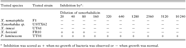
Table 2. In vitro antibiotic activity on Xenorhabdus and Photorhabdus of various dilutions of a xenorhabdicin suspension obtained from X. nematophila F1

Fig. 1. Fitness of Steinernema carpocapsae with antagonistic bacteria and Xenorhabdus nematophila co-injected into the insect. The darker bars represent the results obtained with variant 1 cells and the lighter ones the results obtained with variant 2 cells. The reference number of all the antagonistic bacteria co-injected into the insect is given below the corresponding data. (A) Parasitic success. (B) Distribution of the reproductive rate.
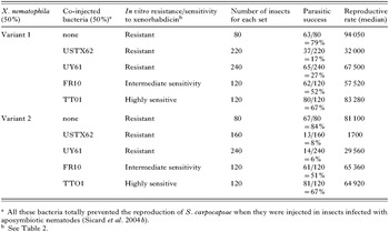
Table 3. Effect of antagonistic bacteria on the fitness of Steinernema carpocapsae in the presence of Xenorhabdus nematophila
 You have
Access
- 25
- Cited by