Introduction
Almost 300 lacrimal sac tumours have been reported, mostly primary epithelial tumours, of which 75 per cent were malignant.Reference Stefanyszyn, Hidayat, Pe'er and Flanagan1, Reference Parmar and Rose2 Small cell carcinoma (also known by its World Health Organization classification of ‘small cell carcinoma, neuroendocrine type’) is likewise rare in the head and neck region; this site accounts for 0.5–1 per cent of all small cell carcinomas, including those in the lung.Reference Peres-Ordonez, Barnes, Eveson, Reichart and Sidransky3, Reference Walenkamp, Sonke and Sleijfer4 Extrapulmonary small cell carcinoma is thought to derive in some way from multipotential stem cells which have the capacity for divergent differentiation.Reference Renner5 Although some differences may exist in the clinical behaviour of tumours arising from different subsites, it is known that small cell carcinoma of the head and neck region is aggressive and has a propensity for distant metastasis and/or regional recurrence after treatment.
Here, we present the first reported case of primary small cell carcinoma of the lacrimal sac, which was treated successfully with chemo-radiotherapy.
Case report
A 67-year-old Japanese woman was referred to our department with a two-month history of left canthal swelling, epiphora and occasional nasal bleeding.
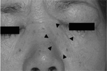
Physical examination revealed a 2.5 cm, firm, nontender mass in the region of the left medial canthus (Figure 1). There was no diplopia or blurred vision.
Clinical photograph of the left medial canthus showing the firm, nontender, approximately 2.5 cm mass.

Nasal endoscopy revealed an irregularly surfaced, dark brown, readily bleeding tumour in the left inferior meatus.
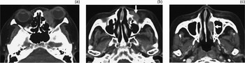
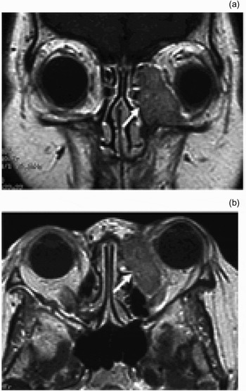
An enhanced computed tomography (CT) scan revealed a weakly enhancing soft tissue mass mainly located in the left lacrimal sac. The mass extended into the orbit, with bony erosion, and also into the nasolacrimal duct and the inferior meatus (Figure 2). However, the mass did not extend into the ethmoid or maxillary sinuses. A gadolinium-enhanced magnetic resonance imaging (MRI) scan revealed a weakly and homogeneously enhancing tumour which displaced the eyeball laterally and anteriorly (Figure 3). A whole body CT scan revealed no cervical lymphadenopathy and no evidence of distant metastasis, including pulmonary.
Axial, contrast-enhanced computed tomography images showing an enhancing soft tissue mass (arrow) located mainly in the left lacrimal sac, and extending into the orbit, nasolacrimal duct and inferior meatus.

(a) Coronal and (b) axial, T1-weighted, gadolinium-enhanced magnetic resonance imaging scans showing a weakly and homogeneously enhancing tumour (arrow) displacing the eyeball laterally and anteriorly.

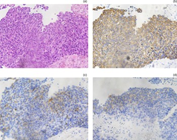
Histopathological analysis of a biopsy specimen from the inferior meatus revealed small cells with scant cytoplasm, a high nuclear:cytoplasmic ratio, nuclear moulding, finely granular chromatin, necrosis and mitosis (Figure 4). The tumour cells were immunohistochemically positive for cytokeratin (an epithelial marker) and cluster of differentiation 56 glycoprotein, and partially positive for synaptophysin (a neuroendocrine marker). These findings confirmed the diagnosis of primary small cell carcinoma of the lacrimal sac.
Photomicrographs showing histological and immunohistochemical findings. (a) The tumour is composed of small cells with scant cytoplasm, a high nuclear:cytoplasmic ratio, nuclear moulding, finely granular chromatin, necrosis and mitosis (H&E; ×200). The tumour cells were immunohistochemically positive for (b) cytokeratin (an epithelial marker) and (c) cluster of differentiation 56 glycoprotein, and partially positive for (d) synaptophysin (a neuroendocrine marker) (×200)

The patient was treated with four cycles of chemotherapy, together with concurrent radiotherapy. The systemic chemotherapy regimen comprised 60 mg/m2 cisplatin administered (together with adequate hydration) via a 2-hour infusion on day one, and 75 mg/m2 etoposide administered via a 2-hour infusion on days one to three. The chemotherapy was repeated in cycles of 21 days. The patient underwent 54 Gy of irradiation to the primary site, with a 1 cm margin. A four-MV linac X-ray was used with a single anterior port, with a conventional fractionation schedule of 2 Gy per day, five times a week. The patient experienced the following toxicity: grade two alopecia and radiation dermatitis, and grade three neutropenia, according to the National Cancer Institute (NCI) common toxicity criteria (CTCAE v3.0).Reference Trotti, Colevas, Setser, Rusch, Jaques and Budach6
After treatment, CT scanning revealed only a small amount of scar formation in the left lacrimal sac. The patient was followed up with yearly sinonasal, neck and chest CT scans. Late radiation toxicity comprised only keratitis in the left eye. There was no evidence of recurrence or metastasis over five years' follow up.
Discussion
Primary carcinomas of the lacrimal sac are rare. To the best of our knowledge, 81 patients with primary lacrimal sac carcinoma were reported in the English language literature from 1990 to 2009; their median age was 64 years (range, 26 to 94 years).Reference Stefanyszyn, Hidayat, Pe'er and Flanagan1, Reference Parmar and Rose2, Reference Pe'er, Hidayat, Ilsar, Landau and Stefanyszyn7–Reference Anderson, Wojno and Grossniklaus9 Of these patients, 30 (37 per cent) had squamous cell carcinoma, 15 (19 per cent) adenocarcinoma, 14 (17 per cent) transitional cell carcinoma, eight (10 per cent) adenoid cystic carcinoma and six (7 per cent) mucoepidermoid carcinoma. No reports of primary small cell carcinoma of the lacrimal sac have previously been published.
In 42 of the reported patients with lacrimal sac carcinoma, the presenting signs and symptoms were as follows: epiphora in 33 (79 per cent), recurrent dacryocystitis in 28 (67 per cent), lacrimal sac mass in 23 (55 per cent) and nasal bleeding in nine (22 per cent).Reference Stefanyszyn, Hidayat, Pe'er and Flanagan1, Reference Parmar and Rose2, Reference Pe'er, Hidayat, Ilsar, Landau and Stefanyszyn7–Reference Liu, Lin, Kao, Kau, Tsai and Hsu14 However, some of these symptoms were missing in most patients, resulting in delayed diagnosis. Lacrimal sac carcinoma is commonly diagnosed at a late stage, and usually only after tumour biopsy or excision.
Nasal endoscopy is invaluable in demonstrating intranasal extension of the tumour, which may be along the nasolacrimal duct. Axial and coronal CT scans of the paranasal sinuses and orbit can help define the extent of the tumour, as well as any evidence of bony destruction of the lacrimal fossa. In a series of 74 cases of malignant lacrimal sac tumours, 66 (81.9 per cent) exhibited direct extension into the orbit, nasolacrimal duct or paranasal sinuses.Reference Ni, D'Amico, Fan and Kuo15 In our patient, CT and MRI scanning revealed that the tumour was mainly located in the region of the left lacrimal sac, and did not extend into the ethmoid or maxillary sinuses. The tumour extended into the orbit and also into the inferior meatus via the nasolacrimal duct, as described in previous reports of lacrimal sac carcinoma.Reference Salib and Afoakwah8
Small cell carcinoma is also known as small cell neuroendocrine carcinoma, poorly differentiated neuroendocrine carcinoma, and ‘small cell carcinoma, neuroendocrine type’. It is a high grade carcinoma composed of small- to intermediate-sized cells resembling those of small cell carcinoma of pulmonary origin.Reference Peres-Ordonez, Barnes, Eveson, Reichart and Sidransky3 The histological hallmarks of the tumour comprise necrosis, large numbers of apoptotic cells, a high mitotic rate and lack of neurofibrillary stroma. Immunohistochemically, the tumour cells express at least one neuroendocrine marker, such as synaptophysin, cluster of differentiation 56 glycoprotein or chromogranin, as well as cytokeratins as an epithelial marker. Our patient's pathological findings were consistent with the definition of primary small cell carcinoma of the lacrimal sac, and this was therefore the diagnosis.
Of all reported cases of small cell carcinoma (intra- as well as extrapulmonary), only 0.5–1 per cent have occurred in the head and neck.Reference Walenkamp, Sonke and Sleijfer4 Head and neck small cell carcinoma most commonly affects the larynx, but can also develop in the salivary glands and paranasal sinuses.Reference Nagao, Gaffey, Olsen, Serizawa and Lewis16
Although the clinical behaviour of tumours at different subsites may differ somewhat, small cell carcinoma of the head and neck is known to be aggressive and to have a propensity for regional and/or distant spread.Reference Weng, Chu, Liu and Chen17 Because distant metastasis occurs early and often, many investigators have suggested that small cell carcinoma should be considered a ‘systemic’ disease from the outset of treatment.
• This paper describes a case of primary small cell carcinoma of the lacrimal sac
• Small cell carcinoma originating in the head and neck region is reported to be highly aggressive, and carries a poor prognosis
• This patient was treated successfully with chemo-radiation therapy
Therapeutic management can include surgery, radiotherapy, chemotherapy or a combination of these modalities. Because of the rarity of the tumour and the extremely small number of published single institution series and case reports, there are currently no firm treatment recommendations or guidelines. In patients with small cell carcinoma of the head and neck, the greatest improvement in survival is achieved by systemic chemotherapy. Aguilar et al. Reference Aguilar, Robbins, Stephens, Dimery and Batsakis18 reported that two-year survival rates of patients treated with local therapy alone consisting mainly of radiotherapy and of those with radiotherapy in combination with chemotherapy were 10 and 52 per cent, respectively. Baugh et al. reported that patients who received chemotherapy survived longer than those who did not, with comparative median survival rates of 19 and 11 months, respectively.Reference Baugh, Wolf, Beals, Krause and Forastiere19 A wide range of chemotherapeutic agents has been used, but platinum-based chemotherapy regimens have become the mainstay of treatment, usually in combination with etoposide.Reference Barker, Glisson, Garden, El-Naggar, Morrison and Ang20
Accordingly, for our patient we selected chemotherapy consisting of cisplatin and etoposide, plus local radiotherapy. Fortunately, she was completely cured, with no recurrence or metastasis for five years.
Conclusion
We report a rare case of primary small cell carcinoma of the lacrimal sac. The patient was treated with chemotherapy (comprising cisplatin and etoposide) in combination with concurrent radiotherapy, and was completely cured.