Introduction
Middle-third helical rim defects typically arise secondary to trauma or oncological resection, and pose a challenging reconstructive problem. For defects of less than 2.5 cm in width, several options exist, including wedge resectionReference Tanzer1 or Antia–Buch helical chondrocutaneous advancement flaps.Reference Antia and Buch2 The Antia–Buch procedure is a technique for reconstructing defects up to 2 cm, whereby the anterior skin and cartilage of the helical sulcus is incised, the posteromedial skin is undermined, the helix is freed from its medial attachments and is advanced to cover the defect. To cover larger defects, the helix may need to be advanced in both directions, along with a V-Y advancement of the crus helicis.Reference Scott and Klaassen3 Small middle- and lower-third helical rim defects can be closed with chondrocutaneous rotation flaps using the antitragus, antihelix or lobule.
For larger defects greater than 2 cm in size, staged procedures have been traditionally used for reconstruction. Converse's tunnel technique is a two-staged procedure involving the use of a prefabricated flap.Reference Converse4 In the initial stage, an auricular cartilage graft taken from the contralateral ear is tunnelled under the post-auricular skin adjacent to the helical rim defect. The second stage is performed three weeks later, by taking the anteriorly based skin flap along with the cartilage graft and insetting it into the helical rim defect.Reference Converse4
An alternative two-stage procedure is Dieffenbach's flap,Reference Dieffenbach5 which involves harvesting contralateral auricular cartilage and suturing it to the helical rim defect, then elevating post-auricular skin, and advancing and covering the cartilage graft. The pedicle is subsequently divided at three weeks and inset. Alternatively, tubed bipedicled flaps may be used and are typically staged over three procedures.
Herein, we present the option of single-stage helical rim reconstruction using a post-auricular bipedicled flap with an ipsilateral conchal cartilage graft.
Case report
This technique may be performed under local or general anaesthesia. The helical rim defect is measured, and markings are made on the posterior ear surface and post-auricular skin that parallel this, for elevation of the bipedicled flap. If required, a conchal graft harvest is planned according to the defect size, and is marked on the post-auricular surface to be harvested from the ipsilateral ear. The skin on the posterior ear and post-auricular surface are incised and elevated in a supra-perichondrial/subcutaneous plane until the bipedicled flap is mobile, extending from the post-auricular skin to the skin over the helical rim defect. Cartilage may then be harvested from the conchal bowl once the flap is elevated, and is secured to the defect using size 5-0 absorbable sutures. The flap is re-draped onto the post-auricular surface and inset with size 4-0 and 5-0 absorbable sutures. A dental roll bolster is sutured into the post-auricular fold to provide support, and the wounds are treated with topical Polyfax™ (polymyxin) ointment and skin glue. A head bandage is applied to provide further support. Both the head bandage and dental roll are removed three weeks following surgery.
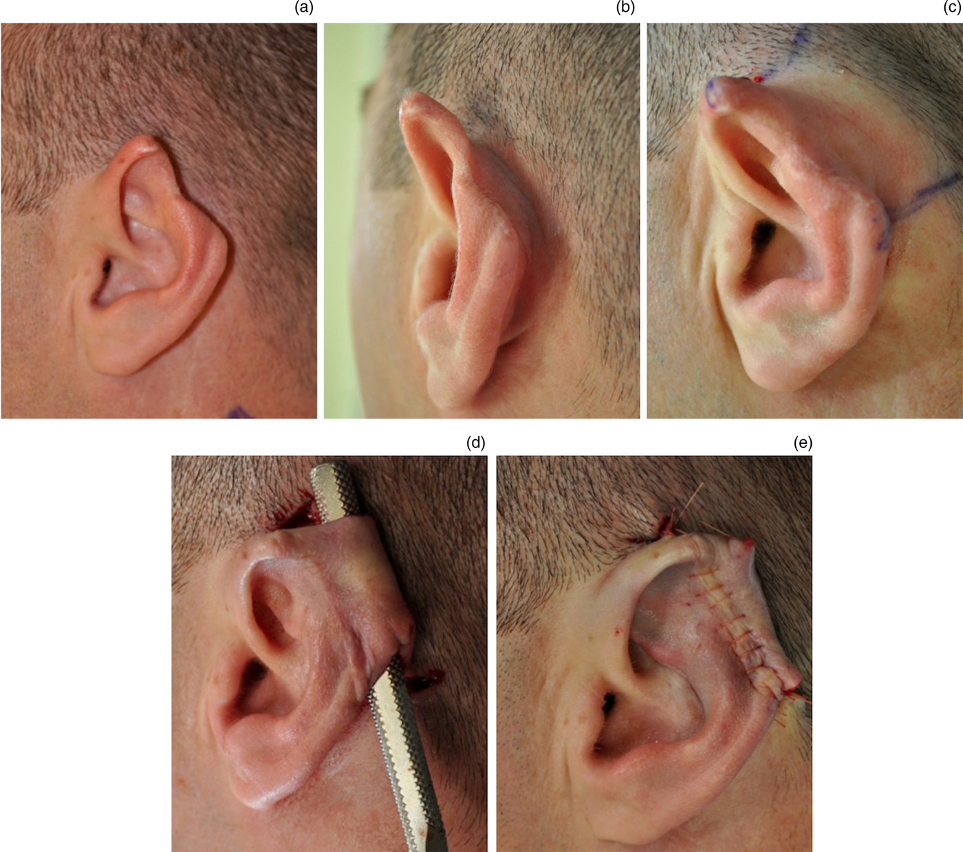
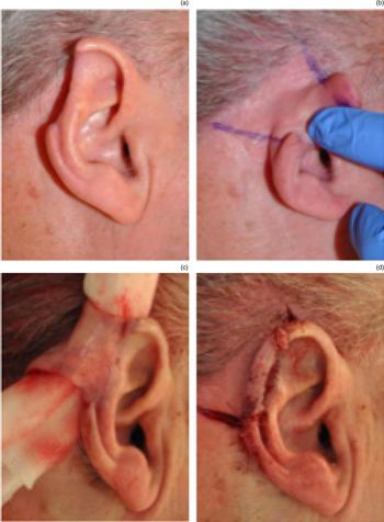
We illustrate this technique with two case studies. Figure 1 illustrates a 40-year-old male with a 2.5 cm helical rim defect from a previously sustained human bite to the left ear. Figures 2 and 3 illustrate a 74-year-old male who previously had a right ear malignant melanoma and underwent a 1 cm excision of the helical rim. Both patients were treated with our single-stage technique utilising a bipedicled flap and ipsilateral conchal cartilage graft.
Single-stage helical rim reconstruction following traumatic human bite. (a & b) Pre-operative helical defect; (c) marking of post-auricular bipedicled flap; (d) elevation of bipedicled flap from post-auricular skin; and (e) flap contoured into helical rim and post-auricular sulcus.

Single-stage helical rim reconstruction following melanoma excision. (a) Pre-operative helical rim defect; (b) pre-operative marking of bipedicled flap on post-auricular skin; (c) elevation of bipedicled flap; and (d) flap inset to helical rim and post-auricular sulcus.

(a–c) Final result achieved for melanoma excision patient in Figure 2, five years following reconstruction.

Discussion
Full thickness defects of the helical rim can be reconstructed using several methods, depending on their size and position. To date, techniques for helical rim reconstruction have involved staged procedures and cartilage grafts harvested from the contralateral ear. However, this condemns patients to multiple operations to reconstruct the helical rim. Furthermore, cartilage grafts taken from the contralateral ear subjects the patient to potential donor site morbidity. In order to avoid the aforementioned drawbacks, we present an option of a single-stage procedure using an ipsilateral graft harvest to achieve reconstruction of middle-third helical rim defects greater than 2 cm.
The single-stage tubed pedicle flap illustrated herein provides a robust technique for reconstructing the middle-third helical rim, which preserves the post-auricular sulcus and provides a good cosmetic result. The head bandage and dental roll bolster were maintained for three weeks to protect the contoured cartilage and the definition of the post-auricular sulcus. Premature removal of the dental roll bolster results in tenting of the post-auricular flap, blunting of the post-auricular sulcus and generally sub-optimal results.
To our knowledge, this is the first description of a single-stage helical rim reconstruction technique. Application of this technique to post-traumatic and oncological resection defects has revealed aesthetic outcomes that are comparable to the previously described staged procedures. Our single-stage technique also results in a minimal reduction in helical size and asymmetry. The use of a single-stage and limiting to ipsilateral graft harvest are also well tolerated by the patient, and offer advantages over traditional staged reconstruction of large helical rim defects.
Competing interests
None declared