Introduction
Brugada syndrome is an inherited arrhythmogenic disorder characterised by coved-type ST-segment elevation in the right precordial leads in structurally normal hearts and is associated with an increased risk of sudden death.Reference Brugada and Brugada 1 The syndrome is genetically and clinically heterogeneous and presents typically in the fourth or fifth decade of life.
Since its first description in 1992, significant progress has been made to characterise the syndrome in the adult population.Reference Wilde, Antzelevitch and Borggrefe 2 , Reference Antzelevitch, Brugada and Borggrefe 3 Although in the initial description, three out of the eight patients were children, subsequent studies have revealed that the prevalence of Brugada syndrome in the paediatric population is low (0.0098%) compared with the adult population (0.14–0.7%).Reference Hermida, Lemoine, Aoun, Jarry, Ray and Quiet 4 Nevertheless, in recent years, there has been growing evidence in the literature of earlier onset of the disease.
The genetic basis of Brugada syndrome involves abnormalities in the sodium channel current in the cardiac cell. The human gene SCN5A encodes the main sodium channel, an integral membrane protein found primarily in the cardiac muscles. This protein mediates the fast influx of sodium ions across the cell membrane, generating the initial upstroke of the cardiac action potential. Voltage-gated sodium channels are responsible for the generation and propagation of the action potential in different excitable tissues of the heart. Several mutations in the SCN5A gene have been implicated in different inherited arrhythmogenic cardiac syndromes including Brugada syndrome and long QT syndrome type 3. In Brugada syndrome, SCN5A mutations lead to a loss-of-function phenotype that manifests as cardiac conduction disease.
The aim of this publication was to summarise the manifestations of Brugada disease from newborns to adolescents, based on a literature review and using descriptive cases from our extensive paediatric population.
The cases presented belong to the Brugada syndrome database at our institution, including a paediatric cohort of almost 500 children belonging to 415 different families. The diagnosis of Brugada syndrome for all of the illustrative cases was based on an electrocardiogram documenting a coved-type ST-segment elevation ⩾2 mm in one or more precordial leads from V1 to V3, either occurring spontaneously or in the presence of a sodium channel blocker drug.
It is our hope that going beyond the asymptomatic electrocardiographic pattern will allow the reader to understand the more severe presentations and the related risks in the paediatric patient population.
Rhythm disturbances and conduction anomalies
In the normal heart, sodium channels are responsible for the progression of the electrical potential from the sinus node, through the atrial myocardium, along the atrioventricular node, across the intra-ventricular conduction system including the Purkinje fibres, and finally throughout the ventricular myocardium. Intrinsic anomalies in the SCN5A gene can lead to abnormal electric activity at these multiple structures of the conduction system and manifest clinically as different rhythm disorders. The clinical heterogeneity of sodium channel mutations can be explained at least partially in the characteristics and degree of the channel dysfunction. Complete absence of SCN5A activity is lethal in mice models.Reference Papadatos, Wallerstein and Head 5 Along the spectrum from normal to complete absence of activity, several degrees of disease can manifest during the first few decades of life. In this first section, we present a description of the different electrical manifestations of the Brugada syndrome during childhood.
Atrial disease. Several studies have demonstrated major differences in the characteristics of the SCC5A channels in the atrial and ventricular myocardium, suggesting that the impact of some sodium mutations may be greater in the atria than in the ventricle, predisposing to the development of earlier atrial arrhythmias.Reference Kusano, Tanimaya and Nakamura 6 , Reference Francis and Antzelevitch 7
Atrial muscle involvement can manifest as sinus node dysfunction and can present as the initial clinical sign of Brugada syndrome during the first decade of life. Children with Brugada syndrome present more frequently with sinus node dysfunction compared with older subjects (7.5% in children versus 1.5% in adults). Sinus node dysfunction can also manifest as sinus bradycardia, sinus arrest, tachycardia–bradycardia syndrome, or even asystole. Congenital sick sinus syndrome has been associated with mutations in the sodium channel gene resulting in severely impaired function.Reference Benson, Wang and Dyment 8 Chronotropic incompetence, characterised by the inability of the heart rate to respond to exercise and reach at least 85% of the maximum age-predicted heart rate, is also a manifestation of sinus node dysfunction related to this disease.
The sodium channel plays a major role in the action potential onset. Action potentials in the cells of the sinus node are mainly calcium channel dependent. The sodium channel current encoded by SCN5A is responsible for the propagation of the electrical current. Even if the sodium channel is absent inside the node, its role in the perinodal area allows the transmission of the action potential to the rest of the atria.Reference Nof, Gikson and Antzelevich 9 Sinus bradycardia seems to result from the inability of the current to conduct from the sinus node to the adjacent atrial myocardium, resulting clinically in sinoatrial block.Reference Letsas, Korantzopoulos and Efremidis 10 , Reference Sumiyoshi, Nakazato and Tokano 11
Clinically, the P-R interval reflects the transmission of the sinus impulse within the atrial and nodal tissue. There is a normal age-related prolongation of the P-R interval during the first decade of life. The average normal P-R interval extends from 100 ms before the 1st month of life to 150 ms in adolescence.Reference Davignon, Rautaharju, Boisselle, Soumis, Mégélas and Choquette 12 Loss of function of the sodium channels can manifest as widening of the P wave and prolongation of the P-R interval. Prolonged atrial conduction has been identified as the most common manifestation of Brugada syndrome in children. Moreover, the combination of atrial conduction delay and ventricular arrhythmias is a marker for sodium channel mutations in children.Reference Conte, Dewals and Sieira 13
Supraventricular tachyarrhythmias in the form of atrial fibrillation and atrial flutter have been reported to occur in 7.5% of children with Brugada syndrome. In addition, in 75% of patients presenting with documented atrial arrhythmias, there is evidence of simultaneous sinus node dysfunction at the time of diagnosis.Reference Makiyama, Akao and Tsuji 14
Atrial flutter can be the first manifestation of abnormal atrial electrical conduction in an otherwise asymptomatic patient (Case I). It has been current practice in many paediatric cardiology centres to attempt pharmacological cardioversion of atrial flutter using amiodarone. The risk of giving a strong sodium channel blocker medication to a patient with an underlying sodium channel mutation includes prolonged asystole requiring external pacing and persistent and severe ventricular arrhythmias. Therefore, in every case, it is important to obtain a 12-lead electrocardiogram in sinus rhythm to confirm normal conduction before exposing children to sodium channel blocker therapy.
At the more severe end of the spectrum, progressive disorganisation in the microstructure of the myocardium leads to more chaotic manifestations of atrial arrhythmias. Atrial fibrillation represents the clinical activity of a severely fibrosed atrium with profound electrical impairment. In most paediatric cases, atrial tachycardias are associated with sinus node dysfunction, manifesting as bradycardia–tachycardia syndrome. This variant of sick sinus syndrome, in which slow and fast rhythms alternate, mimics the atrial disease typical of patients at older ages.
The clinical spectrum of atrial compromise in paediatric Brugada syndrome ranges from asymptomatic sinus bradycardia to atrial standstill. The clinical progression of the disease from bradycardia to atrial non-excitability suggests the presence of external factors that contribute to the expression of the complete phenotype.
Case I
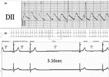
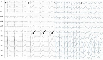
A 7-year-old boy with a first episode of typical atrial flutter was treated initially with sotalol for 48 hours without success (Fig 1a). He underwent a direct-current cardioversion under sedation followed by sinus arrest of 6.5 seconds and junctional bradycardia at 30 bpm requiring treatment with isoproterenol. The 12-lead electrocardiogram in sinus rhythm was normal, but the telemetry showed evidence of sinus node dysfunction with severe bradycardia and sinus pauses of >3 seconds (Fig 1b). The family history was negative and both parents had normal electrocardiograms. An ajmaline test resulted in a typical Brugada pattern immediately followed by ventricular tachycardia, initially monomorphic and later degenerating into a polymorphic ventricular tachycardia. It is important to note that the ajmaline had been discontinued at 0.7 mg/kg, as soon as the electrocardiogram reached diagnostic standards. An isoproterenol infusion was started and the patient recovered sinus rhythm within minutes. The patient’s family refused an implantable cardioverter defibrillator, and therefore he received an epicardial atrial pacemaker for sinus node dysfunction (Fig 2). Genetic test resulted positive for a SCN5A mutation.
( a ) (above) Lead II long tracing at admission in the emergency department showing a typical atrial flutter with “saw-tooth” pattern with negative flutter waves in II. ( b ) (below) Telemetry image showing a sinus pause of 3.16 seconds.

12-lead electrocardiogram (ECG) during ajmaline test. (a) Baseline normal ECG. (b) ECG at a dose of ajmaline of 0.7 mg/kg. Note the appearance of Brugada type-I ECG with T wave alternates in leads V1 and V2 (arrows). Infusion of ajmaline was discontinued at this point. (c) Rhythm disorganisation and irregularity. (d) Episode of polymorphic ventricular tachycardia 2 minutes after ajmaline challenge.

Conduction system disease. An incomplete right bundle block with a QRS<80 ms is a frequent normal finding in the right precordial leads (V1–V2) for children; however, diffusely prolonged QRS complexes, complete right bundle branch block, and left posterior hemiblock can be clinical expressions of Brugada syndrome (Case II).
Progressive conduction system disease is a phenotype resulting from certain SCN5A mutations. There are cases in the literature describing rate-dependent atrioventricular block associated with homozygous SCN5A missense mutations.Reference Lupoglazoff, Cheav and Baroudi 15
Loss-of-function SCN5A mutations can also manifest as infra-Hisian conduction disease. Suppression of the sodium channel function may be associated with profound conduction disturbance and bradyarrhythmic phenotypes expressed in the early years of life. Conduction abnormalities in Brugada syndrome have been associated with an increased risk of ventricular arrhythmias in children.Reference Kanter, Pjeiffer and Hu 16
Case II
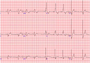
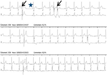
An 8-year-old patient with a history of familial symptomatic Brugada syndrome – father and paternal uncle – presented with different rhythm abnormalities at an early age. The arrhythmias included sinus bradycardia with frequent junctional escape rhythm, intra-atrial conduction delay resulting in first-degree atrioventricular block, complete right bundle branch block, and left anterior hemiblock. A 24-hour Holter monitor showed frequent ventricular premature beats of two morphologies. He had a positive ajmaline test at 11 years of age. He remains asymptomatic with biannual clinic visits and annual Holter monitor studies (Figs 3 and 4).
12-lead electrocardiogram illustrating several conduction abnormalities in the patient in Case II: note the sinus bradycardia with occasional junctional escape beats, the intra-atrial conduction delay with a biphasic P wave in leads I and II, first-degree atrioventricular block with a P-R interval of 198 ms, and the presence of complete right bundle block and left anterior hemiblock resulting in a superior QRS axis.

24-hour Holter showing isolated premature ventricular contractions or ventricular couplets of two different QRS morphologies.

Ventricular disease. Ventricular tachycardia is an uncommon arrhythmia in people <18 years of age with a structurally normal heart, and has an incidence of 0.6 per 100,000 patient-years.Reference Roggen, Pavlovic and Pfammatter 17
Electrical vulnerability of the myocardium is a distinctive feature of Brugada syndrome. The mutation in the gene encoding for the sodium channel reduces the inward sodium channel current and disrupts the delicate ion balance of the cardiac cell. The depression or loss of the action potential in specific areas of the right ventricle epicardium creates a transmural voltage gradient responsible for the ST-elevation.Reference Yan and Antzelevitch 18 Moreover, extrasystolic activity related to phase 2 re-entry acts as a trigger for ventricular tachycardia and ventricular fibrillation.
The arrhythmia most frequently leading to sudden death in Brugada syndrome is ventricular fibrillation or polymorphic ventricular tachycardia, initiated after a short coupled ventricular extrasystole. The arrhythmia typically occurs at night or at rest during the day. Among children, fever is the most frequent trigger.
Monomorphic ventricular tachycardia triggered by fever has also been reported in the literature in infants with SCN5A mutation.Reference Kim, Kyung and Kang 19 Beta-blockers have been shown to control these monomorphic arrhythmias related to fever in infants.Reference Chockalingam, Rammeloo and Postema 20
Ventricular monomorphic or polymorphic arrhythmias in children with normal hearts demand a comprehensive evaluation for potential aetiologies. In those cases associated with sinus node dysfunction, intra-atrial or intra-ventricular conduction delay or a positive family history for sudden death, Brugada syndrome must be considered in the differential diagnosis.
Paediatric clinical entities
In this section, we present a description of the different paediatric entities that have been confirmed as related to Brugada syndrome; however, we emphasise that some are anecdotal reports and should not definitively guide current paediatric practice.
Prenatal arrhythmias and recurrent fetal loss
Sodium channel mutations associated with long QT syndrome have been reported to result in repetitive stillbirth, severe ventricular arrhythmias, and fetal heart failure.Reference Miller, Estrella and Myerburg 21 , Reference Baruteau and Scheleich 22 However, SCN5A loss-of-function mutations have not been shown to manifest clinically in the antenatal period to date.
Sudden infant death syndrome
Sudden infant death syndrome is the sudden unexplained death of a child <1 year of age. 23 It is the most common cause of death in infants between the age of 1 month and 1 year.Reference Moon, Horne and Hauck 24 It occurs usually during sleep, at night, and without previous warning. The exact cause of death is unknown, but many possible causes have been proposed including suffocation and prematurity. Research into the clinical and genetic basis of sudden infant death syndrome has identified that a proportion of deaths are due to a primary rhythm disorder.Reference Gutheroth 25 , Reference Schwartz, Priori and Bloise 26 Recently, SCN5A mutations have been identified in sudden infant death syndrome cases.Reference Wedekind, Smits and Schulze-Bahr 27 Postmortem molecular analysis has clearly identified SCN5A mutations in 10–15% of sudden infant death syndrome victims.Reference Ackerman, Siu and Stuner 28
Investigation using our database identified eight infant deaths below the age of 1 year: all died suddenly during sleep at night. There were two sets of siblings belonging to two different families and one set of first-degree cousins. In most of the families in which sudden infant death syndrome occurred, we could identify other adult members who had suffered from sudden death as well.
Current literature suggests that SCN5A mutations do present symptomatically in the newborn period.Reference Wedekind, Smits and Schulze-Barh 29 , Reference Skinner, Chung and Montgomery 30 , Reference Priori, Napolitano and Giordano 31 Recently, a nationwide study from Denmark demonstrated that 12% of cases of sudden infant death syndrome presented mutations in the sodium channel complex genes.Reference Winkel, Yuan and Olesen 32 Paediatric electrocardiographic and Holter monitor screening as well as genetic investigation of offspring from affected families are likely to identify individuals at risk and improve family counselling.
Severe syncope and aborted sudden death
Sudden cardiac death is defined as death occurring within an hour of the onset of the symptoms. In children, the incidence is 1.3 per 100,000 person-years.Reference Winkel, Holst and Theilade 33 Sudden cardiac death in the young is caused by a variety of factors including structural and arrhythmogenic disorders. Brugada syndrome has been estimated to account for up to 20% of unexplained sudden death without structural heart disease in adults.Reference Antzelevitch, Brugada and Borggrefe 34 Our group has reported several cases of Brugada syndrome associated with sudden death in children.Reference Conte, Dewals and Sieira 13
In all, 50% of symptomatic Brugada syndrome children present with severe syncope or sudden cardiac death as the initial manifestation of the disease. Syncope can start early in patients with Brugada syndrome, even during the first few years of life. Episodes can be repetitive and are usually at rest, especially during fever.
Before adolescence, there is no gender difference in symptomatic patients. This contrasts with the male preponderance in adult life and corroborates the role of testosterone in the pathogenesis of the disease. The prevailing explanation is that hormonal changes taking place during puberty interact with the flow of the ion currents that are abnormal in Brugada syndrome.Reference Di Diego, Cordeiro and Goodrow 35 In addition, recent data suggest that adult men with Brugada syndrome have higher levels of testosterone than age-matched healthy men.Reference Shimizu, Matsuo and Kokubo 36 Moreover, testosterone has been claimed to shorten the action potential in the right ventricular epicardium, facilitating the onset of ventricular arrhythmias.
At an earlier age, a variant of syncope is the breath-holding spell, which occurs in ~5% of the otherwise healthy paediatric population. Previous literature has suggested that severe breath-holding spells, especially if repetitive and atypical, should raise concern for an underlying rhythm abnormality.Reference Allan and Gospe 37 We have identified three patients – two siblings and one unrelated – with severe breath-holding spells, including loss of consciousness and sphincter control, which were later diagnosed as familial Brugada syndrome cases. We present Case III as an example.
Case III
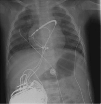
This boy presented with severe breath-holding spells that started at 6 months of age, but also presented with syncope without crying at 10 months. After extensive multidisciplinary investigation, he had a positive ajmaline test at 11 months and received an epicardial implantable cardioverter defibrillator. Genetic testing revealed a heterozygous one-base pair transmembrane mutation in position 3695 of the SCN5A coding sequence, associated with Brugada syndrome.Reference Kapplinger, Tester and Alders 38 This missense mutation was also present in the asymptomatic father, who tested positive for ajmaline as well. The patient is currently 5 years old and had one appropriate shock for ventricular fibrillation 7 months after implantation, no inappropriate shocks, and no implantable cardioverter defibrillator complications to present (Fig 5).
Chest radiography after implantation of an implantable cardioverter defibrillator with an epicardial coil, a bipolar sensing lead, and an abdominal generator.

Adolescent patients may present with repetitive loss of consciousness. A prodrome of severe syncope can present clinically with atypical manifestations, including brief episodes of seizures and unexplained abnormal behaviour. In these cases, close follow-up is needed, and sometimes the implantation of an internal loop recorder can help reach the diagnosis.
Unusual symptoms during fever and febrile seizures
The exact mechanism of the electrocardiographic changes during fever has not been clearly identified in Brugada syndrome.Reference Dumaine, Towbin and Brugada 39 The underlying cause could be an imbalance between the de-polarising and re-polarising currents during the early re-polarisation phase of the action potential.
Benign febrile seizures are a common phenomenon during the first 6 years of life. The seizures are usually triggered by a rapid rise in body temperature, generally in association with a viral disease. The episodes are brief and the prognosis is good.Reference Waruiru and Appleton 40
Atypical febrile seizures have been reported to be associated with sodium channel mutations.Reference Skinner, Chung and Nel 41 A fever-triggered ventricular arrhythmia can easily mimic a febrile seizure event. As performing an electrocardiogram is not routine following an event of febrile seizure, this association could be more frequent than suspected; however, due to the low incidence of Brugada syndrome in the young, electrocardiographic screening should only be considered in cases of atypical febrile seizures, suspicion of arrhythmia – tachycardia out of proportion to the body temperature and weak pulses – or a positive family history suggesting Brugada syndrome.
Evaluation of children
Symptomatic child
Diagnostics
It is rare that a child is the first symptomatic member of a family. In the case of a rhythm abnormality or clinical event in the paediatric age group, the first diagnostic step consists of an electrocardiogram of the patient and first-degree family members. In cases of a normal baseline 12-lead electrocardiogram, it is important to set a plan involving the family and the paediatric and emergency departments to obtain new tracings in the event of fever.
Holter monitors are inexpensive, non-invasive studies that can bring to light unknown manifestations of the disease. Especially in the paediatric group, they can exclude sinus node dysfunction and pauses during less-active periods of the day.
Starting from the age of 6 or 7 years, a treadmill exercise test can unmask unknown chronotropic incompetence as a manifestation of sinus node dysfunction.
Provocative testing with sodium channel blockers is the standard diagnostic method to unmask the Brugada electrocardiographic pattern in patients with suspected Brugada syndrome. A consensus on the age to start testing children and the safety of the different drugs used is not yet well established. In our centre, the protocol consists of an ajmaline infusion at a dose of 1 mg/kg (maximum of 50 mg) administered intravenously during a period of 5 minutes. Ajmaline is considered the best option to unmask Brugada syndrome as the short half-life is desirable in the case of an induced arrhythmia. In many centres, flecainide and procainamide are used instead. Flecainide has a prolonged effect and requires longer post-test cardiac monitoring. On the other hand, procainamide dissociates rapidly from the sodium channel, resulting in a lower level of blockade.
Before administration of ajmaline, precordial leads – V1 upper and V2 upper – are placed one intercostal space above the standard V1 and V2 positions, respectively.Reference Bayes de Luna, Brugada and Baranchuk 42 This lead placement makes the Brugada pattern more noticeable during the test. False-negative tests can result from omitting these high-placed leads. Electrocardiograms are recorded and analysed at baseline and at 1-minute intervals throughout the test.
Owing to the higher blockade property of ajmaline, it is our policy to stop the drug with the appearance of the diagnostic electrocardiographic pattern, or when the QRS prolongation reaches ⩾30% of baseline length. This is especially relevant in symptomatic young children or those presenting with manifestations of electrical conduction disease, as the risk of ventricular events is 10 times higher (10 versus 1%).Reference Conte, Dewals and Sieira 13 Although there have been many literature reports, the exact incidence of life-threatening ventricular arrhythmias in patients with Brugada syndrome has not been well established.Reference Rolf, Bruns and Wichter 43 , Reference Veltmann, Wolpert and Sacher 44 There is no evidence that patients who developed ventricular arrhythmias during the provocative test are at risk for lethal clinical events in the future.
A test is considered positive in the case of J point elevation ⩾2 mV with coved ST-elevation in at least two precordial leads.Reference Wilde, Antzelevitch and Borggrefe 2 Following the test, follow-up electrocardiograms are performed every 5 minutes until the P-R interval and QRS duration return to normal.
Every effort should be made to prepare a safe environment before starting the study, including life-support equipment and experienced paediatric professionals; two external defibrillators should be available in the room and set to an appropriate charge based on the patient’s weight (dose of 2 J/kg). An isoproterenol infusion should be prepared beforehand and ready to administer in case of sinus bradycardia, sinus pauses, atrioventricular block, or ventricular arrhythmias. Experienced paediatric surgical backup in case of prolonged ventricular arrhythmias requiring extracorporeal support is mandatory.
It is unusual to perform an electrophysiological test in young patients with syncope as the first diagnostic step. Most episodes of transient loss of consciousness in the paediatric age group are vagally mediated and do not require interventions, other than changes in lifestyle and anti-gravitational measures. Nevertheless, in selected patients in whom the initial evaluation rules out a vagal cause, either by severity or presentation, or in whom clinical manifestations include electrical disease, an electrophysiology test should be considered in case of a positive provocative test. The study needs to assess the different levels of potential conduction abnormality, including measurement of the baseline A-H and H-V interval and the Wenckebach cycle length. Sinus nodal function is tested by measuring the sinus node recovery time and the corrected sinus node recovery time and comparing results with paediatric standards.Reference Samson, Jolma and Zamora 45 , Reference Yabek, Jarmakani and Robert 46 Finally, atrial and ventricular stimulation protocols without and with isoproterenol are performed.
Genetic test
Mutations in the SCN5A gene account for 20–30% of the cases of Brugada syndrome. Inheritance of the sodium channel mutations is autosomal dominant. Genetic testing should be performed initially in the affected individual.
Cascade screening of at-risk family members is recommended after identification of a proband. Screening of asymptomatic paediatric family members should also include a detailed clinical evaluation, an electrocardiogram, and a genetic counselling meeting.
Genotype-positive children should be closely followed-up to identify any possible clinical manifestations. It is common sense to assume that these children are at higher risk of developing Brugada syndrome, but there are no data for risk stratification at present.
In the event of an unexplained sudden death in a child, a possible inherited cardiac condition should be suspected. In these cases, family evaluation should include genetic testing of the decedent’s blood sample. This process has been termed “molecular autopsy” and involves postmortem DNA analyses of selected genes responsible for the primary arrhythmia disease.Reference Semsarian, Ingles and Wilde 47 In cases where it is impossible to test blood from the victim, a sample of hair could be used instead. The current recommendations for molecular autopsy are included in the international Heart Rhythm Society, United States of America/European Heart Rhythm Association guidelines. The focus is on direct DNA sequencing of four genes including SCN5A and the genes responsible for long QT type 1 and long QT type 2 and catecholaminergic polymorphic ventricular tachycardia.Reference Ackerman, Priori and Willems 48
Implantable cardioverter defibrillator
Indications for an implantable cardioverter defibrillator remain the most difficult aspect of the follow-up in children with Brugada syndrome. Implantable cardioverter defibrillators in young children have a high incidence of complications including inappropriate shocks, lead fractures, and need for early reoperation.Reference Stephenson, Batra and Knilans 49 Moreover, psychological stress and impact on quality of life should also be considered.
It has been our policy to implant epicardial defibrillators for secondary prevention in symptomatic patients with Brugada syndrome below the age of 12 years. Above 12 years of age, a transvenous system is implanted. In selected cases, a transvenous system can be implanted at a younger age if the body constitution permits (Case IV). In patients with potential growth and transvenous systems, it is important to allow extra lead in the form of a loop in the right atrium that can stretch during growth and prevent dislodgment.
Case IV
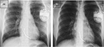
This girl suffered a recovered sudden cardiac arrest at the age of 11 during sleep in 2009. Her sister was awakened by an abnormal respiration sound and discovered the patient unresponsive. She initially received cardiopulmonary resuscitation from a family member. On arrival of the emergency medical team, the external defibrillator tracing showed asystole. A dose of atropine resulted in ventricular fibrillation, which was terminated by direct-current cardioversion. She alternated asystole and polymorphic ventricular tachycardia or ventricular fibrillation until she was stabilised in sinus rhythm after several cardioversions. A complete cardiac screening provided normal results. An ajmaline test was positive and she received a transvenous implantable cardioverter defibrillator. Her genetic screening for SCN5A mutations was negative. At age 14, she received a first inappropriate shock for atrial fibrillation. High doses of beta-blockers and sotalol did not succeed to control the paroxysmal atrial fibrillation, resulting in several shocks. She finally underwent pulmonary vein isolation by means of cryoablation at age 15. Since then, she has had only rare, brief episodes of atrial tachycardia that did not result in shock. There has been no appropriate shock or evidence of ventricular arrhythmias in her follow-up. The family screening demonstrated a positive ajmaline test in her asymptomatic father aged 40 years and asymptomatic 9-year-old brother. The mother and two sisters aged 23 and 19 years, respectively, had negative ajmaline tests (Fig 6).
( a ) Chest radiograph after implantation of a transvenous implantable cardioverter defibrillator at age 11. Note the loop in the coil to allow adaptation to body growth. ( b ) Repeated chest radiography at age 15, note that the coil loop has disappeared.

Our paediatric series consists of 25 implantable cardioverter defibrillators at ages below 16 years: 12 epicardial devices, as previously presented,Reference Conte, Dewals and Sieira 13 and 13 transvenous systems. Indication for implantation in 90% of the patients was severe and/or repetitive syncope or recovered sudden death in case of positive ajmaline test and/or electrophysiology study. The regular practice in our centre is to test the defibrillator threshold at the time of implantation.
Device programming for this age should remain simple. In our centre, we use a single ventricular fibrillation zone above 220–240 bpm according to age and physical activity, to avoid inappropriate shock for sinus tachycardia. Follow-up after implantation consists of visits at 1 and 3 months and then regularly every 6 months. The home-monitoring technology allows data transmission between the patient and the medical provider without the disadvantages – for example, missing school and waiting times – of visiting the healthcare facility.
Taking into account the epicardial implantations of patients younger than 12 years of age, 30% (n=5) presented with ajmaline-induced sustained ventricular arrhythmias; however, after a mean follow-up of 6 years, only a single patient presented with an appropriate shock due to ventricular fibrillation. On the other hand, 30% of the patients (n=4) presented with inappropriate implantable cardioverter defibrillator shocks either for sinus tachycardia (n=1), atrial fibrillation (n=1), or lead dysfunction (n=2).
It is still unclear whether patients with ajmaline-induced ventricular arrhythmias are at high risk for future clinical ventricular events or sudden death. In our experience, from the five children who presented with ventricular tachycardia or ventricular fibrillation during the ajmaline test, none of them developed clinical ventricular arrhythmias or sudden cardiac death during the follow-up.
Quinidine therapy has been used to control ventricular arrhythmias in young patients with Brugada syndrome.Reference Probst, Evain and Gournay 50 , Reference Hermida, Denjoy and Clerc 51 Data on long-term follow-up are needed to assess its efficacy in the paediatric population.
Apart from class IA agents, other drugs including beta-blockers, lidocaine, mexiletine, or magnesium have been tried without any success to prevent ventricular fibrillation in the adult population.Reference Nademanee, Veerakul and Mower 52 The single indication for beta-blocker therapy in Brugada syndrome in children is monomorphic ventricular tachycardia induced by fever.
Follow-up
With a new diagnosis of Brugada syndrome in a family, especially in cases of sudden cardiac death in one of the family members, there is significant suffering and ongoing psychological issues that need to be addressed. In an ideal setting, a multidisciplinary approach is encouraged, to provide clinical and psychological support adapted according to age and needs. Ideally, the team needs to include a paediatric cardiologist as well as a paediatric or adult electrophysiologist and a genetic expert. The family’s primary-care physician should be in close contact with the specialist team.
Families of symptomatic children are counselled to aggressively treat fever with a combination of ibuprofen and acetaminophen, as per class I recommendation in the expert consensus guidelines from the Heart Rhythm Society.Reference Priori, Wilde and Horie 53 Infants are also pre-treated with antipyretics before routine immunisations. If ST-elevation or arrhythmia occurs during fever, we suggest monitoring the child in the hospital during the febrile period.
Parents should also be educated about medications that are associated with increased risk in Brugada syndrome (drug list – www.brugadadrugs.org).
Asymptomatic paediatric screening
In the case of an asymptomatic paediatric relative, the follow-up in our centre includes a 12-lead electrocardiogram every 6 months until adolescence and yearly during adulthood. An electrocardiogram should also be recorded in case of fever at least once during childhood. Yearly Holter monitors are encouraged to identify any subclinical rhythm abnormality.
Screening of the asymptomatic offspring in a family with known Brugada syndrome is an extremely controversial subject. Our experience underscores the importance of performing a thorough evaluation of the family after a diagnosis of Brugada syndrome. The initial clinical investigation should include a personal and family history and a physical examination. In selected cases with a malignant family history, a provocative test could be considered starting at the age of 5 years if the family requests; however, risk stratification with an electrophysiology study and implantable cardioverter defibrillator implantation as primary prevention is not indicated in the asymptomatic child.
In every case, it is of extreme importance that families with a diagnosis of Brugada syndrome learn cardiopulmonary resuscitation. School teachers and sport trainers should also be encouraged to get formal training as well. An automatic external defibrillator should be available at schools and athletic venues.
Sudden cardiac death or sudden infant death syndrome without an underlying known disease is more challenging. In these cases, establishing the cause of death and screening the surviving family members is critically important. The initial evaluation should include a family tree of three generations with a focus on previous sudden or premature deaths, including severe or recurrent syncope, epilepsy, car accidents, and drowning. A detailed description of the event that preceded death may help confirm whether the death was likely due to an arrhythmia.
Discussion
Although SCN5A mutations are present since birth, expression of the phenotype, both electrically and clinically, does not manifest during childhood in the vast majority of cases. Children presenting with SCN5A mutations – genotypic positive – have less electrocardiographic expression than adults. Most of the paediatric cases reported in the literature consist of asymptomatic Brugada syndrome.Reference Probst, Denjoy and Meregalli 54
Nevertheless, there is now enough evidence that the disease can present clinically at a young age.
In symptomatic children, ajmaline provocation test can be safely used as a diagnostic tool to unmask latent Brugada electrocardiographic pattern by qan experienced team with access to advanced life support.Reference Mc Millan, Day and Batsota 55 Even though the prevalence of ventricular arrhythmias related to Brugada syndrome is lower than in the adult population, our policy is to perform an electrophysiology evaluation in selected ajmaline-positive patients with syncope.
The indication for implantable cardioverter defibrillator in the paediatric population with the diagnosis of Brugada syndrome remains challenging. Select patients can benefit from a defibrillator; the implantation and follow-up need to be carried out in a highly specialised centre.
In asymptomatic paediatric family members, one area of controversy is whether to perform a provocative test,Reference Kaski 56 as it can help identify individuals at risk and permit a closer follow-up.
Most paediatric cardiologists are rarely faced with a young patient with Brugada syndrome. There is still no universal consensus and significant practice variation exists among paediatric electrophysiologists as shown in a study by Harris et al.Reference Harris, Miyake and Motonaga 57
In every case, care should be individualised according to the specific clinical presentation, taking into account the family history, genetic data, and the family’s specific preferences.
Acknowledgements
None.
Financial Support
This research received no specific grant from any funding agency, commercial, or not-for-profit sectors.
Conflicts of Interest
None.
Ethical Standards
The authors assert that all procedures contributing to this work comply with the ethical standards of the European Union and belgian guidelines on human experimentation and with the Helsinki Declaration of 1975, as revised in 2008, and has been approved by the institutional committees of the UZ Brussels.