Young-onset hypertension is defined as hypertension diagnosed before the age of 40 years, and it is rare. Young-onset hypertension cases present with essential hypertension characterised by peripheral vascular resistance. Reference Colhoun, Dong and Poulter1,Reference Franklin, Jacobs, Wong, L’Italien and Lapuerta2 Arterial stiffness is closely associated with atherosclerosis and contributes to vascular diseases through endothelial dysfunction and stretching of the vessels. Aortic pulse wave velocity is an indication of aortic stiffness. Reference Zhang, Agnoletti and Protogerou3–Reference Kass5 Pulse wave velocity is defined as the rate at which the pressure wave flows through a vessel part. Reference Willum-Hansen, Staessen and Torp-Pedersen6 Pulse wave velocity measurement is generally considered as a simple, non-invasive, reliable, and reproducible method for determining arterial stiffness. Pulse wave velocity is a strong indicator of future cardiovascular events and all-cause mortality. Reference Sethi, Rivera, Oliveros and Chilton7
The measurement of pulse wave velocity offers an opportunity to identify patients at risk and make timely interventions, such as lifestyle changes and medication therapy. Unlike conventional cardiovascular risk factors, pulse wave velocity is a stable parameter that gradually becomes abnormal and represents vascular ageing. Reference Smulyan, Mookherjee and Safar8 It has been proposed as an independent parameter for the evaluation of individual risk. Reference Townsend, Wilkinson and Schiffrin9,Reference Mancia, Fagard and Narkiewicz10
Several techniques, both invasive and non-invasive, are currently used to assess arterial stiffness. As non-invasive methods, arterial tonometry and ultrasound and MRI-assessed pulse wave velocity measurement are commonly used for the estimation of aortic stiffness. Reference Patel, Janicki and Carew11–Reference DeLoach and Townsend15 These techniques indirectly measure the arterial stiffness of the entire aorta. Reference Slordahl, Piene, Linker and Vik12,Reference DeLoach and Townsend15 The pulse wave velocity measurement using the arterial tonometer is based on the principle of reflectance of the peripheral pulse pressure, but it does not only focus on the compliance of the thoracic aorta but also reflects the wall characteristics of both the abdominal aorta and the carotid and femoral arteries. Reference DeLoach and Townsend15–Reference Hickson, Butlin and Graves17
MRI-assessed pulse wave velocity measurement is based on the measurement of the flow wave in the analysis planes. Unlike other methods, such as tonometry and ultrasound, which are frequently used in clinical trials, MRI assessment is very suitable for evaluating aortic stiffness by focusing on the thoracic aorta, regardless of its geometric shape. Reference Hickson, Butlin and Graves17,Reference Laurent, Cockcroft and Van18 In addition, this method has been well verified compared to invasive pressure recordings. Reference Grotenhuis, Westenberg and Steendijk19
In this study, we aimed to determine whether aortic stiffness played a role in the aetiology of young-onset hypertension by calculating pulse wave velocity using MRI.
Methods
Patients
Between February, 2018 and September, 2019, 20 patients newly diagnosed with young-onset hypertension (patient group) and 20 volunteers (control group) without hypertension (all aged <40 years) were included in the study. The volunteers included in the control group were similar in age, gender, and body mass index to the patient group. Individuals aged over 18 and under 40 years without any other chronic disease other than hypertension and no history of drug use were included in the patient group. Patients with a rheumatologic disease, hyperlipidaemia, or any acute or chronic disease were excluded. Three blood pressure measurements were made in office, with the patient at rest seated for at least 10 minutes. Renal artery doppler USG was performed in all patients to rule out secondary hypertension. All patients underwent adrenal gland evaluation during MRI examination, in which adrenal pathologies were excluded. All patients underwent MR imaging within a maximum of one year from the time of diagnosis. To evaluate aortic stiffness, phase-contrast images were obtained by MRI for the aortic flow in all patients and the volunteers in the control group. Written consent was received from all participants. The study was approved by the local medical ethics committee and was conducted according to the principles in the Declaration of Helsinki.
MRI evaluation of aortic stiffness
The MRI protocol for the pulse wave velocity measurement involved the use of a 1.5 T scanner (Siemens, Erlangen, Germany) to acquire two consecutive non-breath-held, through-plane, velocity-encoded, phase-contrast transverse aortic cine images, one from the aortic arch at the level of pulmonary artery and the other 2 cm above the aortic bifurcation. Image analysis was performed offline by the same analyst blinded to the identity of the patients.
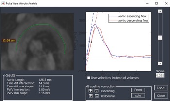
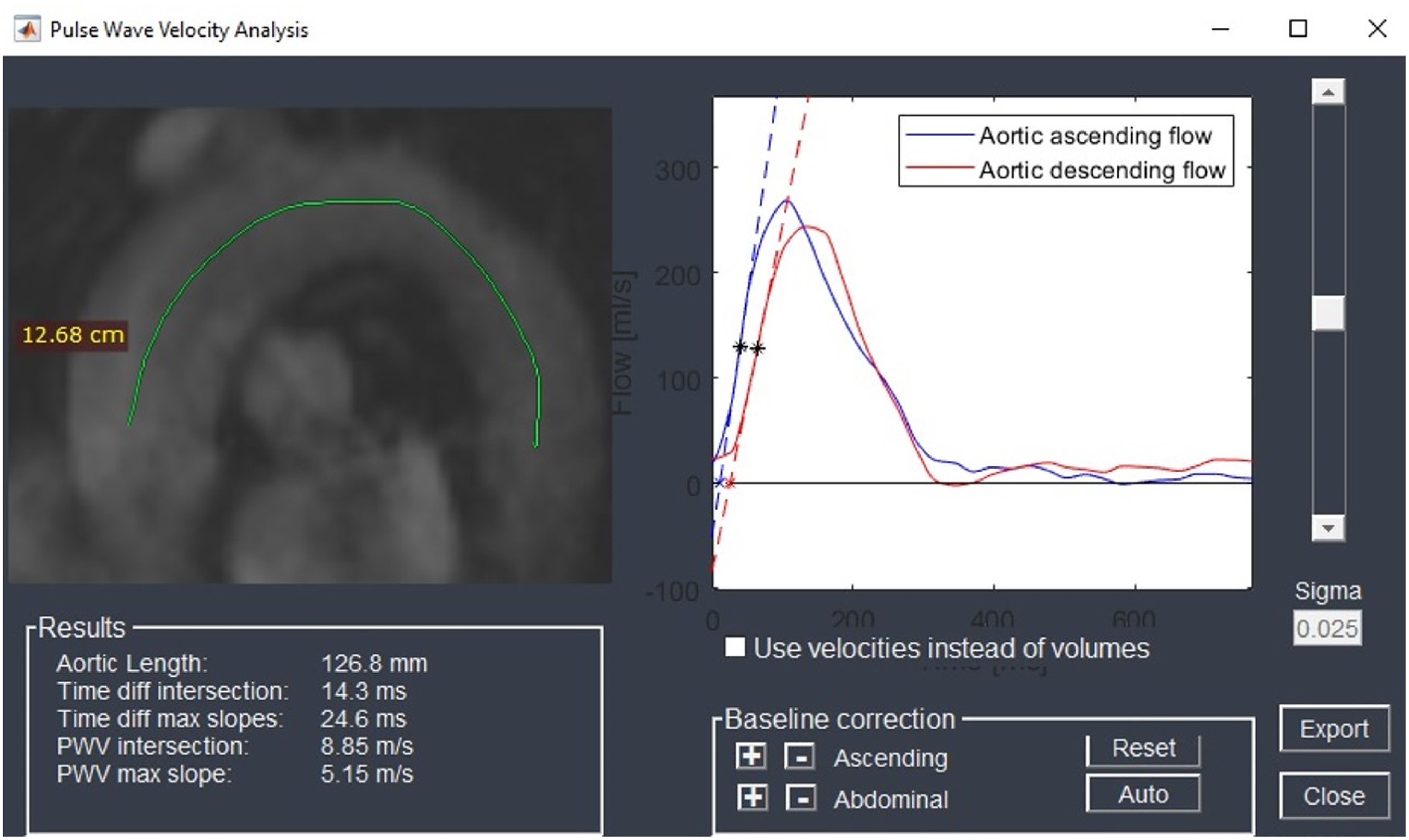
Manual contour drawing for the calculation of pulse wave velocity (the length (Δx) between ascending aorta and descending aorta was measured as indicated by the line) in the aorta. Pulse wave velocity was calculated using the time-to-foot approach implemented through the freely available software Segment cardiac MRI (Medviso, publicly available at https://medviso.com/). For quantitative flow curves, automatic vessel segmentation was performed with manual corrections where needed. Delineations were performed in magnitude images and guided by phase-contrast images where appropriate. Specifically, carotid pulse wave velocity was calculated as x/t (expressed in m/s), where x is the aortic path length between the carotid arch, and t is the time delay between the arrival of the foot of the pulse wave at these locations. Pulse wave velocity estimates were obtained by two independent raters and averaged to generate the final pulse wave velocity values(Fig 1). In our group, the mean aortic arch length was 17.6 ± 3.2 cm. The inter-rater correlation of pulse wave velocity estimates is 0.88, with an r2 of 0.70.
Segment (Medviso) inter face.
Statistical analysis
All data were analysed using the Statistical Package for Social Sciences (SPSS Inc., version 21.0, Chicago, IL, USA). The normally distributed data were expressed in mean and standard deviation, while the data without normal distribution were given as median (25–75%) values. The comparison of the categorical and continuous variables was performed using the chi-square and Mann–Whitney U tests, respectively.
Results
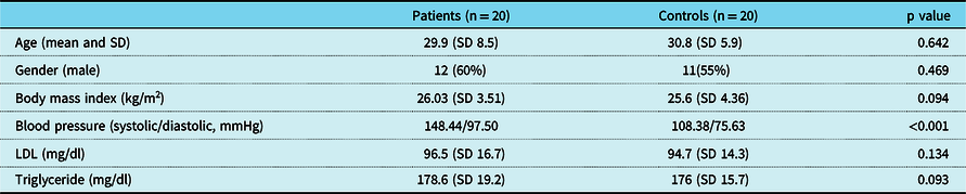
Twelve of the patients with young-onset hypertension were male and eight were female. The mean age of our young-onset hypertension group was 29.9 (18–40) years. All patients underwent MR imaging within a maximum of one year from the time of diagnosis. The mean time between MR imaging and diagnosis was calculated as 25.3 (3–45 days). Of the 20 individuals in the control group, 11 were male and nine were female, with a mean age of 30.8 years (range: 18–39 years). The body mass index was 26.03 (standard deviation 3.51) kg/m2 for the patient group and 25.6 (standard deviation 4.36) kg/m2 for the control group. There was no significant difference between the patient and control groups in terms of age, gender, and body mass index. In the patient group, the mean low-density lipoprotein was 96.5 (standard deviation 16.7) mg/dl and triglyceride was 178.6 (standard deviation 19.2). The mean low-density lipoprotein and triglyceride values of the control group were 94.7 (standard deviation 14.3) and 176 (standard deviation 15.7), respectively.
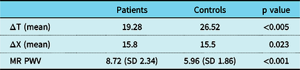
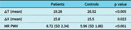
There was no significant difference between the patient and control groups in terms of the low-density lipoprotein and triglyceride values. The mean blood pressure was 148/97 mmHg in the patient group and 108.38/75.63 mmHg in the control group (Table 1). The mean pulse wave velocity was 8.72 (standard deviation 2.34) m/second (range: 7–12.8 m/second) for the patient group and 5.96 (standard deviation 1.86) m/second (range: 4.8–7.1 m/second) for the control group. The pulse wave velocity values were significantly higher in the patient group compared to the control group (p < 0.001) (Table 2).
Demographic features of the patients with young-onset hypertension and the control group
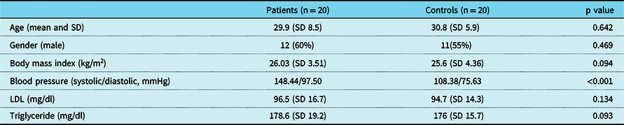
SD: standard deviation; LDL: low-density lipoprotein.
MRI-assessed PWV measurements of the patient and control groups.
Discussion
Hypertension is the most common chronic disease and is one of the major causes of heart failure, stroke, and chronic renal failure. The earlier the onset of hypertension, the longer the exposure time, resulting in a greater risk for cardiovascular events. Although there is not sufficient information about the pathogenesis of young-onset hypertension in the literature, it has been suggested that the main underlying haemodynamic abnormality may be increased peripheral vascular resistance, which causes vascular remodelling in the arteries. Increased arterial stiffness is an important parameter to determine the cardiovascular risk. Reference Laurent, Boutouyrie and Asmar20–Reference Ben-Shlomo, Spears and Boustred22
In addition to increased aortic stiffness being determined by pulse wave velocity measurement, it has been shown in the literature that there is a relationship between aortic stiffness measured by pulse wave velocity and age, ethnicity, and future CVDs. Reference Leung, Dumont, Sandor, Potts and Potts23–Reference Laurent, Cockcroft and Van Bortel26 In a meta-analysis, a 1 m/second increase in pulse wave velocity was reported to be associated with an 11% increase in cardiovascular death. Reference Vlachopoulos, Aznaouridis and Stefanadis27 Pulse wave velocity is an indirect measure of arterial stiffness, and many studies have measured pulse wave velocity using different methods, such as Doppler ultrasonography and tonometry. Reference Mattace-Raso, van der Cammen and Hofman28,Reference Mitchell, Hwang and Vasan29 The most accurate evaluation of aortic pulse wave velocity can be performed by the measurement of intra-arterial pressure. However, this is an invasive modality, and therefore not suitable for widespread clinical use. Reference Lehmann30,Reference Mitchell, Guo and Benjamin31
Tonometry and ultrasound are more commonly used in pulse wave velocity measurement. However, both modalities only provide an estimate of aortic pulse wave velocity due to inadequate acoustic windows and insufficient spatial resolution along the length of the aorta. Reference Leung, Dumont, Sandor, Potts and Potts32,Reference Alecu, Labat and Kearney-Schwartz33 Pulse wave velocity measurement using MRI is widely used as a non-invasive and reliable technique. The most common method of measuring aortic pulse wave velocity is to calculate the aortic length and transit time, i.e., the time it takes for the systolic wave to travel from one reference point within the aorta to another. Reference Maroules, Khera and Ayers34–Reference Kaess, Rong and Larson36
MRI allows for an accurate evaluation of the blood flow rate with sufficient temporal and spatial resolution to investigate the travelling of the aortic systolic flow wave. The true path length of the pulse wave along the aorta can be directly assessed by MRI even in cases of aortic tortuosity, and the regional elastic properties of the aorta can be examined depending on the number of aortic segments studied. Reference Lehmann30,Reference Bolster, Atalar, Hardy and McVeigh37–Reference Grotenhuis, Westenberg and Steendijk39
The presence and severity of hypertension is known to accelerate the increase in aortic pulse wave velocity. Reference Tomiyama, Arai and Koji40 In a 6-year study measuring aortic stiffness as carotid-femoral pulse wave velocity, the presence of hypertension was found to be associated with progression of aortic stiffness compared to normotensive subjects. Reference Benetos, Adamopoulos and Bureau41 Similarly, in our previous tonometry study conducted with young-hypertension cases, we found pulse wave velocity to be significantly higher in patients with hypertension. Reference Gökaslan, Özer Gökaslan, Demirel and Çelik42
Ohyama et al, who followed up hypertension cases of multi-ethnic origin aged 45 years or over for 10 years, measured their aortic stiffness by MRI and reported that blood pressure control was effective in stopping the progression of aortic stiffness. Reference Teixido-Tura and Ambale-Venkatesh43 Van Elderen et al evaluated type I diabetics with normal renal functions in terms of their pulse wave velocity values measured on MRI and found that this patient group had increased pulse wave velocity, independent of renal dysfunction. Reference Van Elderen, Westenberg and Brandts44 Similarly, other researchers showed that the pulse wave velocity increase measured by MRI in patients with type I diabetes was associated with cardiac dysfunction and cerebral small vessel disease. Reference Van Elderen, Brandts and Westenberg45
In another study, the pulse wave velocity measurements were undertaken by MRI in end-stage renal disease cases with decreased arterial compliance and were found to be significantly higher compared to the control group. Reference Zimmerli, Mark and Steedman46
In the current study, the pulse wave velocity values calculated by MRI were found to be increased in hypertensive patients compared to the control group, which is in agreement with the literature. To the best of our knowledge, in the literature, aortic stiffness has not been previously investigated with MRI in patients with young-onset hypertension. Our findings suggest that increased arterial stiffness may also contribute to the aetiology of hypertension that begins at a young age.
The sample size was relatively small, and larger studies are needed to confirm the clinical role of aortic stiffness in cardiovascular risk classification and treatment optimisation. There is also a need for further studies comparing aortic stiffness measured by MRI with other methods.
In conclusion, aortic stiffness may play a role in the aetiology of young-onset hypertension and serve as a non-invasive and reliable screening tool when measured by MRI.
Acknowledgements
We would like to thank the reviewers for their useful comments and suggestions.
Authors’ contributions
C.O.G. and E.D. designed the study. C.O.G and S.G. investigated and supervised the findings of this work. S.S. verified the mathematical methods. All authors discussed the results and contributed to the final manuscript.
Financial support
Research received no specific grant from any funding agency, commercial, or not-for-profit sectors.
Conflicts of interest
None.
Ethical standards
The authors assert that all procedures contributing to this work comply with the ethical standards of the relevant national guidelines on human experimentation and with the Helsinki Declaration of 1975, as revised in 2008, and has been approved by the institutional committees (AFSU ethical commitee).