Implications
Rabbit meat consumption in France is approximately 1.5 kg/person per year, and hence it is worthwhile to improve its nutritional value. This study reports that inclusion of linseed in rabbit diet is a good way to improve this value. Our results will be useful to a future marketing of rabbit meat enriched in n-3 polyunsaturated fatty acids in France.
Introduction
In developed countries, people tend to exercise control over their fat intake. Therefore, one of the main aims of meat research is to produce dietetic and healthy meat. Rabbit meat presents many positive dietetic characteristics such as low lipid and cholesterol levels and high polyunsaturated fatty acid (PUFA) content (Dalle Zotte, Reference Dalle Zotte2002; Hernández, Reference Hernández2008).
In rabbits, as in other monogastrics, it appears that the fatty acid (FA) composition of tissues reflects that of ingested lipids (Maertens, Reference Maertens1998; Mourot and Hermier, Reference Mourot and Hermier2001). Thus, rabbit meat consumption could be healthy for consumers, as manipulation of rabbit diet seems relatively easy in order to increase the level of n-3 PUFA, conjugated linoleic acid (CLA) or vitamin E (Hernández, Reference Hernández2008).
Humans are not able to synthesise n-3 and n-6 PUFA but can synthesise long-chain n-6 and n-3 FAs from dietary precursors (18:2n-6 and 18:3n-3, respectively) that must be supplied by the diet. Numerous epidemiological and clinical studies have documented the health benefits of n-3 PUFA in human nutrition, particularly eicosapentaenoic (EPA) and docosahexanoic (DHA) acids. The best-known effect of these FAs is the control of cardiovascular diseases (Conquer and Holub, Reference Conquer and Holub1998). The key enzymes involved in the formation of these long-chain FAs are Δ6- and Δ5-desaturases, but the conversion is very limited (Alessandri et al., Reference Alessandri, Extier, Astorg, Lavialle, Simon and Guesnet2009). Therefore, one way to increase EPA and DHA contents in the human diet is to produce food, meat in particular, with a high content of these FAs (Ailhaud et al., Reference Ailhaud, Massiera, Weill, Legrand, Alessandri and Guesnet2006).
The relative importance of lipogenic enzyme activities in the liver and adipose tissues is dependent on the age of animals. During growth, the liver is the main site of lipogenesis in young rabbits, but in adults the major site is the adipose tissue (Vézinhet and Nougès, Reference Vézinhet and Nougès1977; Gondret et al., Reference Gondret, Mourot and Bonneau1997; Gondret, Reference Gondret1999).
However, PUFA have a high propensity to oxidise. It is well known that tissue oxidation is one of the main mechanisms by which food quality deteriorates. Lipid oxidation, which can lead to rancidity and colour deterioration, is a major problem in rabbit meat because of its high content of PUFA (Liu et al., Reference Liu, Gai, Gasco, Brugiapaglia, Lussina, Guo, Tong and Zoccarato2009). Indeed, compared with other species, the thiobarbituric acid-reactive substance (TBARS) values observed in rabbit meat are generally higher than values in pork, chicken or in cattle meat (Dalle Zotte, Reference Dalle Zotte2004). However, the positive effect of the diet supplementation of vitamin E on the oxidative stability of lipids in rabbit meat has been shown by Corino et al. (Reference Corino, Pastorelli, Pantaleo, Oriani and Salvatori1999).
The aim of this study was to evaluate the impact of a linseed diet on growth performances, lipogenesis potential, stearoyl-CoA-desaturase (SCD) (that generates monounsaturated FAs (MUFA)) and on meat quality in rabbits.
Material and methods
Animals and diets
Animals used in this experiment were reared and slaughtered in compliance with French regulations for the human care and use of animals in research (certificate of authorisation to experiment on living animals delivered by the French department of Agriculture to M. Kouba).
Twelve Californian × New Zealand 30-day-old weanling mixed-sex rabbits with a body weight of 550 ± 7.3 g were given ad libitum access to a commercial diet formulated for rabbit growth. Six weeks before slaughter at 11 weeks of age, the animals were divided into two homogeneous groups (live weight and sex) with rabbits placed in individual cages. Both groups were fed ad libitum two different diets: a control diet (commercial diet) or a linseed diet containing 40 g of Croquelin® (Combourtillé, France) (a mix of 50% extruded linseeds, 30% wheat bran and 20% sunflower meal) per kg.
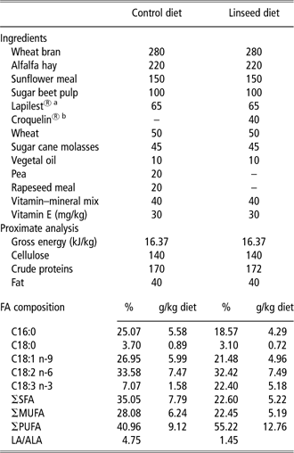
Both diets were isoenergetic (gross energy, 16.37 kJ/kg diet), and contained approximately 17% crude protein and 30 mg of vitamin E/kg diet (Table 1).
Composition (as-fed basis) of experimental diets (g/kg)
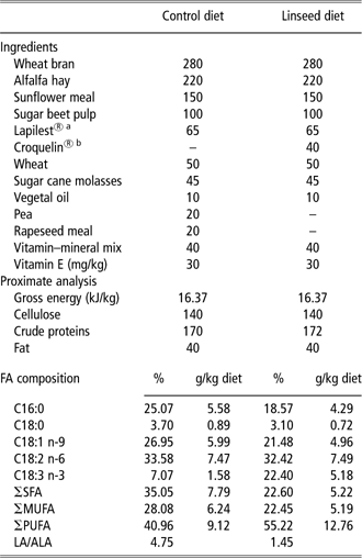
FA = fatty acid; SFA = saturated fatty acids; MUFA = monounsaturated fatty acids; PUFA = polyunsaturated fatty acids; LA = linoleic acid; ALA = α-linolenic acid.
aLapilest®: a mix of beet pulp, grape pulp and seeds, apple marc and sunflower shells.
bCroquelin®: a mix of 50% extruded linseeds, 30% wheat bran and 20% sunflower meal.
During the experimental period, feed intake and live weight were recorded individually each week. At 11 weeks of age, the animals were slaughtered by electric stunning and exsanguination in compliance with French national regulations for commercial slaughtering, but without prior fasting. After slaughter, hot carcasses were weighed and then cut into two halves according to the World Rabbit Scientific Association standard method (Blasco and Ouhayoun, Reference Blasco and Ouhayoun1993). The left half was weighed and cut into three anatomically defined primal cuts, which were also weighed and sampled: shoulder, leg and saddle. Liver and adipose tissues (interscapular and perirenal fat) were weighed, and a sample of the Longissimus dorsi muscle (LD) was taken. Samples of liver, adipose tissues and muscle were immediately assayed for enzyme activities. All the other samples were vacuum-packaged and deep frozen at −20°C pending different chemical analyses.
Meat quality variables
The ultimate pH was measured 24 h post mortem in the LD with a combined glass penetrating electrode (Ingold, Mettler Toledo, Greifensee, Switzerland) and a portable pH meter (WTW 340i, Weilheim, Germany) at the level of the fifth lumbar vertebra in the chilled carcass.
The colour of the LD was recorded 24 h post mortem using a CR-300 Minolta Chroma Meter (Minolta Camera Co., Osaka, Japan), which, at each point, gave the average of the three measurements of lightness (L*), redness (a*) and yellowness (b*).
Chemical analysis of diet, muscle and fats
Diets were analysed for cellulose and crude protein (n × 6.25), according to the Association of Official Analytical Chemists (AOAC, 2006). The gross energy content of the diets was measured using an adiabatic bomb calorimeter (IKA, Staufen, Germany).
The lipids were extracted from samples of liver, Longissimus dorsi muscle, interscapular and perirenal fats and the diets using the method described in Folch et al. (Reference Folch, Lees and Stanley1957) (extraction with a mixture of chloroform–methanol (2 : 1)). Fatty acid methyl esters were prepared with boron trifluoride methanol according to Morrison and Smith (Reference Morrison and Smith1964) and analysed on a Perkin Elmer Autosystem XL gas chromatograph (Waltham, Massachusetts, USA) equipped with fused silica gel capillary column (0.25 mm i.d. ×30 m), filled with stationary phase (80% biscyanopropyl and 20% cyanopropylphenyl), using margaric acid (C17) as internal standard. The chromatography conditions used were: temperature programme from 45°C to 240°C at 20°C to 35°C/min. The injector and detector temperatures were maintained at 220°C and 280°, respectively. Results were expressed as percentages of the total FA content of the diet, liver, muscle and adipose tissues.
The non-polar and polar lipids were separated from the total lipid extracts of the LD on silica cartridges (Sep-Pack, Waters Corporation; Juaneda and Rocquelin, Reference Juaneda and Rocquelin1985).
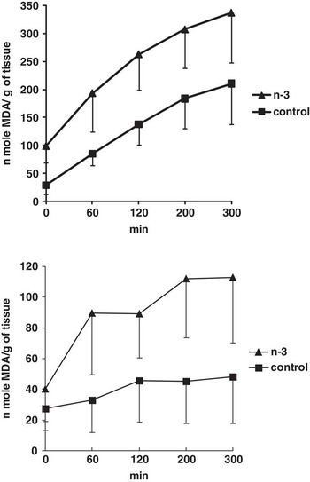
Lipid oxidation in perirenal fat and LD was determined using a modification of the method described by Monahan et al. (Reference Monahan, Buckley, Morrissey, Lynch and Gray1992), by assaying 2-TBARS as reported by Oriani et al. (Reference Oriani, Salvatori, Pastorelli, Pantaelo, Ritieni and Corino2001). TBARS, expressed as nmol malonaldehyde (MDA)/g of tissue, were determined at 0, 60, 120, 200 and 300 min.
Enzyme analysis
Lipogenic enzyme activities
Activities of the lipogenic enzymes were determined in the liver, the Longissimus dorsi muscle, interscapular and perirenal fats. Weighed amounts of tissues (2 g) were homogenised in a buffer containing 0.25 M sucrose and centrifuged at 100 000 g for 40 min at 4°C.
The cytosolic supernatant fraction was collected and was analysed for malic enzyme (ME) activity (EC 1.1.1.40) and glucose-6-phosphate dehydrogenase activity (G6PDH, EC 1.1.1.49) according to procedures outlined by Kouba et al. (Reference Kouba, Hermier and Le Dividich1999). NADPH formation was measured at 37°C by absorbance at 340 nm. Enzyme activity was expressed as μm of NADPH produced per minute per gram of tissue.
The FA synthase (FAS, EC 2.3.1.85) activity was measured as described by Lavau et al. (Reference Lavau, Bazin, Karaoghlanian and Guichard1982). NADPH oxidation was determined at 37°C by absorbance at 340 nm. Enzyme activity was expressed as μm of NADPH used per minute per gram of tissue.
SCD activity
The activity of SCD (Δ9-desaturase, EC 1.14.19.1) was determined in the liver, the LD, and interscapular and perirenal fat homogenates obtained from the samples removed after slaughter. Weighed amounts of tissue were homogenised in a buffer containing 0.25 M sucrose and 0.05 M potassium, and centrifuged at 10 000 × g for 40 min at 4°C. Each incubation mixture for the measurement of SCD contained an aliquot of supernatant, phosphate buffer, MgCl2, ATP, coenzyme A, NADH and [14C]stearic acid, as described by Kouba et al. (Reference Kouba, Hermier and Le Dividich1999). The activity was determined by measuring the conversion of [14C]stearic acid into [14C]oleic acid. One unit of activity was defined as the amount of enzyme that produced 1 nmol of oleic acid per min. Enzyme activity was expressed as unit per gram of tissue.
β-Hydroxyacyl-CoA-dehydrogenase (HAD) activity
The activity level of one mitochondria oxidative marker, reflecting FA β-oxidation, β-HAD (EC 1.1.1.35) was measured in the LD in compliance with the method described by Bass et al. (Reference Bass, Brdiczka, Eyer, Hofer and Pette1969).
Approximately 400 mg of muscle was homogenised and sonicated in 50 vol. (wt./vol.) of ice-chilled 0.1 M phosphate buffer (pH 7.5) containing 2 mM EDTA.
After centrifugation at 1700 × g for 15 min at 4°C, the supernatant fraction was collected and used for β-HAD activity determination. Enzyme activity was measured at 30°C by determination of optical density every 25 s for 5 min using an automatic spectophotometric analyser (Konelab, Thermo Electron Oy /Thermo Electron S. A., Finland). After addition of aceto-acetyl-CoA and NADH, the HAD activity was assessed at 340 nm. One unit of activity was defined as the amount of enzyme that degraded substrate per minute. Enzyme activity was expressed as μm of disappeared NADH/min per gram of fresh muscle.
Statistical analysis
Effects of diets on the different variables were examined with one-way ANOVA using SAS (SAS Institute, 1999). The effect of tissue origin on FA composition, TBARS and enzyme activities was also studied with one-way ANOVA using SAS (1999). The individual rabbit was the experimental unit for analysis of all data.
Results
Chemical composition of the diets
The FA profiles of the two diets differed according to the ingredients (Table 1). As expected, the linseed diet had higher proportions of α-linoleic acid (ALA; 18:3n-3; 22.4% v. 7.1%) and total PUFA (55.2% v. 41%) and lower proportions of saturated (SFA; 22.6% v. 35%) and MUFA (22.4% v. 28.1%) than the control diet. The linoleic acid/α-linolenic acid ratio was decreased in linseed diet (1.45 instead of 4.75). LA (18:2n-6) concentration and proportion were similar in both diets.
Productive performances
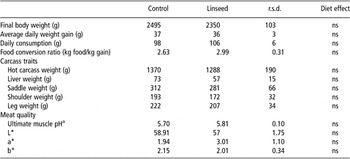
Performances and carcass data are shown in Table 2. Diet had no effect on animal growth, feed intake, carcass weight, liver, saddle, shoulder, leg weights and meat quality.
Effects of diets (n = 6 per diet) on growth performance, carcass traits, feed intake and meat quality

ns = no significant difference (P > 0.05); L* = lightness; a* = redness; b* = yellowness.
apH measured 24 h post mortem in the Longissimus dorsi muscle.
Total lipid content
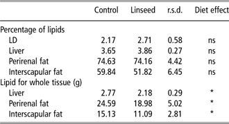
There was no effect of diet on the lipid percentage of the LD, liver, perirenal and interscapular fats (Table 3). The total lipid content deposited in the whole tissue (liver, perirenal and interscapular fats) was decreased by the linseed diet, compared with the control diet.
Effect of diets (n = 6 per diet) on the lipid content (% or for whole tissue) of liver, LD and adipose tissues
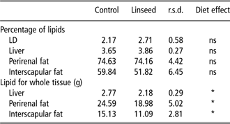
LD = Longissimus dorsi muscle; ns = no significant difference (P > 0.05).
*P < 0.05.
The lipid content of perirenal fat was much higher (P < 0.001) than that of interscapular fat, regardless of the diet.
Lipogenic enzymes, SCD and β-HAD activities
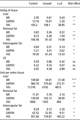
The lipogenic enzyme activities (ME, G6PDH and FAS) are shown in Table 4. The liver presented the highest lipogenic enzyme activities (P < 0.001) in comparison with the muscle and the adipose tissues.
Effects of diets (n = 6 per diet) on ME, G6PDH, FAS activities in liver, adipose tissues and the LD

ME = malic enzyme; G6PDH = glucose-6-phosphate-dehydrogenase; FAS = fatty acid synthase; LD = Longissimus dorsi muscle; ns = no significant difference (P > 0.05).
*P < 0.05, **P < 0.01 and ***P < 0.01.
ME, G6PDH, FAS activities are expressed in μmol NADPH/min per g of tissue.
In liver, and perirenal and interscapular adipose tissues, the linseed diet led to a decrease of the activities of ME, G6PDH and FAS, regardless of the expression of these activities, per gram of tissue or for the whole tissue. However, there was no diet effect on ME, G6PDH and FAS activities in the muscle.
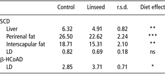
Adipose tissues and liver from rabbits fed the linseed diet had 15% to 22% less SCD activity than rabbits fed the control diet (Table 5). The muscle enzyme activity was not affected by the diet.
Effect of diet (n = 6 per diet) on SCD activity in liver, adipose tissues and LD and on (β-HcoAD activity in the LD
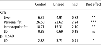
SCD = stearoyl-CoA-desaturase; LD = Longissimus dorsi muscle; β-HCoAD = β-hydroxyacyl-CoA-dehydrogenase; ns = no significant difference (P > 0.05).
SCD activity is expressed in nmol of oleic acid formed/min per g of tissue.
β-HCoAD activity is expressed in μmol of disappeared NADH/min per g of tissue.
*P < 0.05, **P < 0.01 and ***P < 0.01.
In the LD, the linseed diet increased (P < 0.04) the activity of β- HAD (Table 5).
Fatty acid composition
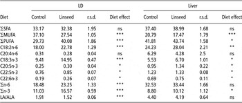
In muscle and liver, the linseed diet decreased MUFA proportion and increased PUFA percentages (Table 6). The SFA proportion was not affected by the diet, in both tissues.
FA composition in the LD and liver (n = 6 per diet)

FA = fatty acid; LD = Longissimus dorsi muscle; SFA = saturated fatty acids; MUFA = monounsaturated fatty acids; PUFA = polyunsaturated fatty acids; ns = no significant difference (P > 0.05); LA/ALA = linoleic acid/α-linolenic acid ratio
*P < 0.05, **P < 0.01 and***P < 0.01.
Results are expressed in percentage of total FA content.
There was an increase in the level of EPA, DPA and DHA in both tissues, with the linseed diet, but the arachidonic acid (ARA) percentage was not affected by the diet in both tissues. The proportions of LA and ALA were much higher in rabbits fed the linseed diet than in rabbits fed the control diet, in both tissues. The linseed diet decreased significantly the LA/ALA ratio in the LD, but not in the liver.
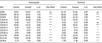
The MUFA proportion was lower and the PUFA level was higher in perirenal and interscapular fats of animals fed the linseed diet (Table 7). The SFA proportion was not affected by the diet. There was an increase in the level of EPA, DPA and DHA, with the linseed diet in both tissues. The percentage of ARA was not affected by the diet in the perirenal fat, but it was decreased by the linseed diet in the interscapular fat. The n-6 and n-3 FA proportions were increased by the linseed diet and the LA/ALA was decreased by the experimental diet in both adipose tissues.
FA composition in interscapular and perirenal fat (n = 6 per diet)

FA = fatty acid; SFA = saturated fatty acids; MUFA = monounsaturated fatty acids; PUFA = polyunsaturated fatty acids; ns = no significant difference (P > 0.05); LA/ALA = linoleic acid/α-linolenic acid ratio.
*P < 0.05, **P < 0.01 and ***P < 0.01.
Results are expressed in percentage of total FA content.
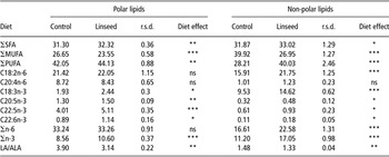
The FA composition of polar or non-polar lipids in the LD is summarised in Table 8. The diet significantly affected the amount of different FA. In polar and non-polar fractions, the percentage of all n-3 FA increased in rabbits fed the linseed diet, whereas the MUFA percentage was lower and the SFA percentage was higher.
Polar and non-polar FA composition of the LD (n = 6 per diet)

FA = fatty acid; LD = Longissimus dorsi muscle; SFA = saturated fatty acids; MUFA = monounsaturated fatty acids; PUFA = polyunsaturated fatty acids; LA/ALA = linoleic acid/α-linolenic acid ratio; ns = no significant difference (P > 0.05).
*P < 0.05, **P < 0.01 and ***P < 0.01.
Results are expressed in percentage of total FA content.
In polar and non-polar lipids, the LA/ALA was decreased by the linseed diet.
Lipid oxidation
Lipid oxidation was significantly higher in the perirenal fat and the LD of rabbits fed the linseed diet than rabbits fed the control diet (P < 0.001; Figure 1).
Effect of diet on the lipid oxidation (nmole malonaldehyde (MDA)/g of fresh tissue) in perirenal fat (top figure) and in Longissimus dorsi muscle (bottom figure).

Discussion
This study showed that the incorporation of n-3 FA in the diet had no significant effect on performances and carcass characteristics. These results were in agreement with previous work in rabbits (Fernandez and Fraga, Reference Fernandez and Fraga1996; Combes, Reference Combes2004; Kouba et al., Reference Kouba, Benatmane, Blochet and Mourot2008) and in pigs (Kouba et al., Reference Kouba, Enser, Whittington, Nute and Wood2003; Guillevic et al., Reference Guillevic, Kouba and Mourot2009).
The muscular ultimate pH and the lightness (b*) value were not affected by the diet, as already demonstrated by Kouba et al. (Reference Kouba, Benatmane, Blochet and Mourot2008).
ME and glucose-6-phosphate dehydrogenase are the main enzymes involved in supplying NADPH for the reductive biosynthesis of FAs (Wise and Ball, Reference Wise and Ball1964; Young et al., Reference Young, Shrago and Lardy1964), and FAS is a multi-enzyme that plays a key role in FA synthesis. The hepatic activities of these lipogenic enzymes were much higher than the activities in adipose tissues and muscle. These results are in general agreement with other studies showing that, at 11 weeks of age (age of slaughtering of commercial rabbits), liver is the major site of lipogenesis (60% of total lipogenesis), with adipose tissue accounting for 35% and digestive tract for 5%. (Gondret et al., Reference Gondret, Mourot and Bonneau1997; Gondret, Reference Gondret1999). Our results also confirmed previous studies showing that, in rabbits, de novo lipogenesis is greater in perirenal adipose than in subcutaneous and interscapular adipose tissues (Gondret et al., Reference Gondret, Mourot and Bonneau1997). When both adipose tissues were considered, the lipogenesis was also lower in the whole interscapular fat than in the whole perirenal fat with the linseed diet.
The supplementation of n-3 PUFA in the diet decreased lipogenic enzyme activities in the liver and in perirenal and interscapular fats. The consequence of the lower lipogenesis with linseed diet was that the total lipid depots in these tissues were also reduced. However, linseed diet had no effect on these enzyme activities in the LD. These results were in agreement with a previous study showing that PUFA inhibit hepatic lipogenesis in rabbit (Corino et al., Reference Corino, Mourot, Magni, Pastorelli and Rosi2002).
SCD generates MUFA from SFA (Bloomfield and Bloch, Reference Bloomfield and Bloch1960; Marsh and James, Reference Marsh and James1962; Gelhorn and Benjamin, Reference Gelhorn and Benjamin1965). To our knowledge, for the first time this study provides findings about SCD activity in the rabbit. This study also shows that this activity is quite low in rabbit liver, as compared to that of chicken liver, but it is quite similar to that of turkey liver (Kouba et al., Reference Kouba, Bernard-Griffiths and Lemarchal1993) and it is higher than that in pig liver (Kouba et al., Reference Kouba, Mourot and Peiniau1997). As for lipogenic enzymes, the SCD activity is the highest in perirenal fat, regardless of the diet. The inhibitory effect of PUFA on SCD activity observed in this study in rabbit liver and adipose tissues has already been demonstrated in pig adipose tissue (Kouba and Mourot, Reference Kouba and Mourot1998; Kouba et al., Reference Kouba, Enser, Whittington, Nute and Wood2003) and more recently in the muscle of beef cattle (Waters et al., Reference Waters, Kelly, O'Boyle, Moloney and Kenny2009). The lack of effect of linseed diet on muscle enzyme activity in the rabbits observed in this study had also been observed in pig muscle (Kouba et al., Reference Kouba, Enser, Whittington, Nute and Wood2003). The decrease of the Δ9-desaturase activity in the liver and the adipose tissues of rabbits fed linseed diet compared with those fed the control diet resulted in an obvious reduction in the MUFA percentage, which is consistent with previous studies in the rabbit (Kouba et al., Reference Kouba, Benatmane, Blochet and Mourot2008) and in the pig (Kouba et al, Reference Kouba, Enser, Whittington, Nute and Wood2003). Results of this study showed that the changes in the MUFA percentage could be attributed, at least in part, to a reduction in Δ9-desaturase activity.
In contrast, β-oxidation, estimated by the β-HAD activity, increased in the muscle of animals fed the n-3 PUFA-supplemented diet. Previous studies have already shown that the enhancement of β-oxidation in adipose tissues of mice fed a diet supplemented in EPA and DHA (Flachs et al., Reference Flachs, Horakova, Brauner, Rossmeisl, Pecina, Franssen-Van Hal, Ruzickova, Sponarova, Drahota, Vlcek, Keijer, Houstek and Kopecky2005). It is well known that PUFA increase transcription of the regulatory μm of FA oxidation, via activation of peroxysome proliferator-activated receptor (PPAR) (Mori et al., Reference Mori, Kondo, Hase, Tokimitsu and Murase2007).
Our results show that the enrichment of the rabbit diet with extruded linseeds, a major source of n-3 PUFA, led to an enrichment of muscle and adipose tissue n-3 PUFA content, and confirmed previous studies in rabbits (Bernardini et al., Reference Bernardini, Dal Bosco and Castellini1999; Dal Bosco et al., Reference Dal Bosco, Castellini, Bianchi and Mugnai2004; Combes and Cauquil, Reference Combes and Cauquil2006; Petracci et al., Reference Petracci, Bianchi and Cavani2009). This conclusion has also been demonstrated in pigs (Enser et al., Reference Enser, Richardson, Wood, Gill and Sheard2000) and in chickens (Crespo and Esteve-Garcia, Reference Crespo and Esteve-Garcia2002).
Our results show more favourable PUFA/SFA and LA/ALA ratios in the muscle and adipose tissues from rabbits fed linseed diet, but not in the liver in which diet had no effect on these ratios. These favourable parameters were already demonstrated in a previous work (Kouba et al., Reference Kouba, Benatmane, Blochet and Mourot2008).
Lipid oxidation in meat, particularly in rabbits, is mainly due to oxidation of PUFA. Indeed, FA composition of rabbit meat is characterised by a high PUFA content. Therefore, feeding these animals with a linseed diet leads to an increase in the level of n-3 PUFA, which could explain the high value of TBARS registered in this study in muscle and fat. This tendency agreed with results obtained by Kouba et al. (Reference Kouba, Benatmane, Blochet and Mourot2008), and more recently by Petracci et al., (Reference Petracci, Bianchi and Cavani2009). Our results show a higher risk of peroxidation of the fat compared with the muscle, which is due to a much higher content of lipids, especially PUFA, in perirenal fat. This result has already been shown on Guizhou minipigs (Yang et al., Reference Yang, Zhao, Xiao, Zhan, Du, Wu and Ma2010). It is well known that extra supplementation of vitamin E in the diet improves the oxidative stability of the meat and leads to an increase of this vitamin in rabbit meat (Dal Bosco et al., Reference Dal Bosco, Castellini, Bianchi and Mugnai2004). It seems that the quantity of vitamin E added to the diets in our study (30 mg/kg) was insufficient to limit the deterioration of meat. However, our results are not in agreement with Dal Bosco et al. (Reference Dal Bosco, Castellini, Bianchi and Mugnai2004). These authors found that feeding an n-3 PUFA-enriched diet significantly lowered the TBARS level in the LD, but they used a large amount of vitamin E in their study (74 or 289 mg/kg).
Conclusion
The results of this study show that performances of rabbits are not affected by diet enriched with n-3 PUFA. The PUFA/SFA and LA/ALA ratios were more favourable with an n-3 PUFA enrichment of rabbit diet. Inclusion of linseed in rabbit diet is a valid means of meeting consumer demand for meat that is nutritionally beneficial.
The results of this study suggest that Δ9-desaturase may be involved in the regulation of MUFA content and hence in the quality of rabbit adipose tissue. A better understanding of this enzyme could offer a way to control meat quality in rabbits.
Acknowledgements
The authors thank Bretagne Lapins for its financial support.