A brief introduction of fatty liver disease (FLD)
FLD results from the impairment of normal processes relevant to synthesis and elimination of liver lipid. In pathology, FLD is manifested by early reversibility of hepatic steatosis (or fatty liver) and further inflammatory stage (or steatohepatitis) (Ref. Reference Wong1). The hepatic steatosis accumulates excess lipid in vesicles composed of triglyceride (TG) fat (Ref. Reference Ganji2). Clinically, hepatic steatosis can be diagnosed by X-ray computed tomography (CT), ultrasound, and definitive liver biopsy (Refs Reference Karlas3, Reference Martin-Rodriguez4, Reference Schwenzer5). The hepatic steatosis is dark on CT and bright on ultrasound. Their combination is thus termed as flip-flop sign (Refs Reference Lee and Park6, Reference Pia, Aresu and Pascalis7, Reference Filippone8). There are some serum markers and cell death parameters to complement the imaging examination for noninvasive assessment of FLD (Refs Reference Lombardi, Pisano and Fargion9, Reference Sowa10). Hepatic steatosis may be caused by alcohol or nonalcoholic factors such as diabetes mellitus, obesity, protein malnutrition, cell toxins and anoxia. Microscopically, the abnormal retention of lipid within hepatocytes displaces the cytoplasm. Microvesicular fatty degeneration is characterised by small fat vacuoles in the cell, commonly induced by tetracyclines, pregnancy, Reye's syndrome and hepatitis C (Refs Reference Saibara11, Reference Yu12, Reference Nepomnyashchikh13). Macrovesicular fatty degeneration usually is because of oversupply of lipid such as alcoholism, obesity, insulin resistance or change of nutritional status (Refs Reference Neto-Ferreira14, Reference Kim15, Reference Zago16, Reference Setshedi17). The breakdown of excess alcohol produces large amounts of chemical energy in the form of NADH [nicotinamide adenine dinucleotide (NAD) + hydrogen (H)], which inhibits fatty acid metabolism and simultaneously increasing the synthesis of fatty acids of the cell (Refs Reference Zhao18, Reference Zivkovic19). Heavy alcohol intake results in lipids being overproduced more than they being utilised. Hepatic steatosis is associated with obesity, diabetes and metabolic syndrome. Nutrient malnutrition can cause a mobilisation of fat from adipocytes and fat overaccumulation in the liver where lipid metabolism occurs. No single mechanism leads to steatosis. Different etiological factors can disrupt normal lipid metabolism via multiple mechanisms and incur lipid accumulation through the cell. These mechanisms are identified upon whether they ultimately cause an overaccumulation of lipid that cannot be disposed quickly enough, or whether they fail to cause lipid breakdown. When hepatic steatosis is further exacerbated, steatohepatitis is developed. During the period of steatohepatitis, apoptotic cell death plays an important role in subsequent liver failure.
Mechanism of autophagy
Autophagy is a cellular process of self-digestion, in which cells capture their own cytoplasm and organelles, and consume them in autolysosomes. Autophagy-related genes (Atgs) are conserved from yeast to mammals and regulate cannibalism of intracellular cytoplasm, and degradation of large molecules (i.e. ubiquitinated proteins) or organelles [i.e. lipid droplets (LDs) and mitochondria]. Autophagic process degrades cellular components that are either deleterious to homeostatic stability (e.g. damaged organelles and microbial invaders) or energy-generated for metabolic activity (Fig. 1). There are three different types of autophagy in mammalian cells, each of which involves the delivery of intracellular cargo to lysosomes for degradation. The predominant form, macroautophagy (often as autophagy), produces vesicles called autophagosomes that encapsulate and distribute cytoplasmic material to lysosomes. Autophagy is the unique mechanism to dispose large structures such as organelles and protein aggregates. Selective autophagy of organelles is defined as different names, that is mitophagy (mitochondria), reticulophagy (endoplasmic reticulum) and lipophagy (lipids) respectively.
Autophagic process degrades cellular components for energy generation, biosynthesis, elimination of foreign microbial invaders, etc.

Autophagy can engulf intact organelles and large molecules into double-membraned vacuoles for degradation. Therefore, autophagy is a membrane-dependent process that is initiated by the formation of a preautophagosomal structure leading to an isolation membrane or phagophore. Autophagy as a continuous process covers a series of steps, including (1) activation, (2) nucleation, (3) elongation, (4) trafficking towards lysosomes and (5) fusion of two membranes (Ref. Reference Yang and Klionsky20). Autophagic process can be roughly classified into the following stages: (a) Activation/initiation. The process begins with the formation of Beclin 1 complex, which gives rise to nascent autophagosome membranes. Autophagosome nucleation requires the Beclin 1 complex to recruit the class III phosphatidylinositol 3-kinase (VPS34) and to generate phosphatidylinositol 3-phosphate (Reference Funderburk, Wang and Yue21). (b) Elongation. Autophagosome membranes are elongated through two ubiquitin-like molecules, Atg12 and Atg8 [or light chain 3 (LC3) in mammals] to work in different conjugation systems. In the first ubiquitin-like reaction, Atg12 covalently connects Atg7, Atg10 and Atg5 successively, which together bind Atg16L1. Atg12 conjugation system forms pre-autophagosomal structure. In LC3 conjugation system, the early reaction is that LC3-II is interacted with Atg4. The cleaved LC3-II is later combined with phosphatidylethanolamine via the catalysation of Atg7 and Atg3. The lipidated LC3-II is associated with newly forming autophagosome membranes. Fresh membranes gather around cargo, encapsulate the cargo in a vesicle, fuse with a lysosome, and subsequently generate an autolysosome. LC3-II is situated on mature autophagosomes until after fusion with lysosomes and is able to be used as a marker to monitor autophagy. (c) Termination. The autolysosomal contents are degraded by diverse enzymes such as proteases, lipases, nucleases and glycosidases. Breakdown products, that is amino acids, lipids, nucleosides and carbohydrates, are released into the cytosol for synthetic and metabolic pathways.
Under physiological conditions, basal autophagy plays a housekeeping role in the elimination of damaged or outdated components that can otherwise become toxic. Autophagic function has particular importance to keep cellular refreshing in quiescent and terminally differentiated cells, since damaged components in those cells are not diluted by cell replication. Breakdown products of autophagy are inputs to cellular metabolism, through which they are used to supplement energy and to build new proteins and membranes. In starvation, autophagy provides a nutrient source and promotes cell survival. As powerful promoter of metabolic homeostasis, autophagy is induced by a broad range of active stressors, for example aggregated proteins, oxidised lipids, damaged organelles, and intracellular pathogens. Autophagy is also able to dispose LDs. Defects in autophagy are linked to metabolic syndrome and FLD. Modulation of autophagy affects the progression of alcoholic and nonalcoholic fatty liver diseases (NAFLD).
Evidence for autophagy to regulate hepatic steatosis
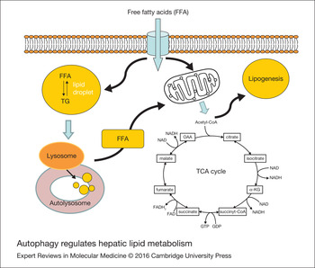
Massive facts demonstrate that autophagy as a regulatory mechanism modulates hepatic steatosis. Evidence for autophagy to regulate hepatic steatosis includes: (i) Selective degradation of LDs through lipophagy (Ref. Reference Singh22). Intracellular lipids are generally stored as TGs in a LD and are surrounded by a phospholipid monolayer (Fig. 2). Autophagy regulates hepatic TG content based on the following observations: (a) Subsequent to inhibition of autophagy by 3-methyladenine targeting mammalian PI3-kinase VPS34, an increased TG contents are detected in hepatocytes cultured in regular medium or supplemented with an unsaturated fatty acid (i.e. oleic acid) (Ref. Reference Singh22); (b) after lysosomal degradation is inhibited by chloroquine (CQ), TG contents are also enhanced in hepatocytes that are fed with supplemental fatty acids (Ref. Reference Mei23); (c) when the autophagy is suppressed through siRNA knockdown of Atg5, TG contents are significantly elevated in fatty acid-treated hepatocytes (Ref. Reference Singh22); and (d) liver-specific Atg7 knockout mice can increase hepatic TG contents either in fed mice or in mice starved for 24 h (Ref. Reference Singh22). (ii) Abundance of liver LDs modulated by free fatty acids (FFA). As a substrate for autophagy, LDs are impounded in autophagosomes that fuse with lysosomes to form autolysosomes. LD components are degraded by lysosomal enzymes. The autophagic function is altered subsequent to changes in nutrient supply. The autophagy can regulate LD metabolism to meet the cell's energy demands. Pathophysiological states can modulate cellular lipid metabolism via autophagic pathway. Autophagy may thus is a novel target for the prevention of abnormal lipid metabolism and accumulation (Ref. Reference Dong and Czaja24). Lipophagic process gets a feedback inhibition after administering FFA in vitro or a high-fat diet (HFD). Moreover, FFA may not only promote LD growth, but impair LD breakdown as well. (iii) Maintenance of energy balance via the intracellular storage and utilisation of lipids. During nutrient deprivation, cellular lipids stored as TGs in LDs are hydrolysed by cytosolic lipases into fatty acids for energy. A further response to starvation is the activation of autophagy. The autophagic process sequesters intracellular organelles (i.e. LDs, glycogen granules and proteins) into double-membrane autophagosomes that are then fused with lysosomes for degradation as cellular energy source. Lipolysis and autophagy share similarities in regulation and function. Autophagy regulates intracellular lipid stores via lipophagy. Autophagic process is actively stimulated during nutrient deprivation. (iv) Failure of lipid metabolism. The failure of lipid metabolism can lead to mechanisms, which would normally utilise or dispose lipids becoming impaired, resulting in the lipid accumulation in the cell. For example: (1) Certain toxins, such as alcohol, carbon tetrachloride and aspirin, interfere with cellular machinery resulted in fundamental deviation of liver lipid metabolism. (2) Protein malnutrition (e.g. Kwashiorkor) brings on a lack of precursor apoproteins, in which lipids normally participated in lipoprotein synthesis therefore become accumulated. (v) Defective autophagy. Dysfunction of autophagy causes an abnormality of lipid metabolism. Following inhibition of autophagy, TG storage in LDs is increased in cultured hepatocytes. In mouse liver with defective autophagy, gross liver volume is enlarged because of lipid overaccumulation. Further analysis reveals that size and number of LDs in hepatocytes as well as levels of hepatic TGs and cholesterol are all elevated. Clinically, the critical role of autophagy in lipid metabolism is also demonstrated by its involvement in lipid accumulation of human diseases, such as metabolic syndrome and alcoholism (Ref. Reference Singh22). In patients with Gaucher's disease, their lysosomes fail to degrade lipids and steatosis arises from the accumulation of glycolipids. (vi) Enhancer and inhibitor of autophagy. Autophagy activity is increased by autophagy enhancer (carbamazepine or rapamycin) and decreased by autophagy inhibitor (CQ) respectively. Both carbamazepine and rapamycin can activate and enhance alcohol-induced macroautophagy in hepatocytes in vitro and in vivo. Degrees of fatty liver injury are able to be further exacerbated by CQ. The protective effect of carbamazepine and rapamycin is reflected by reducing steatosis and improving insulin sensitivity as demonstrated in high fat diet-induced nonalcoholic fatty liver condition. (vii) The production of large LDs characteristic of white adipose tissue. Inhibition of autophagy blocks white adipocyte differentiation. When atg7 gene is deleted in mouse white adipose tissue, those white adipocytes show similar features of typical brown adipocytes. The brown adipose tissue with the rapid energy-burning antagonise essential roles of white adipose tissue (Refs Reference Saely, Geiger and Drexel25, Reference Becerril26, Reference Zhang27).
Autophagy regulates hepatic lipid metabolism. Cellular lipids are surrounded by a phospholipid monolayer, stored as triglycerides (TG) in a lipid droplet (LD). Autophagy can degrade LDs through lipophagy. Pharmacological inhibition of autophagy increases TG contents in hepatocytes as demonstrated in vitro and in vivo studies, respectively. The breakdown products of lipophagy are used to generate energy and provide a source for lipogenesis.

Autophagy regulates lipid metabolism that mediates the development of FLD via comprehensive mechanisms. There is ample evidence to support that the autophagy regulates LD accumulation in hepatocytes. Autophagic function of liver is implicated in the development of steatohepatitis. The steatohepatitis, a deteriorative change of hepatic steatosis, are characterised by increased storage of lipids in LDs and can progress to chronic liver injury and its complications, for example fibrosis and hepatocellular cancer. Limiting LD storage can protect hepatocytes against nonalcoholic steatohepatitis through mechanism such as the oxidation of excessive lipid. Moreover, alcohol increases autophagy that limits the accumulation of lipid in LDs and the development of a fatty liver in an acute binge alcohol model (Refs Reference Zhang27, Reference Singh and Cuervo28, Reference Singh29). The enhancement of autophagy does not lead to a change in long-lived protein degradation, but prefers to lipophagy as proven by the increased number of LD-contained autophagosomes in alcohol-fed animal livers, suggesting a selective movement of lipid into the autophagic compartment for degradation. Numbers of mitochondria engulfed in autophagosomes are also increased, suggesting another selective autophagic response to dispose alcohol-damaged mitochondria. Augmentation of autophagy by rapamycin decreases alcohol-induced steatosis in vivo, whereas inhibition of autophagy increases lipid contents (Refs Reference Singh and Cuervo28, Reference Singh29). These results need to be further examined in the chronic animal model of alcoholic liver injury, though previous data showed that LD accumulation was regulated by autophagy in human alcoholic livers. Above findings also suggest that the variable degree of alcoholic liver disease (ALD) in patients with similar amount of excessive alcohol intake may be because of the difference of individual autophagy. Perhaps, genetic variation in autophagic function regulates the development of steatosis in human alcoholic and nonalcoholic steatohepatitis.
Molecular regulation of autophagy during hepatic lipid metabolism
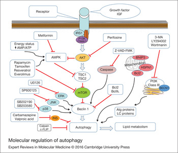
The autophagy is highly regulated by a genetic programme as well as by cellular integration regarding growth factor stimulation, hormonal receptor activation, stress response, nutrient availability and energy status through an elaborate array of signalling pathways (Fig. 3). These regulatory factors are implicated in different stages, such as: (a) activation and nucleation, (b) elongation of the membrane and formation of the autophagosome and (c) trafficking towards lysosomes and fusion of membranes (Ref. Reference Yang and Klionsky20). The autophagic process is mediated by: (i) genetic factors. A number of critical genes have been identified in recent years, in which gene products modulate distinct steps in the induction or progression of autophagy (Refs Reference Yang and Klionsky20, Reference He and Klionsky30). The lysosomal termination defects during autophagy cause clinical diseases such as Niemann–Pick disease type C (a fatal inherited lipid storage disorder with severe neurodegeneration and liver dysfunction) and Pompe disease (a lysosomal storage disorder because of deficiency of acid alpha-glucosidase) (Refs Reference Maetzel31, Reference Lim, Li and Raben32). The role of liver-specific autophagy that regulates TGs in LDs has also been demonstrated through conditional deletion of FAK-family interacting protein of 200 kDa (FIP200). Of note, defective autophagy itself is not enough to cause fatty liver. The compromised autophagy may serve as a potential “second hit” that aggravates the progression from reversible steatosis to inflammatory stage or steatohepatitis using inflammatory cell infiltration and liver fibrosis as hallmarks. (ii) atgs. The autophagic process is coordinated by a complicate network composed of known 32 Atgs and their protein products. Atg1 complex of yeast (or ULK1 in mammals) is an essential positive regulator of autophagosome formation (Ref. Reference Kuma and Mizushima33). When ULK1 and another complex member (homologue of yeast Atg13) are phosphorylated, mammalian target of rapamycin complex 1 (mTORC1) blocks the initiation/activation of autophagy. The autophagy is inhibited by the ULK1 complex binding to the mTORC1 as sufficient nutrients are available in the cell. In starvation, mTORC1 is dissociated from the ULK1 complex to trigger autophagosome nucleation and elongation. The mTORC1 pathway is a major intracellular hub for integrating autophagy-related signals (Ref. Reference Efeyan and Sabatini34). Mammalian Atg2 has two homologues Atg2A and Atg2B, which are required for autophagosome formation and regulation of LD dispersion (Ref. Reference Velikkakath35). Atg3 conjugation enzyme interacts with atg8 and catalyses the combination of Atg8 and phosphatidylethanolamine (Ref. Reference Yamada36). Atg3 is recruited to join LC3 lipidation process (Ref. Reference Fujita37). Atg4B acts on the pro-form of LC3B to convert it into cytosolic form LC3-I. The mammalian free LC3-I (or Atg8 homologues) is further converted into phosphatidylethanolamine-conjugated and autophagosome-membrane associated LC3-II form, which is an initiating step of autophagic response (Refs Reference Kabeya38, Reference Kabeya39, Reference He40, Reference Satoo41). Atg4D can likewise work on other Atg8 homologues and play a similar to that of Atg4B. Lipid conjugation of LC3-I results from a comprehensive action of Atg7 and Atg3 activities. Hepatocyte-specific Atg5 knockout results in defective function of autophagy in mouse hepatocytes. Atg5 knockout in mouse livers can cause much severe inflammation, fibrosis and tumourigenesis (Ref. Reference Ni42). Atg8 mediates conjugation reaction of microtubule-associated protein-1 LC3. Atg12 is conjugated to Atg5 by Atg7 and Atg10 groups to form a functional complex of Atg5–Atg12–Atg16L1. Atg32 interacts with Atg8 and Atg11 to produce selective mitochondrial autophagy (Refs Reference Kanki43, Reference Okamoto, Kondo-Okamoto and Ohsumi44). (iii) Forkhead box O (FOXO) transcription factors. Autophagy is regulated by FOXO transcription factors, since their activation leads to transcription of atg genes (Refs Reference He and Klionsky30, Reference Webb and Brunet45). FOXO has been demonstrated to be able to regulate the expression of autophagy genes in mouse skeletal muscle. Inhibition of mTORC2 by siRNA knockdown of rictor increased FOXO3 nuclear localisation and autophagy in skeletal muscle (Ref. Reference Zhou46). However, future studies are needed to determine whether mTORC2 would also be important in other tissues in addition to skeletal muscle. (iv) Mitophagy. Hypoxia and activation of hypoxia-inducible factors induce the transcription of mitophagy-specific genes and activates function of mitophagy (Ref. Reference Zhang47). There are elaborate mechanisms regulating selective autophagy of organelles. Atg32 is localised in yeast mitochondria as a receptor and is interacted with Atg8 and Atg11 to perform selective autophagy (Refs Reference Kanki43, Reference Okamoto, Kondo-Okamoto and Ohsumi44). Mammalian autophagy of depolarised mitochondria protects cells from toxic reactive oxygen species (ROS). This process is initiated by Pink1-dependent mitochondrial translocation of Parkin. Subsequent ubiquitination of mitochondrial proteins and recruitment of p62 bring mitochondria to autophagosomes (Refs Reference Geisler48, Reference Plun-Favreau49). The disposal of damaged mitochondria by autophagy has two homeostatic roles: (a) limiting oxidative damage and (b) maintaining a functional mitochondrial pool. Treatment with rapamycin inhibits N-acetyl-p-aminophenol (APAP)-induced necrosis in mouse livers, likely mediated by the induction of mitophagy. This observation has clinical importance for its therapeutic potential, because many patients with APAP-overdose have already suppressed the metabolism phase as they occur in the emergency room. Mitophagy may diminish the formation of mitochondria-derived ROS and the release of pro-cell death factors from mitochondria. Indeed, APAP-induced ROS are repressed by rapamycin, but elevated by CQ treatment (Ref. Reference Ni50). In addition, Efavirenz also induces mitophagy as a cell survival mechanism (Ref. Reference Apostolova51). (v) Gglucagon. Glucagon as predominant hormone of the fasted state triggers hepatic autophagy. Glucagon is the first hormone known to simulate autophagy. The accordant effect of glucagon is elicited at intermediate amino acid levels. The autophagic effect of glucagon is modulated by a hormone-induced depletion of intracellular glycine, alanine, glutamate and glutamine. The glutamine is the most potent activator (Ref. Reference Schworer and Mortimore52). The glucagon receptor activates its effector adenylate cyclase via the coupled G-protein. Resultantly, both cyclic adenosine monophosphate (cAMP) and downstream protein kinase A can modify effects of glucagon. Consistently, β-agonists such as epinephrine can also activate adenylyl cyclase and has comparable effects on autophagy induction. These stimulatory effects on autophagy are liver-specific. Reversely, they inhibit autophagy and protein degradation in the muscle. Adrenergic receptor activation, which has much alike the role of glucagon and activates adenylate cyclase, promotes cAMP production and stimulates liver autophagy as well. Autophagy of hepatic glycogen appears to be regulated by similar mechanisms (Ref. Reference Yin, Ding and Gao53). (vi) Phosphatidylinositol 3-kinase (PI3K)/Akt signalling pathway. PI3K/Akt signalling pathway is activated subsequent to growth factors. PI3K signalling network plays critical roles in the regulation of cell survival and intracellular trafficking. Akt is able dramatically to reduce the AMP/adenosine triphosphate (ATP) ratio and suppresses adenosine 5′-monophosphate-activated protein kinase (AMPK) activity (Ref. Reference Tao54). The AMPK can impede mTOR signalling. mTORC1 complex regulates the mammalian uncoordinated-51-like protein kinase complex, including ULK1, Atg13, Atg101 and RB1CC1. The AMPK is thereby thought of as a positive regulator of autophagy in response to AMP levels. The autophagy is also positively stimulated by the Beclin 1 complex that consists of Beclin 1, class III phosphatidylinositol-3-kinase (VPS34 or PI3KC3) and Atg14L or UVRAG. The phosphatidylinositol-3-phosphate from the activation of Beclin 1 complex initiates autophagosomal nucleation. (vii) Insulin. Insulin is a master hormone of the fed state to inhibit autophagic process in mammals. The autophagy contributes to both insulin secretion and physiological sensitivity to the hormone. In liver, defective autophagy leads to insulin resistance as demonstrated in obese mice (Ref. Reference Yang55). When the decreased hepatic autophagy is restored through retroviral expression of atg7, the insulin resistance is relieved. A declined autophagy in obese animals may also be a reason of difficult weight-loss (Ref. Reference Yang55). Moreover, the ageing process gradually weakens autophagic capability and reduces total caloric expenditures (Ref. Reference Rabinowitz and White56). There are potential causative links between autophagy decline and the decreased energy demand for self-regeneration. (viii) Starvation. Starvation activates autophagic response in the first 24 h, which can be found in liver, pancreas, kidney, skeletal muscle, and heart (Ref. Reference Mizushima57). Autophagy supports mammalian cell survival because of a severe deficiency in nutrients. Lymphocytes shrink their cell volume ~50% over 3 months to maintain energy balance through autophagy (Ref. Reference Lum58). Glycogen and LDs are inner storage of cellular nutrients, whereas other cellular components have a dual function. Autophagy is a key process to bridge cellular energy needs over nutrient supply. Sometimes, calorie restriction promotes mammalian cell survival. Starvation induces autophagy through the inhibition of a multiprotein complex mTORC1 (Ref. Reference Jung59). Abundant nutrients or growth factors activate the mTORC1 signalling pathway. mTORC1 inhibits autophagy through the negative regulation of a large substrate complex composed of ULK1, Atg13, Atg101 and FIP200 (Refs Reference Mizushima60, Reference Chan61, Reference Hosokawa62, Reference Ganley63, Reference Jung64, Reference Wong65, Reference Hara66). (ix) Energy depletion. Loss of energy charge stimulates autophagy and inhibits mTORC1 partially through activation of the AMPK, leading to the activation of ULK1 (Refs Reference Jung59, Reference Mihaylova and Shaw67, Reference Kim68). Other autophagic regulators contain Beclin 1 complex associated with VPS34, and some stimulatory or inhibitory proteins such as ATG14L, UVRAG, Ambra1 and Rubicon (Ref. Reference He and Levine69). In response to pro-autophagic stimuli, the increased production of phosphatidylinositol-3-phosphate by Beclin 1 complex regulates autophagosome formation (Refs Reference He and Levine69, Reference Itakura70). The Beclin 1 complex is suppressed by the PI3K/Akt pathway as well as by an interaction with antiapoptotic Bcl-2 family proteins (Refs Reference Pattingre71, Reference Wang72). The cellular energy depletion can cause a series of expression of regulatory proteins. These autophagic regulators modulate autophagic process. Final products of autophagy are utilised to energy generation or self-regenerating components. (x) Caffeine. Caffeine consumption is associated with a low risk of NAFLD that recently has become the major cause of liver disease worldwide (Ref. Reference Sinha73). An experimental study demonstrated that caffeine could increase lipid uptake in lysosomes and dramatically alleviated hepatosteatosis in mice fed a HFD. In liver, caffeine stimulates β-oxidation and activates autophagy–lysosomal pathway, which results in a significant decrease of intrahepatic lipid content. Moreover, caffeine can downregulate target gene expression of rapamycin signal to stimulate hepatic autophagic flux. Caffeine-induced autophagy plays a lipolytic role in mammalian livers, which may be beneficial to the improvement of NAFLD (Ref. Reference Sinha73). (xi) Circadian regulation. Circadian autophagy rhythm can regulate lipid metabolism and further modulates the pathogenesis of NAFLD. C/EBPb is a mediator between hepatic autophagy and lipid metabolism and is rhythmically expressed in hepatocytes. Both circadian and nutritional signals regulate C/EBPb expression. When rhythmical expression of hepatic C/EBPb is interrupted through adenoviral RNAi treatment, circadian regulation of autophagy rhythm is disrupted subsequent to an absence of functional liver clock. Autophagy is regulated by the C/EBPb-mediated biological clock and maintains a circadian nutrient homeostasis (Ref. Reference Ma, Panda and Lin74).
Molecular regulation of autophagy. Several chemicals have showed their regulatory roles on the autophagic process, which may further modulate hepatic lipid metabolism. Moreover, some high-throughput screenings for small molecules such as miRNA and siRNA that regulate autophagy have been conducted and are undergoing, which may develop new therapeutics to benefit patients with fatty liver disease.

Cross-talks between hepatic autophagy and apoptosis
Excessive fat is accumulated in liver tissue, which can induce lipotoxicity and hepatocyte apoptosis. Lipotoxicity is a cytotoxic condition because of the accumulation of lipid intermediates in nonadipose tissue. Excessive FFA-mediated lipotoxicity plays an important role during the pathogenesis of NAFLD. Differential effects of unsaturated and saturated fatty acid-induced lipotoxicity had been identified, which can further regulate hepatic autophagy and apoptosis. OA (oleic acid) is an unsaturated fatty acid that facilitates biosynthesis of TG-enriched LDs and stimulates autophagy dependent on ROS and PI3-kinase pathway. However, saturated PA (palmitic acid) inhibits autophagy through caspase-dependent Beclin 1 cleavage and significantly induces apoptosis, which also suggests there is a cross-talk between apoptosis and autophagy. Moreover, a HFD-induced hepatic steatosis/apoptosis was associated with defective function of liver autophagy. LDs-triggered autophagy may be a protective mechanism against saturated fatty acid-mediated hepatotoxicity/lipotoxicity (Ref. Reference Mei23). Cross-talks between autophagy and apoptosis are important machinery in pathogenesis of FLD. Antiapoptotic Bcl-2 proteins can suppress autophagy via an interaction with Beclin1 complex. p53 protein and caspase activation also modulate autophagic process at certain sites. p53 protein regulates the expression of Bcl-2 family proteins, for example Bax and Bid. The activation of caspases has a dual role, which can promote autophagy (e.g. Atg4D) or inhibits autophagy (e.g. Beclin 1 and Atg3). Atg12 and Atg5 cleavage products as pro-apoptotic mediators can counteract antiapoptotic Bcl-2 family proteins. Collectively, Atg function is regulated by caspase, calpain, c-FLIP, Bcl-2, p53, etc. Cross-talks between autophagy and apoptosis play a critical role during the pathogenesis of liver injury, which has been well summarised in author's another review (Ref. Reference Wang75). Additionally, an induction of CXC chemokine receptor 3 in dietary-induced steatohepatitis was associated with upregulation of hepatic pro-inflammatory cytokines, activation of NF-kappaB, and autophagosome–lysosome impairment (Ref. Reference Zhang76). Chaperone-mediated autophagy modulates signalling pathway between hepatic lipid metabolism and inflammatory response (Refs Reference Das77, Reference Schneider, Suh and Cuervo78).
Modulation of autophagy as therapeutic targets affects the progression of FLD
Autophagy as a degradation process can dispose LDs in the liver cell. Thus, pharmacological modulation of autophagy may be a potential approach to alleviate fatty liver injury (Fig. 4). Several chemicals have showed their effects on the improvement of FLD resulting from both alcoholic and nonalcoholic conditions. Moreover, some high-throughput screens for small molecules that act on autophagy have been conducted and are undergoing, and these will certainly generate new therapeutic approaches to benefit patients with FLD (Ref. Reference Lin79).
Modulation of autophagy as therapeutic targets is implicated in the progression of FLD resulting from both alcoholic and nonalcoholic factors. Pharmacological modulation of autophagy may be a novel approach to alleviate fatty liver injury.

Pharmacological promotion of autophagy can alleviate alcoholic fatty liver injury
ALD initiates an early steatosis. The reversible steatosis may be further aggravated into steatohepatitis in heavy alcohol drinkers (approximately 30%), and then fibrosis and cirrhosis (Refs Reference Zakhari and Li80, Reference Ding, Manley and Ni81, Reference Gao and Bataller82). Genetic factor determines the individual susceptibility to alcohol intoxication. Defensive capacity of liver cells against detrimental effects of alcohol is also person-dependent. The activated autophagy is a protective mechanism to attenuate acute ethanol-induced steatosis and liver injury. Acute ethanol-induced autophagy can selectively target damaged mitochondria and LDs in ethanol-treated primary hepatocytes (Refs Reference Ding, Manley and Ni81, Reference Ding83). When ethanol was given via intraperitoneal injection (acute) or by a 4-week oral feeding regime (chronic) in C57BL/6 mice, autophagy enhancers (carbamazepine or rapamycin) and autophagy inhibitor (CQ) were utilised to test the role of autophagy during liver injury. Autophagy could be activated following both acute and chronic ethanol treatment. The activation of autophagy altered degree of hepatic steatosis and blood levels of TGs, liver enzyme, glucose and insulin. Carbamazepine and rapamycin could promote ethanol-induced cellular autophagy in the liver. Fatty liver injury was aggravated by CQ, whereas it was alleviated by carbamazepine. In another study, rapamycin-induced autophagy can entirely inhibit acute alcohol-caused steatosis. Rapamycin may suppress lipogenesis via the blockage of mTORC1 pathway as well. The rapamycin has less potent inhibition on mTOR as compared with Torin 1 and does not regulate lipogenesis gene expression (Ref. Reference Peterson84). Therefore, autophagy would play a very critical role in alcoholic steatosis. mTOR inhibitors can be potential drugs for prevention or treatment of ALD, considering they are able to stimulate autophagy and to interrupt lipogenesis. Presently, the mechanism by which chronic alcohol abuse modulates the autophagic process remains unclear. It is well known that long-term alcohol consumption disrupts intracellular vesicular movement and delivery of proteases to lysosomes. Both protein trafficking and lysosomal functions within the hepatocyte are damaged by chronic alcohol exposure, which represses autophagic response (Ref. Reference Donohue85). Notably, a global activation of hepatic autophagy can degrade lipids, proteins and organelles. It may be beneficial to alleviate liver injury, but not specific to remove an overaccumulation of liver lipids. A perfect protective mechanism against hepatic steatosis needs the upregulation of autophagy specific for lipophagy. Alcohol abuse causes mitochondrial damage and accumulation of LDs. In order to prohibit alcohol-induced hepatic toxicity, the priority will develop therapeutic drugs that can upregulate autophagy selectively targeting mitochondria and LDs (Ref. Reference Ding83). Future studies will focus on how selectively to enhance the role of individual autophagy for therapeutic purpose. Small molecules such as miRNA and siRNA are able to modify autophagic signalling pathway. Regulatory roles of these small molecules for autophagy/FLD are under investigation now.
Pharmacological promotion of autophagy can alleviate nonalcoholic fatty liver injury
Pathological characteristic of NAFLD is manifested by the accumulation of fat in the liver in the absence of alcohol consumption. Currently, NAFLD is the most common chronic liver disease. Its prevalence is approximately 20–30% in adults, and increases to approximately 80% in patients with obesity and diabetes (Refs Reference de Alwis and Day86, Reference Neuschwander-Tetri87, Reference Malhi and Gores88). NAFLD is usually accompanied by insulin resistance and metabolic syndrome. NAFLD can increase the risk for development of fibrosis, cirrhosis and HCC (Ref. Reference Malhi and Gores88). At present, there is no effective treatment available for NAFLD. In pre-clinical study, C57BL/6 mice were given with high fat diet for 12 weeks. Protective effects of carbamazepine and rapamycin in reducing steatosis as well as in improving insulin sensitivity were demonstrated by HFD-induced fatty livers (Ref. Reference Chang89). Rapamycin as an mTOR inhibitor induces autophagy. Pharmacological role of rapamycin can improve fatty liver through induction of lipophagy. Rapamycin-sensitive mTORC1 negatively regulates autophagy by interacting with the ULK1–Atg13–FIP200 complex in mammalian cells (Ref. Reference Mizushima60). Rapamycin treatment positively stimulates the co-localisation of LDs with LAMP1 (lysosome-associated membrane protein type 1) or LDs with LC3-positive autophagosomes, which significantly reduces LD number and oleic acid-elevated TG level (Refs Reference Singh22, Reference Mei23). Rapamycin also attenuates HFD-induced fatty liver injury in a mouse model (Ref. Reference Chang89). Rapamycin-treated mice have reduced body weight, low serum leptin and insulin levels as compared with control mice (Ref. Reference Chang89). Another example is liver-specific deletion of raptor, a key component of the mTORC1 complex. Raptor knockout mice, like rapamycin treatment, are highly resistant to HFD-induced hepatic steatosis and weight gain (Ref. Reference Gao and Bataller82). mTORC2 activated by growth factors directly phosphorylates AKT to allow the maximal activation of AKT (Refs Reference Sarbassov90, Reference Mammucari91). AKT then phosphorylates tuberous sclerosis complex1/2 (Fig. 3). mTOR positively regulates SREBP-1 by controlling the nuclei location of Lipin 1 (Ref. Reference Peterson84). The suppression of mTOR by Torin 1 results in dephosphorylation of Lipin 1. The nuclear localised and dephosphorylated Lipin 1 decreases SREBP-1-mediated lipogenesis by reducing the abundance of nuclear SREBP-1 protein (Ref. Reference Peterson84). Insulin has been shown to activate AKT, which increases expression of lipogenic genes by inducing nuclear accumulation of SREBP-1 (Ref. Reference Porstmann92). The suppression of mTOR has two obvious benefits, for example: (i) induction of lipophagy and (ii) inhibition of lipogenesis mediated by SREBP pathway. Thus, mTOR targets may be a novel approach to develop therapeutic drugs for FLD. Moreover, mTOR inhibitors can be useful for treating both NAFLD and alcoholic FLD, since they can simultaneously work on different sites of autophagy and lipogenesis. In addition, metformin can activate both PRKA-MTOR-ULK1 and SIRT1-FOXO signalling pathways. A recent study using siRNA-technique has confirmed that metformin is able to relieve hepatic steatosis through SIRT1/FOXO-mediated autophagic induction (Ref. Reference Song93). Except for autophagy machinery, other investigation on NAFLD also shows the updated evidence. By employing rat model of NAFLD induced by choline-deficient/L-amino acid-defined (CDAA)-diet, the treatment with MIYAIRI 588 can significantly improve liver lipid deposition and substantially decrease the TG content, insulin resistance, and hepatic inflammatory indexes. Probiotic MIYAIRI 588 stimulates the activation of hepatic AMPK and AKT and upregulates the expression of lipogenesis- or lipolysis-related proteins (Ref. Reference Endo94). When male Sprague–Dawley rats are fed a HFD for 4, 12 and 24 weeks, animals can replicate the progression of human NAFLD. A downregulation of enoyl-coenzyme A hydratase (ECHS1) exacerbates hepatic steatosis, which is validated in rat models as well as in patients’ liver simples. ECHS1 is a new potential target for therapeutic interventions of NAFLD (Ref. Reference Zhang95). Recently, TM6SF2 is found to be a new regulator of liver fat metabolism influencing TG secretion and hepatic LD content (Ref. Reference Mahdessian96).
Dual effects of autophagy on lipid metabolism
Autophagy plays a crucial role in liver lipid metabolism. Autophagic defects or its suppression can accelerate a fast development of steatosis. Autophagy contributing to the pathogenesis of FLD has been demonstrated in autophagy-defective mice (Ref. Reference Singh22). Actually, a compromise in hepatic autophagy is the basis for excessive accumulation of LDs following an exposure to high concentration of alcohol. Moreover, pharmacological stimulation of autophagy improves hepatotoxicity and steatosis in the alcoholic fatty liver (Ref. Reference Ding83). Autophagy activation is beneficial for hepatocyte proliferation and liver repair. However, an enhancement of the autophagy in hepatic stellate cells favours their activation and consequently triggers liver fibrosis (Ref. Reference Thoen97). Inversely, an inhibition of autophagy leads to the accumulation of LDs in hepatic stellate cells, resulting in decreased fibrogenesis (Ref. Reference Hernandez-Gea98). Fucoidan from Fucus vesiculosus can inhibit autophagy of hepatic stellate cells and the formation of extracellular matrix, probably through the downregulation of transforming growth factor beta 1/Smads pathways. Fucoidan thus is a potential therapeutic agent for liver fibrosis (Ref. Reference Li99). Hepatic stellate cells instigated by lipopolysaccharide produce many cytokines, such as IFNβ (interferon beta), TNFα (tumour necrosis factor alpha) and IL6 (interleukin 6). These cytokines strongly inhibits DNA synthesis and induces apoptosis of hepatocytes. Blockade of autophagy significantly augments hepatocyte apoptosis (Ref. Reference Dangi100). In addition, the autophagic system is able to mobilise hepatic lipids that are utilised by viruses to favour their replication. Previous studies had demonstrated that autophagy was a host defence mechanism against most viral invasion, but activation of autophagy could also be a favourite event in the replication of dengue virus. During viral replication, dengue virus needs a high level of intracellular ATP to satisfy the energy requirement. The autophagic process degrades LDs and releases FFA, which just provide abundant ATP molecules via β-oxidation (Ref. Reference Heaton and Randall101). Further studies will determine which subgroup of hepatotropic virus needs lipophagy for energy requirement during viral replication and whether blockage of the autophagic system can selectively affect virogenesis, not normal cell metabolism. Furthermore, dual roles of autophagy are remarkably observed in liver cancer development. Autophagy can promote tumour cell survival at certain stages, but can also stimulate tumour cell elimination at other stages.
Summary and future study
Autophagy is required for normal development of adipose tissue and pancreas. Autophagic process as an organ-specific target has been implicated in the development of therapeutic intervention against human metabolic disorders that is obesity and metabolic syndrome. Autophagy is an essential means of refreshing and remodelling cells and maintains metabolic homeostasis. The autophagy provides energy and internal nutrients involving lipid metabolism in liver. As such, the autophagy is a potential contributor to the formation of hepatic steatosis. Some questions still deserve immediate attention. Is there preferential degradation of a LD subset by lipophagy? What is the exact mechanism that the lipophagy selectively targets LDs? What determines the lipophagy threshold that switches a stimulatory into an inhibitory effect on FFA? What are possible roles of lipophagy on metabolic defects that often co-exist in patients with obesity, diabetes, and hyperlipidaemia? Autophagic activity can also induce a dual effect subsequent to degradation of lipids. Thus, both activation and inhibition of autophagy hold promise for the improvement of FLD that can be induced by either alcoholic or nonalcoholic factors, dependent upon different cell types. The strategy of cell type-specific autophagy has to be considered in future study.
Financial support and/or conflicts of interest
The author has nothing to disclose.
Acknowledgements and funding
CHOI research grant.