Tuberous sclerosis, an extremely variable genetic multisystem disorder, is characterised by hamartomas in several organs including the brain, heart, skin, eyes, kidney, lung, and liver. The affected genes are TS1 and TS2, encoding “hamartin” and “tuberin” proteins. Cardiac rhabdomyomas are the main features of the disease and usually asymptomatic. Reference Curatolo, Bombardieri and Jozwiak1
Primary cardiac tumours are rare in children with a prevalence of 0.0017 to 0.28 in autopsy series and majority of them are benign. Reference Mahani, Lu, Rigsby, Krishnamurthy, Dorfman and Agarwal2 Rhabdomyoma is the most common cardiac tumour during childhood and tuberous sclerosis is associated with cardiac rhabdomyomas in one-third of patients. Reference Vaughan, Veugelers and Basson3 Cardiac lipomas are rare lesions and occur generally in adults and commonly asymptomatic. Reference Uzun, Wilson, Vujanic, Parsons and De Giovanni4 Fat-containing cardiac lesions may associate with tuberous sclerosis in children, but true cardiac lipomas are very rare in this population.
Herein, we present a very rare association of intracardiac lipoma with tuberous sclerosis diagnosed via cardiac MRI and aim to take attention to usefulness of cardiac MRI in diagnosis of cardiac masses.
Case
A Fourteen-year-old male patient was referred to paediatric cardiology department for echocardiographic evaluation. Physical examination revealed motor and mental retardation, millimetric papillary skin lesions, cafe au lait lesions, and nonspecific midsystolic murmur. He had first applied to hospital with generalised tonic-clonic convulsions at 1 year. Brain MRI was performed because of recurrent seizures at 18 months age, and bilateral cortical temporo-fronto-occipital lesions which were hyperintense in T1 and hypointense in T2 were detected and considered as tubers seen in tuberous sclerosis.
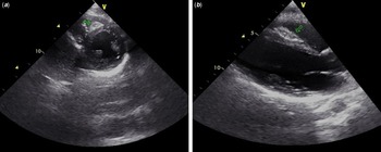
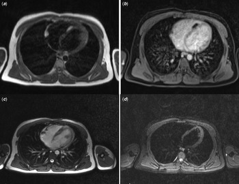
Echocardiography revealed a fusiform mass lesion on right midmuscular region of the interventricular septum, which had hyperechogen edges and a hypoechogen centre, with dimensions 26.5 to 10.5 mm (Fig 1). On apical region of the interventricular septum, a second smaller lesion was seen. Cardiac MRI showed two lesions on interventricular septum, one on midmuscular and one on apical septum, with dimensions of 23 × 9 mm and 15 × 4 mm, respectively, which were hyperintense in T1 and hypointense in T2 sections. On fat suppression sequence, the masses appeared hypointense, non-enhancing, and these MRI findings were compatible with a benign cardiac lipoma (Fig 2).
Echocardiografic view of the mass lesion, ( a ) short axis, ( b ) long axis.

Cardiac MRI images. ( a ) T1-weighted image and ( b ) vibe fat suppression image with contrast. (c) T2-weighted image TrueFISP single shot and (d) trim fat suppression image.

Discussion
Cardiac rhabdomyomas are closely associated with tuberous sclerosis complex and affect approximately half of the patients, and most of them are asymptomatic. However, rarely they may cause arrhythmia, non-immune hydrops, or even death prenatally. Large lesions may rarely cause obstruction of cardiac outflow or embolic events. Reference Curatolo, Bombardieri and Jozwiak1 In clinical practice, an echocardiogram and electrocardiography should be performed on patients with tuberous sclerosis for evaluation of possible rhabdomyomas and arrhythmia, in infantile period. Our patient was consulted to paediatric cardiology when he was 18 months old, but because of the parent’s inconsistency to regular follow-up and consultation, echocardiography could not be performed in infancy. So that first echocardiographic evaluation and diagnosis of intracardiac lesion was delayed.
Cardiac lipomas are rare benign primary cardiac neoplasms of mature adipose tissue and composed of mature collections of adipocytes. Reference Vaughan, Veugelers and Basson3 They are more common in adults. Most lipomas are epicardial; however, they can involve all three layers of the heart, including myocardium and endocardium. Reference Mahani, Lu, Rigsby, Krishnamurthy, Dorfman and Agarwal2 The incidence of cardiac lipoma is reported as 8 to 12% of primary cardiac tumours, whereas true lipoma, particularly in the interventricular septum, is extremely rare. Reference Gulati, Sharma, Kothari, Juneja, Saxena and Talwar5,Reference O’Donnell, Abbara and Chaithiraphan6 These tumours vary in size, ranging from extremely small tumours to massive epicardial masses and size impacts clinical condition. They are usually asymptomatic but may cause symptoms due to pericardial effusion, arrhythmia, or intracardiac obstruction. Reference Vaughan, Veugelers and Basson3 Lipomas are usually solitary, but multiple lipomas have been reported associated with some CHDs, tuberous sclerosis or sometimes sporadically in normal hearts. Reference Mahani, Lu, Rigsby, Krishnamurthy, Dorfman and Agarwal2 There have been reports of cardiac lesions considered as angiomyolipomas or fat-containing lesions in patients with tuberous sclerosis. In a recent study, more than one-third (35%) of adolescents and adults with tuberous sclerosis were found to have cardiac lesions with signal characteristics consistent with fat-containing lesions. Reference Shaaya, Hirshberg and Rabe7 There are also some reports of lipomatous lesions, diagnosed by CT and MRI in tuberous sclerosis. Reference Jabir and Al-Hyassat8 But true cardiac lipomas are very rare. As a very rare association with tuberous sclerosis, the lipomas diagnosed in our patient were not solitary, non-obstructive, and well limited in interventricular septum and asymptomatic, so there was no indication for a surgical extraction of the lesion. Therefore, histological diagnosis could not be performed for our case.
Echocardiographic assessment of our patient revealed a well-bounded fusiform lesion with echogenic borders and less echogenic centre. Transthoracic echocardiography may have some limitations including operator dependency, poor quality of acoustic window in some patients, and limited capability to differentiate cardiac lesion characteristics. Reference Gulati, Sharma, Kothari, Juneja, Saxena and Talwar5 In echocardiographic examination, true cardiac lipomas are encapsulated, containing neoplastic fat cells, and homogeneously echogenic. Reference Ward, Kadoch, Jacobi, Lopez, Salvo and Cham9 Because of the limitations of echocardiography, cardiac MRI is usually needed to describe the characteristics of the lesion and is a useful diagnostic technic that can differentiate fat-containing tumours from other non-fatty lesions. Reference Vaughan, Veugelers and Basson3 Main characteristics of lipomas are homogeneous high-signal intensity on T1-weighted sequences, mildly hyperintense on T2-weighted sequences, loss of signal on fat suppression sequences, and no enhancement on first-pass perfusion sequences. Reference Mahani, Lu, Rigsby, Krishnamurthy, Dorfman and Agarwal2 We also performed cardiac MRI for our patient, and it revealed two lesions on interventricular septum, which were hyperintense in T1 sections. On fat suppression sequence, the masses appeared hypointense, non-enhancing, and these MRI findings were compatible with a benign cardiac lipoma. As these findings were sufficient for diagnosis, no further tests were performed, and clinical follow-up was planned for our patient as he was asymptomatic.
In conclusion, most of the cardiac lesions associated with tuberous sclerosis are rhabdomyomas, but rarely other cardiac lesions of different nature can be seen. True cardiac lipomas are very rarely in association with tuberous sclerosis although other fat-containing lesions like angiomyolipomas are some more frequent. Echocardiography should be used as screening test for cardiac lesions in tuberous sclerosis but cardiac MRI is needed to determine nature of the lesion, rhabdomyoma, lipoma, etc.
Financial support
The authors report that they did not benefit from any donation and received no specific grant from any funding agency or from commercial or not-for-profit sectors.
Conflicts of interest
None.
Ethical standards
An informed consent was obtained from patient’s parents. The authors declare that all procedures contributing to this work comply with the ethical standards of the relevant national guidelines on human experimentation and with the Helsinki Declaration of 1975, as revised in 2008, and has been approved by the institutional committees.