Introduction
Emotion recognition in humans is considered to be an innate ability that largely generalizes at least for a specific set of basic emotions across different cultures. The importance of correct identification of emotional expressions for healthy individuals has been frequently emphasized in the literature. In general, accurate emotion recognition is deemed to be necessary for successful human interaction. However, certain types of cerebral pathology have been shown to impair patients’ abilities to correctly identify emotions and to process affective information.
Deficits have been observed in patients showing a number of different neurodegenerative conditions and the deterioration in emotion recognition abilities has been attributed to global cognitive impairments due to pathological processes rather than direct functional deficits (e.g. Kumfor et al., Reference Kumfor, Sapey-Triomphe, Leyton, Burrell, Hodges and Piguet2014). Notwithstanding, evidence from a number of studies suggests an important role of the prefrontal cortex for processing of emotional stimuli, thus making damage to this brain region a likely candidate for potential impairments (e.g. Vandekerckhove et al., Reference Vandekerckhove2014). Further evidence suggests that neurodegenerative processes impair emotion recognition. Moreover, Alzheimer's disease (Hargrave et al., Reference Hargrave, Maddock and Stone2002) has been reported to lead to impairments in FER, although recent replications have not been able to show such an effect (Kumfor and Piguet, Reference Kumfor and Piguet2013; Kumfor et al., Reference Kumfor, Sapey-Triomphe, Leyton, Burrell, Hodges and Piguet2014).
Since Alzheimer's disease is a progressive condition which typically develops insidiously, related cognitive impairments may be expected to develop in a similar manner. In this vein, potential FER impairments of patients with differing severity of cognitive deficits need attention. Specifically, patients’ less severe forms of cognitive impairment (i.e. patients suffering from preclinical Alzheimer's disease) can be classified according to well-established criteria into patients reporting SCD (Jessen et al., Reference Jessen2014) and MCI (Petersen and Negash, Reference Petersen and Negash2008). Increased cerebral pathology has been reported in MCI and subjective cognitive impairment (SCI) as compared to normal controls (e.g. Vos et al., Reference Vos2013; Jessen et al., Reference Jessen2014; Sun et al., Reference Sun, Yang, Lin and Han2015). Classification into these categories typically takes place according to neuropsychological criteria, where SCD patients are characterized by SCI that is too subtle to be detected in standardized psychometric tests (Jessen et al., Reference Jessen2014). MCI is a clinical condition defined by the presence of cognitive impairment in the context of essentially intact acitivities of daily living. Usually, MCI groups are subtyped according to diagnostic criteria for patients with non-aMCI who are impaired in attention, executive functioning, and language but not memory whereas patients with amnestic MCI show impairments in memory with or without cognitive deficits in attention, executive functioning, and language (Petersen and Negash, Reference Petersen and Negash2008; Pusswald et al., Reference Pusswald2013).
Although specific impairments of emotion recognition are comparatively well established in Alzheimer's disease, examinations of FER in prodromal Alzheimer's disease is still in its infancy. Indeed, a recent review of FER in MCI found compromised FER in MCI compared to normal aging in six studies (McCade et al., Reference McCade, Savage and Naismith2011). A recent study found a relationship between FER and psychosocial disability and caregiver burden in patients with MCI (McCade et al., Reference McCade, Savage, Guastella, Hickie, Lewis and Naismith2013).
Besides deficits due to neurological impairments, FER has been shown to be altered in depressed individuals. Typically, FER performance in individuals with a history of depression has been shown to be decreased in some, but not all emotional expressions (Anderson et al., Reference Anderson2011). Furthermore, in previous studies intelligence test performance has been positively linked to FER (Sullivan and Ruffman, Reference Sullivan and Ruffman2004). To the authors’ knowledge, to date there has been no investigation of differences in FER between SCD and MCI compared to healthy aging. In the present study, we examine FER in three groups of patients suffering from SCD, non-amnestic, or amnestic MCI compared to a large age-equivalent sample of healthy controls. We expect to observe increasing FER deficits between groups as pathology increases. Specifically, higher FER performance was expected to be observed for controls than for SCD, non-aMCI, and aMCI samples. We note that potential differences between non-aMCI and aMCI samples would not likely be due to increasing pathology but may be seen as expressions of impairments in different central nervous structures. Moreover, we provide evidence for potentially moderating influences of age, sex, number of educational years, self-reported depressive symptoms, and premorbid IQ test performance.
Methods
Participants
Patients were recruited from the Vienna Conversion to Dementia Study, a prospective cohort study at the Medical University of Vienna. In all, 137 patients (SCD and MCI) contacting the memory clinic for assessment of perceived cognitive problems and 138 healthy controls were recruited. All healthy controls had scores > 26 in the Mini-Mental State Examination (MMSE; Folstein et al., Reference Folstein, Folstein and Hugh1975) thus meeting common criteria of cognitively healthy samples. Patients were categorized into groups exhibiting SCD (n = 68; mean age = 68.96; SD = 7.51; 42f; MMSE median = 29, inter quartile range = 28–30), non-aMCI (n = 44; mean age = 73.00; SD = 7.92; 29f; MMSE median = 28, inter quartile range = 27–29), and aMCI (n = 25; mean age = 69.32; SD = 7.63; 12f; MMSE median = 28, inter quartile range = 27–29) according to current diagnostic criteria (Petersen & Negash, Reference Petersen and Negash2008; Jessen et al., Reference Jessen2014). A sample of 138 healthy volunteers of about the same age range (mean age = 66.49; SD = 10.49; 89f; MMSE median = 29, inter quartile range = 28–30) was recruited from senior citizen community meetings and through advertisements in hospitals and at general practitioners. Preceding inclusion in either of the present samples, all participants underwent neuropsychological and medical screenings. This procedure was necessary to exclude participants meeting the following criteria: (i) participants suffering from neurological diseases (e.g. epilepsy, stroke); (ii) current psychiatric disorders; (iii) substance abuse; (iv) history of severe head injury; (v) substantial physical illness (e.g. cardiovascular diseases).
Materials
Vienna emotion recognition task – short form (vert-k)
The VERT-K consists of 36 colored pictures of faces either expressing anger, disgust, fear, happiness, sadness (i.e. basic emotions), or showing neutral facial expressions in six pictures respectively. The stimuli are based on Caucasian actors only and comprise an equal number of male and female pictures presented on black background. Stimulus pictures are presented in randomized order on a computer screen and participants are instructed to choose emotions to the corresponding facial expressions (for a detailed description, see Derntl et al., Reference Derntl, Kryspin-Exner, Fernbach, Moser and Habel2008). Scores are based on the total number of correct responses.
Beck depression inventory ii (bdi ii; Hautzinger et al., Reference Hautzinger, Keller and Kühner2006)
The BDI II is a 21-item self-report measure for assessment of depression. Participants need to indicate in each item which of four statements that reflect more or less depressive states applied to them during the past two weeks. Scores were calculated with higher scores indicating higher depression.
Vocabulary test (wst; Schmidt and Metzler, Reference Schmidt and Metzler1992)
The vocabulary test is a widely used Rasch-calibrated measure of crystallized intelligence in German language. In 42 items, participants have to indicate one meaningful word in presence of five distractor words. Scores of the vocabulary test have been considered to provide a useful estimate of premorbid IQ because test performance appears to be largely unaffected by age or degenerative central nervous processes.
Neuropsychological assessment
All participants were assessed on the Neuropsychological Test Battery Vienna (NTBV). The NTBV is a multidimensional test battery comprising the six domains attention, executive functions (interference, lexical verbal fluency, planning, and non-verbal fluency), language, and memory. First, attention was assessed by means of the Alterskonzentrations test, a geriatric alertness test (Gatterer, Reference Gatterer2008), the digit symbol test of the German version of the revised Wechsler Adult Intelligence Scale (Tewes, Reference Tewes1994), Trail Making Test B (TMT; Reitan, Reference Reitan1979), and the subtest symbol counting of the cerebral insufficiency test (Lehrl and Fischer, Reference Lehrl and Fischer1997). Second, interference was assessed by the Stroop tasks of the Nürnberger Alters Inventar (Oswald and Fleischmann, Reference Oswald and Fleischmann1997), a cognitive test battery for geriatric samples, and the interference task of the cerebral insufficiency test (Lehrl and Fischer, Reference Lehrl and Fischer1997). Third, lexical verbal fluency was tested with the phonematic verbal fluency test (Goodglass and Kaplan, Reference Goodglass and Kaplan1983). Fourth, for assessment of planning and non-verbal fluency, the Five-Point Test (Regard et al., Reference Regard, Strauss and Knapp1982), the Maze subtest of the Nürnberger Alters Inventar (Oswald and Fleischmann, Reference Oswald and Fleischmann1997), and the TMT A (Reitan, Reference Reitan1979) were used. Fifth, the domain language was tested with the semantic verbal fluency tasks (Goodglass and Kaplan, Reference Goodglass and Kaplan1983) and a modified version of the Boston Naming Test (Morris et al., Reference Morris1989). Finally, episodic memory was assessed by means of the Verbal Selective Reminding Test (Lehrner et al., Reference Lehrner, Gleiss, Maly, Auff and Dal-Bianco2006).
Procedure
Upon agreeing on participation, volunteers participated in a clinical interview in which potential reasons for study exclusion were determined. If all exclusion criteria were ruled out, participants were invited to individual neuropsychological assessments taking place at the memory clinic of the Vienna General Hospital. Confidentiality of data was ensured and participants provided written informed consent. Testing took place by a trained researcher in a quiet location and lasted for about two hours. All assessments were administered as pen-and-paper tests with the exception of the VERT-K which was administered on a computer. The present study protocol adheres to the guidelines as laid out in the Helsinki Declaration and the study was approved by the Ethics Committee of the Medical University of Vienna.
Statistical analysis
A series of one-way analyses of variance was performed to assess group differences in regard to performance in emotion recognition tasks and neuropsychological assessments. Bonferroni-corrected post-hoc analyses were used for pair-wise group comparisons. Effect sizes were interpreted in accordance with Cohen's (Reference Cohen1988) classification of small, medium, and large effects. Furthermore, theory-guided multiple hierarchical linear regressions were performed to assess influences of diagnosis (controls vs. SCD vs. aMCI vs. MCI), age, sex, number of educational years, premorbid intelligence test performance, and depression inventory scores on performance on emotion recognition tasks. All analyses were performed in SPSS 21.0 and the statistical open-source software package R 3.1.1 (R Development Core Team, 2014).
Results
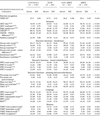
Patients and controls differed significantly from each other in emotion recognition performance (F(3, 271) = 6.09; p = 0.001). Bonferroni-corrected post-hoc tests revealed a large significant effect between controls and the aMCI group (p = 0.001; d = −0.83) but not SCD and non-aMCI groups (p = 1.0; d = −0.15 and p = 0.065; d = −0.46, respectively), although effect sizes showed higher performance of controls. Moreover, the SCD group differed significantly from aMCI (p = 0.021; d = −0.65) but not non-aMCI (p = 0.736; d = −0.30). There was no significant difference between non-aMCI and aMCI groups (p = 0.735; d = −0.36). Of note, even though differences failed to reach nominal significance most likely due to low sample sizes, medium-sized effects were observed for non-aMCI versus SCD and aMCI comparisons. Descriptive statistics for the VERT-K, neuropsychological variables and results from analyses of variance with post-hoc tests are provided in Table 1.
Group differences in emotion recognition and neuropsychological variables by domain

Note. SCD = Subjective cognitive decline; non-aMCI = non-amnestic mild cognitive impairment; aMCI = amnestic mild cognitive impairment; AKT = Alterskonzentrations test (geriatric alertness test); BNT = Boston naming test; PWT = Phonematische Wortflüssigkeit (phonematic verbal fluency); SWT = Semantische Wortflüssigkeit (semantic verbal fluency); TMT = Trail Making Test; VERT-K = Vienna emotion recognition task – Short Form; VSRT = Verbal selective reminding test; Bonferroni-corrected significant pairwise differences: a = Controls versus SCD; b = Controls versus non-aMCI; c = Controls versus aMCI; d = SCD versus non-aMCI; e = SCD versus aMCI; f = non-aMCI versus aMCI.
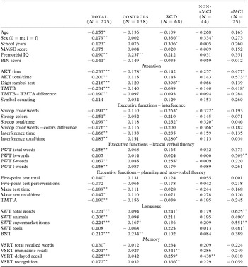
Results of our correlational analyses presented several points of interest. First, for the total sample, two-tailed Pearson correlations revealed a negative relationship between age and emotion recognition and higher performance for women (Table 2). Second, both premorbid IQ and number of school years were positively associated with emotion recognition performance, thus linking emotion recognition performance to general cognitive ability and education. Third, better performance in neuropsychological tests was unequivocally linked with higher FER performance. Finally, there was no significant relationship between MMSE scores and emotion recognition.
Total and diagnosis-specific correlations of emotion recognition performance (VERT-K) with demographics, premorbid IQ, Mini-Mental State scores, BDI scores, and neuropsychological subtests

Note. SCD = subjective cognitive decline; non-aMCI = non-amnestic mild cognitive impairment; aMCI = amnestic mild cognitive impairment; AKT = Alterskonzentrations test (geriatric alertness test); BDI = beck depression inventory; BNT = Boston naming test; PWT = Phonematische Wortflüssigkeit (phonematic verbal fluency); MMSE = Mini-Mental State Examination; SWT = Semantische Wortflüssigkeit (semantic verbal fluency); TMT = trail making test; VERT-K = vienna emotion recognition task – Short Form; VSRT = verbal selective reminding test. * = p < 0.05, ** = p < 0.01, *** = p < 0.001.
Correlational results for specific subgroups according to diagnosis were broadly similar, yielding predominantly identical signs and similar strength of correlation coefficients. However, in contrast to results of the total sample, a considerable number of the subgroup-specific correlations failed to reach nominal significance which may be attributed to the smaller sample sizes (see Table 2 for detailed results).
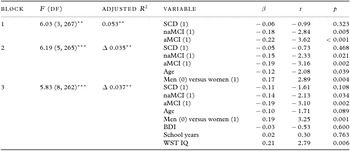
For the multiple hierarchical regression analysis, diagnosis types were dummy-coded and entered into a first block, yielding significant negative effects on emotion recognition of aMCI and non-aMCI but not SCD groups when referenced to controls. Entering age and sex of participants in a second block improved model fit significantly, indicating better performance of younger individuals and women. Model fit further improved when depression scores, number of educational years, and premorbid intelligence test performance were entered in a third block (Adjusted R 2 = 0.125). However, of the additional predictors only premorbid intelligence test performance contributed significantly to variance explanation showing a positive effect and age did not reach significance anymore (Table 3).
Hierarchical Regression on Emotion Recognition Scores (VERT-K)

Note. n = 273; SCD = subjective cognitive decline; naMCI = non-amnestic mild cognitive impairment; aMCI = amnestic mild cognitive impairment; BDI = beck depression inventory; WST = Wortschatztest (vocabulary test); Dummy coded variables are referenced to Controls (0); all VIFs < 1.7; Significant changes in R 2 indicate best model fit of model 3; ** = p < 0.01; *** = p < 0.001.
Discussion
In all, our results show substantially decreasing emotion recognition performance according to severity of diagnosis. Performance of controls was invariably higher than the performance of patients with SCD who showed higher performance than non-aMCI patients who in turn yielded better performance than aMCI patients. These observations were consistent with our expectations that patients with more severe cognitive impairment would show higher impairment in emotion recognition tasks as well. In this vein, it needs to be acknowledged that increases in brain pathology can only account for the differences between healthy controls, SCD, and MCI patients but not non-aMCI and aMCI patients. Rather, differential FER performance of aMCI compared to non-aMCI patients may be more likely attributed on one hand to deficits in limbic structures (i.e. for aMCI) or neocortical structures (i.e. for non-aMCI) on the other hand (e.g. He et al., Reference He, Farias, Martinez, Reed, Mungas and DeCarli2009).
We note that not all of the pairwise comparisons reached nominal significance. However, with the exception of controls versus SCD samples all effect sizes showed non-trivial effects favoring groups with lower pathology. Nonetheless, it cannot be ruled out that differences of type rather than severity of pathology may be responsible for differences between the patient groups (see above). Particularly differences between non-aMCI and aMCI patients are not necessarily due to pathology severity but may instead be linked to differences in affected brain structures (i.e. regional variability appears to be considerably more pronounced in non-aMCI than aMCI; e.g. Grundman et al., Reference Grundman2004).
These results are consistent with previous accounts of impaired emotion recognition in patients suffering from MCI (McCade et al., Reference McCade, Savage and Naismith2011). Thus, the progressive FER impairment from controls over SCD to MCI patients may be interpreted as an effect of progressive global cognitive impairment. Consistent with previous studies (e.g. Kumfor et al., Reference Kumfor, Sapey-Triomphe, Leyton, Burrell, Hodges and Piguet2014), these findings may be interpreted as evidence for emotion recognition as a result of global cognitive impairment (i.e. particularly frontal lobe damage) rather than a specific functional deficit.
Alternatively, these deficits might be associated with impairments in a number of executive functions. In this vein, our correlational analyses indicated largely negative associations between emotion recognition performance and performance in neuropsychological tests (i.e. reflecting specific deficits in cognitive functioning) within the total sample as well as within subsamples (although several correlational results failed to reach nominal significance due to small sample sizes). For instance, it has been demonstrated that language skills may be helpful in interpreting emotional expressions (Allender and Kaszniak, Reference Allender and Kaszniak1989).
In our sample linguistic abilities were more impaired in MCI than SCD groups. Similarly, impaired memory of patient samples in our present study might have led to impaired recall of emotional expressions and consequently lower recognition performance. Such an interpretation conforms to recognition deficits in patients suffering from memory deficits of differing severity (Hargrave et al., Reference Hargrave, Maddock and Stone2002; Snowden et al., Reference Snowden, Austin, Sembi, Thompson, Craufurd and Neary2008). Moreover, impaired attentional processes were evident in patients in the present study which have as well been shown to contribute to deficient emotion recognition performance (Kohler et al., Reference Kohler, Bilker, Hagendoorn, Gur and Gur2000).
It has been suggested that structural aspects of the limbic system may be responsible for FER performance differences between patients and healthy controls (Bio et al., Reference Bio, Soeiro-de-Souza, Garcia Otaduy, Machado-Vieira and Moreno2013). Thus, it is conceivable that volumetric and perhaps functional differences particularly in the amygdala or hippocampus between our control and patient groups may play a role in explaining the patient FER deficits. However, we were not able to directly assess this in the present study because we did not assess neuroimaging data.
Sex differences in our data indicated better performance of women than of men in the overall sample. However, within-group differences according to diagnoses revealed significant advantages for women in SCD and non-aMCI patients only. Although the direction of mean differences was identical for controls and aMCI patients, standardized mean differences for controls were negligible (d = 0.004). Advantageous results for women in recognizing subtle emotions have been previously described (e.g. Hoffmann et al., Reference Hoffmann, Kessler, Eppel, Rukavina and Traue2010) however, there are also studies which do not indicate sex differences in general emotion recognition (e.g. Hoheisel and Kryspin-Exner, Reference Hoheisel and Kryspin-Exner2005). Thus, although sex-specific performance differences have been previously described in general population samples, this is the first time, that sex differences were found in a sample of patients with SCD and non-aMCI.
We observed that premorbid IQ predicted FER performance positively. To our knowledge, this is the first time that better emotion recognition of individuals with higher IQ was observed in patients with SCD and MCI. This finding is interesting because premorbid IQ test scores have been shown to remain comparatively unaffected by neurological impairments and thus provide estimates for cognitive ability preceding the onset of cognitive declines. Therefore, these results may be interpreted as indicative of better emotion recognition in high-cognitive ability individuals. An alternative interpretation may be that participants with higher IQ scores have a larger cognitive reserve thus perhaps compensating for FER deficits. Either way, the observed positive correlation conforms to previous research indicating associations between intelligence test performance and emotion recognition in healthy samples (Sullivan and Ruffman, Reference Sullivan and Ruffman2004).
It needs to be acknowledged that in the present study, not all investigated emotion recognition score differences between diagnostic groups (i.e. Controls vs. SCD vs. non-aMCI vs. aMCI) reached nominal significance. However, observed differences showed the expected decrease of test performance with increasing sample pathology. Effect strengths were substantial, showing non-trivial effects for all comparisons except for control and SCD participants. Moreover, we investigated performance on overall FER only but did not assess effects on the specific basic emotions. This approach was preferred because the VERT-K allows concise and economical assessment of global emotion recognition scores. However, owing to the brevity of the VERT-K subscales and expected reliability decreases within single subscales we focused on overall emotion recognition only.
Similarly, the large number of correlation coefficients that have been calculated in the present study will have led to an increase of type-1 error probability. However, the similar signs and strengths of coefficients in all groups as well as the large (i.e. above-chance probability) number of significant coefficients corroborates robustness of our findings. Finally, it should be recognized that SCD and MCI patients in the present study may likely have been affected from quite different forms of underlying cerebral pathology. Since no neuroimaging data about such differences were available in the present study, more fine-grained assessment of conceivable effects of the differing pathology was not possible.
In all, we demonstrated in the present study substantial differences in FER performance between patients showing SCD, non-amnestic, and amnestic mild cognitively impaired patients, as well as healthy controls. These differences were substantial and FER impairment was observed to be more severe in groups with more severe neurological deficits. These findings can be interpreted as evidence for emotion recognition deficits as a result of global cognitive impairment between patients and controls and specific functional deficits between MCI groups.
Conflict of interest
None.
Description of the authors’ roles
JL designed the study and supervised the data collection. RAW and NR collected the data. JL, IKE, DM, SK, EA, PDB, and GP contributed to writing the paper. JP designed and carried out the statistical analysis and wrote the first draft of the paper.