Infective endocarditis is a microbial infection of the endothelial surface of the heart, predominantly the heart valves, that is associated with high mortality and morbidity. Reference Ferrieri, Gewitz and Gerber1 It typically occurs when bacteria, or other microorganisms, enter the bloodstream in patients with abnormal or denuded endothelium.
Congenital heart disease (CHD) and rheumatic heart disease (RHD) are the main predisposing cardiac conditions in children with endocarditis in low- and middle-income countries. Reference Sadiq, Nazir and Sheikh2 Central indwelling catheters, through their ability to introduce bacteria into the blood stream and potential to cause endocardial erosion, is also a risk factor. Reference Baltimore, Gewitz and Baddour3 Endocarditis has also been identified in patients with no predisposing cardiac conditions, particularly in association with Staphylococcus aureus bacteraemia. Reference Valente, Jain and Scheurer4
The diagnosis of endocarditis is approached as a syndrome using the Modified Duke Criteria that classifies it as “definite”, “possible” and “rejected” according to major and minor clinical criteria. Reference Baltimore, Gewitz and Baddour3 In an analysis of 100 consecutive patients in the Duke University database with “definite” endocarditis according to pathological criteria, 76 had clinical criteria for “definite” endocarditis and 24 had clinical criteria for “possible” endocarditis, while none had criteria for “rejected” endocarditis. Reference Durack, Lukes, Bright and Service5
Surgery is an important part of the management of these patients. Achieving a low mortality rate, a low embolic rate and no residual valvular dysfunction is the ideal outcome. There was a significant reduction in mortality and systemic embolisation in adult patients who had early surgery compared with delayed surgery in The Early Surgery versus Conventional Treatment for Infective Endocarditis (EASE) study. Reference Kang, Kim and Kim6 Findings from a retrospective review of the surgical management of children at Texas Children’s Hospital were that early surgery is associated with a low risk of embolisation, recurrence and operative mortality. Reference Shamszad, Khan, Rossano and Fraser7
At our facility we follow the 2002 and updated 2015 American Heart Association guidelines for the management of endocarditis in children. Reference Ferrieri, Gewitz and Gerber1,Reference Baltimore, Gewitz and Baddour3 However, in the absence of clinical trials in children, these recommendations are based on case series and expert opinion, with low levels of scientific evidence. Reference Baltimore, Gewitz and Baddour3 In patients who are surgically managed for valvular endocarditis, it is our policy to achieve valve repairs as far as possible. The decision to replace or repair a valve is made at the time of surgery based on the viability of the valve. Outcomes of the management of these children in our settings are not known, making it difficult to assess the treatment strategies used and optimise the management of this complex disorder. We therefore aimed to describe the profile of infants and children with infective endocarditis at our facility and assess their outcomes to assist in optimal decision-making.
Methods
Study population
A retrospective chart review of children, from birth to 14 years of age, diagnosed with endocarditis at Red Cross War Memorial Children’s and Tygerberg Hospitals in Cape Town, South Africa, from January 2013 to December 2017 was conducted. These two tertiary-level government hospitals serve the public sector of the Western Cape Province of South Africa.
Patients were identified through a search of the paediatric cardiology unit database using the search terms “Infective Endocarditis, Endocarditis and Vegetation”. This was augmented with cross-referencing echocardiograms, ward and surgery record books, and a search of discharge summaries using the relevant ICD-10 code (I33.0). Each case was classified using the Modified Duke Criteria. Patients meeting criteria for “possible” endocarditis (without a suitable alternate diagnosis) and “definite” endocarditis were included for analysis. Patients meeting criteria for “rejected” endocarditis were excluded. Information was entered and stored on the University of Cape Town-supported research electronic data capture (REDCap) database.
Demographic details and basic patient characteristics, including human immunodeficiency virus status, were entered into the database. Echocardiographic findings prior to and after the completion of treatment were recorded. We used the most recent American Heart Association/American College of Cardiology classification to quantify valve regurgitation or stenosis as mild, moderate or severe in standardised echo reports. Reference Nishimura, Otto and Bonow8 The causative organism, if identified, was recorded. Complications and embolic phenomena were described. The type of intervention performed (medical and surgical), the time interval from diagnosis until surgery as well as the type of surgical intervention were noted.
Statistical analysis
Data were analysed using Stata 14.2 (StataCorp, Texas, United States of America). Continuous variables have been expressed as medians with interquartile ranges, and categorical variables are expressed as absolute number frequencies and percentages. The change in classification of valvular dysfunction from pre- to post-treatment was analysed using the McNemars-Bowker test of symmetry. 9
Results
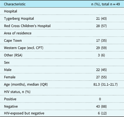
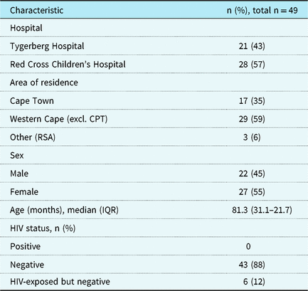
Over a 5-year period from January 2013 to December 2017, 49 new cases of infant and childhood endocarditis were identified, the equivalent of approximately one new case every 5–6 weeks. Patient characteristics are depicted in Table 1. The in-hospital mortality rate was 20% (n = 10). According to the Modified Duke Criteria, 63% (n = 31) were classified as “definite” and 37% (n = 18) were classified as “possible” endocarditis.
Patient characteristics.
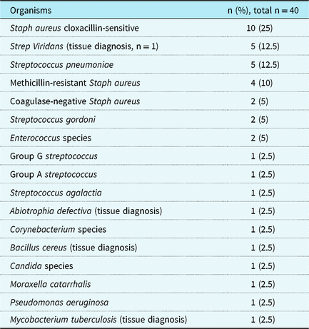
In 82% (n = 40) of the reviewed cases, a causative organism was identified, whereas culture-negative endocarditis occurred in 18% (n = 9). Gram-positive organisms were the most common type of causative organism. Staphylococcus aureus was the commonest organism cultured (Table 2), occurring in 33% (n = 16) of patients; 44% (n = 7) of those patients had surgery. The case fatality rate was 37.5% (n = 6). Out of the 10 in-hospital deaths, 60% (n = 6) were associated with S. aureus.
Organisms and their frequency of occurrence.

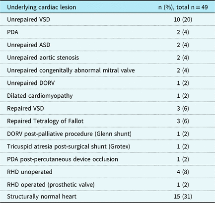
Table 3 indicates the underlying cardiac lesions. Unrepaired CHD occurred in 41% of cases; the most frequently identified lesion, unrepaired ventricular septal defect, was identified in 20% (n = 10) of cases. RHD was the predisposing condition in 10% (n = 5) of cases. In one patient, endocarditis occurred after mitral valve replacement for RHD. Repaired or previously operated CHD occurred in 18% (n = 9), while 31% (n = 15) of patients had structurally normal hearts. Six per cent (n = 3) of patients had a central venous catheter and 6% (n = 3) had dental caries.
Underlying conditions.

VSD = ventricular septal defect; PDA = patent ductus arteriosus; ASD = atrial septal defect; DORV = double outlet right ventricle.
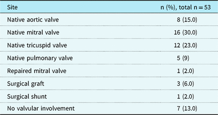
There were 53 sites involved, with more than one site occurring in four patients. The list of sites involved is shown in Table 4. Left-sided valvular endocarditis (47%) occurred more frequently than right-sided valvular endocarditis (32%).
Sites of endocardial infection.

Embolic phenomena and complications
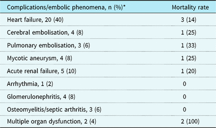
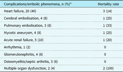
Congestive heart failure occurred in 41% (n = 20) of patients with a mortality rate of 14% (n = 3). All patients with multi-organ dysfunction died. Pulmonary embolus occurred in three patients with a mortality rate of 33%, and cerebral embolus occurred in four patients with a mortality rate of 25% (Table 5). Three out of 26 patients (11.5%) who had surgery had embolic phenomena prior to the operation.
Complications and associated mortality.
* Data not available on one patient who was transferred to another facility.
Surgery
Surgery was performed in 53% (n = 26) of patients, and medical therapy alone was used in 45% (n = 22). Information on management could not be obtained for one patient who had been transferred to another facility. Time interval from admission to surgery was a median of 20 days (interquartile range, 9–47 days). Twenty-three per cent (n = 6) had tricuspid valve repair, 15% (n = 4) had mitral valve repair, 11.5% (n = 3) had aortic valve repair, one had a removal of infected graft and tricuspid valve repair, and one had both aortic and tricuspid valve repair. Fifteen per cent (n = 4) had mitral valve replacement with a mechanical valve, 8% (n = 2) had aortic valve replacement with a mechanical valve. One patient had aortic valve replaced by pulmonary autograft (Ross procedure).
Treatment outcomes
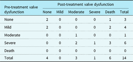
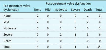
Valvular regurgitation and valvular stenosis were grouped together as valve pathology resulting from endocarditis and classified as mild, moderate or severe valvular dysfunction. Patients were classified according to the most severe category of valve dysfunction. Table 6 is a description of the number of patients who underwent medical treatment in each classification of valvular dysfunction prior to and at the completion of treatment. The symmetry test was not significant (p = 0.08) and the marginal homogeneity test was borderline significant (p = 0.05), indicating a marginal shift in the classification of valvular dysfunction pre- and post-treatment. Six of 14 patients treated medically for valvular endocarditis died in hospital (mortality rate = 43%).
Outcomes of valve dysfunction in those who had medical treatment.
Symmetry test p = 0.08 and marginal homogeneity test p = 0.05.
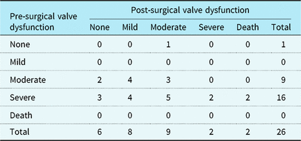
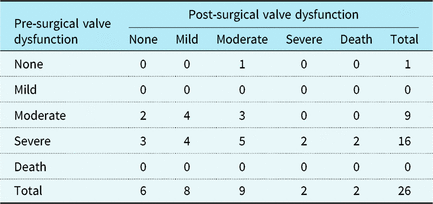
Table 7 is a description of the number of patients who underwent surgery in each classification of valvular dysfunction prior to surgery and before discharge. Both the symmetry test and the marginal homogeneity test are highly significant, thus indicating a shift in the classification of valvular dysfunction pre- and post-treatment (p = 0.007 and 0.002). Two of 26 patients managed surgically died postoperatively (post-operative mortality rate = 7.7%).
Outcomes of valve dysfunction in those who had surgery.
Symmetry test p = 0.007 and marginal homogeneity test p = 0.002.
Overall 6% (n = 3) of patients had severe residual valvular dysfunction, 24% (n = 12) had moderate residual valvular dysfunction, and 16% (n = 8) had mild residual valvular dysfunction at the completion of their treatment.
Discussion
Although infective endocarditis is a rare condition in children, challenges remain in the diagnosis and management of these patients. We have shown a high in-hospital mortality rate of 20%, and despite a significant reduction of valvular dysfunction with treatment, >40% of patients were left with some degree of residual valvular dysfunction – the long-term impact of which is not apparent in this review. In total, the need for surgery was high, with more than half of patients undergoing surgical treatment.
The cases of endocarditis in this review were identified at the two government hospitals in the province but did not include Christian Barnard Netcare Hospital (a private facility). Approximately 83% of South Africans use public healthcare facilities, making this a good representation of childhood endocarditis in the province. 10
A reduction in mortality rate in children with endocarditis from 35% to 20% over the last 40 years at our facility may be accredited to the introduction of echocardiography in 1982, which improved diagnostic capabilities, allowing for prompt treatment. Reference Hugo-Hamman, Md and Human11 Echocardiography is central to the diagnosis and management of endocarditis with a sensitivity of up to 95% in detecting vegetations in children with “definite” endocarditis. Reference Penk, Webb, Shulman and Anderson12 The clinical usefulness of echocardiography is limited however, as not all vegetations are detected and not all patients have criteria for “definite” endocarditis. Reference Evangelista and Gonzalez-Alujas13 The use of Modified Duke Criteria in children has limitations as well. Reference Ferrieri, Gewitz and Gerber1,Reference Tissieres, Gervaix, Beghetti and Jaeggi14 In our analysis, a third of the patients had criteria for “possible” endocarditis. Refinement of the Duke Criteria and alternate methods of diagnosing endocarditis are necessary. Reference Tissieres, Gervaix, Beghetti and Jaeggi14 Molecular techniques for the identification of organisms in patients with culture-negative endocarditis have been developed, but are far better suited for detecting organisms on tissue specimens compared with blood samples. Reference Fournier, Thuny and Richet15 Tissue characterisation cardiac MRI, not yet available at our facility, has been shown to be an effective method for detecting endocardial inflammation in patients with endocarditis. Reference Dursun, Yılmaz and Yılmaz16 Further studies are required to validate the utility of this method.
None of the patients in this review were infected with the human immunodeficiency virus. This finding, supported by the analysis of childhood endocarditis in Leuven, Belgium, Reference Kelchtermans, Grossar and Eyskens17 suggests that human immunodeficiency virus infection and an immune-compromised state does not increase the risk of developing endocarditis in children. The hypothesis that a damaged or denuded endothelium in the presence of bacteraemia is the dominant risk factor holds true for most cases, but does not account for endocarditis that occurred in patients with structurally normal hearts. It is interesting to note that this occurred twice as many times in our analysis compared with that previously described at our facility (31% versus 14%). Reference Hugo-Hamman, Md and Human11 Increasing use of central venous catheters in patients in neonatal and paediatric intensive care units may partly explain this.
Our facility follows the South African Heart Association guidelines, which are adapted from the European Society of Cardiology guidelines, for antibiotic prophylaxis for the prevention of endocarditis following dental procedures. Reference Habib, Lancellotti and Antunes18 They recommend restricting the use of antibiotics to certain high-risk lesions (cyanotic heart disease and patients with prosthetic material). Reference Jankelow, Cupido and Zühlke19 They also emphasised the need for prophylaxis in patients with RHD, Reference Jankelow, Cupido and Zühlke19 an important recommendation given the persistent finding of RHD as a significant predisposing condition at our facility over the last few decades. Reference Hugo-Hamman, Md and Human11 The large number of patients with underlying unrepaired ventricular septal defects (20%) suggests that these patients would also likely benefit from antibiotic prophylaxis for dental procedures.
Staphylococci and streptococci are the most common organisms isolated in children with endocarditis. Reference Shamszad, Khan, Rossano and Fraser7,Reference Kelchtermans, Grossar and Eyskens17 S. aureus endocarditis is a severe form of the disease accounting for 60% of the in-hospital deaths. Just over one-third of patients with S. aureus endocarditis died, and almost half were managed surgically. Its presence necessitates early surgical intervention. Reference Chu, Cabell and Benjamin20
Residual valvular dysfunction typically results in ventricular dysfunction, which may contribute to mortality outside of the treatment period. Reference Shamszad, Khan, Rossano and Fraser7 Despite the significant pre- to post-treatment reduction in valvular dysfunction, a portion of patients had ongoing valve dysfunction after treatment. Patients who underwent antibiotic therapy exclusively showed only a marginal improvement in valvular dysfunction with a high mortality rate (43%), while patients who had surgery showed a significant improvement in valvular dysfunction and a far lower mortality rate (7.7%). Surgery is an important part of the management of these patients, and one of its life-saving benefits is the ability to decrease the infectious load. Reference Baltimore, Gewitz and Baddour3
The EASE trial clearly showed better short- and long-term outcomes in terms of embolic phenomena, congestive heart failure, and death in adult patients with large, left-sided vegetations who had surgery within 48 hours of diagnosis. Reference Kang, Kim and Kim6 There was a high rate of valve replacements in the early surgery group (75%) compared with a slightly lower rate in the conventional management group (63%). Reference Kang, Kim and Kim6 The median interval from diagnosis to surgery in the review from Texas Children’s Hospital was 3 days (interquartile range, 1.0–7.5 days), and half of the patients had valve replacements. Reference Shamszad, Khan, Rossano and Fraser7 The median interval from diagnosis to surgery in our facility was longer (20 days; interquartile range, 9–47 days), and 27% of patients underwent valve replacement surgery, compared with 58% who had valve repair surgery. The post-operative mortality rate was, however, higher in our facility (7.7% versus 2%). Reference Shamszad, Khan, Rossano and Fraser7 A mechanical valve replacement in a child has a life-long risk of thromboembolism and need for anticoagulation and monitoring. Reference Choudhary, Talwar and Airan21 In patients in whom a mechanical valve replacement is inevitable and the social/geographical factors preclude ongoing surveillance, decisions are made in the combined cardiothoracic-cardiology meeting at our facility to treat the patient medically.
Limitations of the study
This was a retrospective study of a relatively small number of patients. This study focused on short-term outcomes and did not capture complications or mortality that may have occurred after the treatment period under review. A prospective registry would provide better clinical evidence to guide decision-making with regard to indications for surgery, optimal timing of surgery, and long-term outcomes of these patients.
Conclusion
Infective endocarditis is a rare, heterogeneous disease. However, it is associated with a high morbidity and mortality. Confirming a definite diagnosis was not possible in at least a third of the cases, highlighting the need for novel techniques for detecting endocardial infection. Early surgery has better outcomes than delayed surgery, but with higher rates of valve replacement. In order to attain the lowest overall mortality rate, the lowest post-operative mortality rate and the highest rate of valve repair, the ideal timing for surgical intervention appears to be between 3 and 20 days.
Acknowledgements
Thank you to the Children’s Heart Disease Research Unit for assistance with this study, and Dr Paul Human for assistance in acquiring data from the surgical database.
Financial Support
W.B. is funded by the MRC UK; L.J.Z. is funded by the MRCSA, NRFSA and MRCUK.
Conflicts of Interest
None.
Ethical Standards
The authors assert that all procedures contributing to this work comply with the ethical standards of the relevant national guidelines on human experimentation (Human Research Ethics Committee of the University of Cape Town and University of Stellenbosch) and with the Helsinki Declaration of 1975, as revised in 2008, and has been approved by University of Cape Town (Human Research Ethics Committee reference number 539/2018) and the University of Stellenbosch (Human Research Ethics Committee reference number N19/01/017).
Contributions
M.L.W. devised the study, conducted the study, wrote the first draft and edited all subsequent drafts. W.B. set up the electronic database and provided statistical analysis. S.R.P. assisted with development of protocol, formatting and editing. G.A.M.C. supervised the project and edited all drafts of the manuscript. B.F. assisted with identifying cases, data capturing and editing all drafts of the manuscript. J.B.L. supervised the project from protocol development stage, identified patients, reviewed echocardiograms and revised all drafts of the manuscript. L.J.Z. supervised the project from protocol development to the final editing. All authors revised the drafts and approved the final version of the manuscript.