Implications
The water buffalo has economic importance for milk production and its derivatives. This study shows the impact of subclinical mastitis on the buffalo milk composition, and on the expression pattern of genes associated with the immune system. Research into the genes of the immune system contributes to the better knowledge about the action mechanism of this system that is activated in animals with mastitis. More information about this disease can aid to avoid economic losses, improve the quality of dairy food, and decrease the negative effects on animal welfare.
Introduction
Buffalo milk has a high total solids (TS) content, with an average protein content of 3.91 ± 0.61% and fat content of 6.87 ± 0.88% (Tonhati et al., Reference Tonhati, Cerón-Muñoz, Oliveira, Duarte, Furtado and Tseimazides2000). These characteristics ensure a high yield in the processing of its derivatives. However, milk composition and production can be significantly affected by mastitis. Changes in milk composition caused by mastitis can make the milk less suitable for consumption and processing, with considerable economic losses to the dairy industry (Rainard and Riollet, Reference Rainard and Riollet2006). Mastitis is characterized by inflammation of the mammary gland resulting from metabolic and physiological changes, trauma or invasion of pathogens (Oviedo-Boyso et al., Reference Oviedo-Boyso, Valdez-Alarcón, Cajero-Juárez, Ochoa-Zarzosa, López-Meza, Bravo-Patino and Baizabal-Aguirre2007). Several types of microorganisms have been associated with mastitis including mostly bacteria, but also fungi and yeast (Petzl et al., Reference Petzl, Zerbe, Günther, Yang, Seyfert, Nürnberg and Schuberth2008).
In buffaloes, studies demonstrated that the pathogens involved in mastitis are coagulase-negative Staphylococci (Moroni et al., Reference Moroni, Rossi, Pisoni, Bronzo, Castiglioni and Boettcher2006), Staphylococcus spp., Streptococcus spp. and Corynebacterium sp. (Moura et al., Reference Moura, Rangel, Melo, Borba, Lima Júnior, Novaes, Urbano and Andrade Neto2017). According to (El-Khodery and Osman, Reference El-Khodery and Osman2008), coliform bacteria (Klebsiella pneumoniae and Escherichia coli) was the most common pathogen followed by Staphylococcus aureus, Streptococcus uberis and Streptococcus agalactiae. Similarly, in cattle, S. aureus, S. agalactiae, S. uberis, Streptococcus dysgalactiae, and the coliforms are major mastitis pathogens and Corynebacterium bovis and coagulase-negative Staphylococci are minor mastitis pathogens (Reyher et al., Reference Reyher, Haine, Dohoo and Revie2012).
Mastitis is either clinical or subclinical, the former of which can be easily diagnosed, whereas, with subclinical mastitis, there are no clinical symptoms or visible changes in milk or udder appearance, even though the immune system responds by sending defense cells to combat the pathogen. The milk somatic cell count threshold of 200,000 cells/ml in buffalo, can be used as an indirect measure to distinguish the udder health and milk quality with (>200,000 cells/ml) or without (<200,000 cells/ml) subclinical mastitis (Dhakal, Reference Dhakal2006). Control strategies that depend on the early and accurate detection of subclinical mastitis are needed since early detection can reduce the negative effects of the disease and lead to higher cure rates. The predominant defense in the early stage of mastitis is the innate immune system, also known as non-specific immunity. The appropriate action of this defense mechanism promotes the rapid elimination of most pathogens (Sordillo and Streicher, Reference Sordillo and Streicher2002).
Pathogens have cell wall structures, known as pathogen-associated molecular patterns. The pathogen-associated molecular patterns are recognized by specific receptors, called toll-like receptors (TLRs), including TLR-2 and TLR-4 that are the most studied and well-characterized receptors of innate immunity (Sharma et al., Reference Sharma, Leyva, Schenkel and Karrow2006; Zähringer et al., Reference Zähringer, Lindner, Inamura, Heine and Alexander2008; Fonseca et al., Reference Fonseca, Cardoso, Higa, Giachetto, Brandão, Brito, Ferreira, Guimarães and Martins2015a), and are expressed on mammary epithelial cells and leukocytes present in milk (Wellnitz and Bruckmaier, Reference Wellnitz and Bruckmaier2012). The TLR-2 in concert with TLR-1 or TLR-6 forms heterodimers to recognize an extended variety of ligands (Yu et al., Reference Yu, Wang and Chen2010). Toll-like receptor 2 recognizes lipoteichoic acid, peptidoglycans and lipoproteins from gram-positive bacteria, whereas TLR-4 recognizes the lipopolysaccharide a component of gram-negative bacteria (Ibeagha-Awemu et al., Reference Ibeagha-Awemu, Lee, Ibeagha, Bannerman, Paape and Zhao2008).
After the recognition occurs, several effector molecules and defense mediators are released or activated (Oppenheim et al., Reference Oppenheim, Biragyn, Kwak and Yang2003), including antimicrobial peptides and specialized chemical mediators (cytokines). Proinflammatory cytokines such as tumor necrosis factor alpha (TNF-α) and interleukin 1 beta (IL-1β) trigger an inflammatory response and mediate the recruitment of neutrophils from the bloodstream to the site of infection to eliminate the invading pathogens (Oviedo-Boyso et al., Reference Oviedo-Boyso, Valdez-Alarcón, Cajero-Juárez, Ochoa-Zarzosa, López-Meza, Bravo-Patino and Baizabal-Aguirre2007). In addition to TNF-α and IL-1β, several cytokines classified as ILs have been found in mammary glands, including IL-8. Gene product of lactoferrin (LTF) is an antimicrobial peptide produced by epithelial cells, macrophages and neutrophils, which binds to free iron ions present in milk, thus preventing the growth of bacteria that use this ion as a metabolic cofactor (Carneiro et al., Reference Carneiro, Domingues and Vaz2009).
The aim of this study was to verify the effects caused by subclinical mastitis on the components of milk and to evaluate the level of expression of the TNF-α, IL-1β, LTF, IL-8, TLR-2 and TLR-4 genes in the milk somatic cells of healthy buffaloes and those with subclinical mastitis. To our knowledge, this is the first study on the expression of these genes in milk cells of buffaloes. The changes identified in gene expression levels will contribute to improving the function of the buffalo immune system in the presence of subclinical mastitis.
Material and methods
Animals
Milk samples were collected from 24 Murrah buffaloes in the first third of lactation, from a farm located in Registro, São Paulo State, Brazil, supplied by the buffalo milk control program of the College of Agriculture and Veterinary Sciences of São Paulo State University (FCAV – UNESP). Twelve samples were collected from healthy animals and 12 from animals with subclinical mastitis.
Sampling
The milk samples were collected from one quarter, directly from the buffalo teats, in the morning at the beginning of milking, following the routine procedure for buffalo cows. Prior to the collection, the buffalo’s teats were washed with running water and dried with a paper towel immediately. To determine which animals were positive or negative for subclinical mastitis we used the California Mastitis Test (CMT). The following criteria for the California Mastitis Test were used: (−) there was no sign of infection, that is, the solution did not show precipitation, (+) positive for mastitis and presented with very viscous liquid and coagulated particles or presented a gelatinous and completely coagulated sample.
After the CMT, aliquots of milk from each animal were stored in flasks containing bronopol as a preservative and sent to the Milk Clinic at Luiz de Queiroz College of Agriculture of University of São Paulo (ESALQ-USP), Piracicaba, São Paulo State, Brazil, for somatic cell counting by flow cytometry in a SomaCount 300® (Bentley Instruments Inc.). In addition to the somatic cell score (SCS), the contents of fat, protein, lactose, TS and solids-not-fat (SNF) were measured by infrared absorption (Bentley Instruments Inc., Chaska, MN, USA). The final sampling was used for RNA extraction, for which approximately 150 ml of milk from each animal was divided into three sterile tubes, which were transported on disposable ice in isothermal containers.
The SCS was used to classify the groups for RNA extraction, in which ⩽161 × 103 cells/ml was considered negative (animals without subclinical mastitis) and ⩾860 × 103 cells/ml was considered positive (animals with subclinical mastitis). All of the milk samples were analyzed for their microbiological profiles, and those with subclinical mastitis presented the following pathogens: S. dysgalactiae, S. uberis, C. bovis, S. agalactiae.
Gene expression
Immediately after collection, 50 ml of milk was centrifuged at 12,000 × g for 10 min at 4°C to isolate the somatic cells. Approximately 100 mg of the precipitate was stored in 1 ml of Trizol® (Invitrogen, Carlsbad, CA, USA), then homogenized and frozen at −80°C for subsequent RNA extraction. Total RNA was extracted with the Trizol® reagent according to manufacturer recommendations. All materials were sterile and free of RNase or were previously treated with RNaseZAP® (Invitrogen) to prevent RNA degradation. After 1 week, the material was thawed in the refrigerator for 4 h, then was briefly homogenized and centrifuged at 12,000 × g for 12 min at 4°C. The supernatant was transferred to a new tube and incubated for 5 min at room temperature. Next, 200 µl of chloroform was added and the samples were shaken before incubation for an additional 3 min at room temperature, followed by centrifugation at 12,000 × g for 15 min at 4°C. The aqueous phase was collected and transferred to a new tube and an equal volume of isopropanol was added. The samples were homogenized by inversion, incubated for 10 min at room temperature, and centrifuged at 12,000 × g for 10 min at 4°C. The supernatant was discarded, and the precipitate was washed with 1 ml of 75% ethanol. The samples were again centrifuged at 7500 × g for 5 min and the supernatant was discarded. The pellet was dried for 30 min at room temperature, resuspended in RNase-free ultrapure water, and incubated in a water bath at 55°C to 60 for 15 min. The samples were then stored in a freezer at −80°C.
The concentration and purity of RNA were measured in a NanoDrop 1000 spectrophotometer (ThermoFisher Scientific, Carlsbad, CA, USA) at 260 and 280 nm, and in a Qubit® 2.0 Fluorometer (Invitrogen). All samples were treated with the TURBO DNA-free™ Kit (Invitrogen, Carlsbad, CA, USA) and the absence of contamination of the samples with genomic DNA was confirmed in a Qubit® 2.0 Fluorometer (Invitrogen) using the Qubit dsDNA Assay Kit (Invitrogen).
The first complementary DNA strand (cDNA) was synthesized using the SuperScript IV First-Strand cDNA Synthesis Reaction Kit (Invitrogen) according to manufacturer recommendations. SuperScript IV can be used for cDNA synthesis of any type of RNA and is highly efficiency even with degraded and limited RNA. For the assay, 1 μl of oligo(dT)20 primer (50 μM), 1 μl of deoxyribonucleoside triphosphates (10 mM) and 2 μl of total RNA were mixed and RNase-free ultrapure water was added to reach a volume of 13 μl. The reaction was incubated for 5 min at 65°C and kept on ice for at least 1 min. Next, 4 μl of 5X SSIV buffer, 1 μl of ribonuclease inhibitor, 1 μl of dithiothreitol (10 mM) and 1 μl of (200 U) SuperScript IV were added. The mixture was incubated at 55°C for 10 min and at 80°C for an additional 10 min. The synthesized cDNA was stored at −20°C for future use.
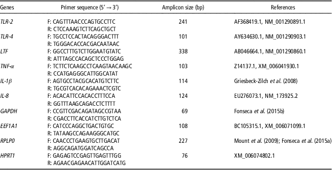
The primers used in the amplification reactions for the study of gene expression were constructed using gene sequences obtained from GenBank (htpp://www.ncbi.nlm.nih.gov/genbank/) (Table 1).
Sequence of forward (F) and reverse (R) primers for the study of gene expression in the milk of buffalo with and without subclinical mastitis

TLR-2 = toll-like receptors 2; TLR-4 = toll-like receptors 4; LTF = lactoferrin; TNF-α = tumor necrosis factor alpha; I-1β = interleukin-1 beta; IL-8 = interleukin-8; GAPDH = glyceraldehyde-3-phosphate dehydrogenase; EEF1A1 = eukaryotic translation elongation factor 1 alpha 1; RPLP0 = ribosomal protein large P0; HPRT1 = hypoxanthine phosphoribosyltransferase 1.
The primers for the TLR-2, TLR-4, LTF, TNF-α, IL-8 genes and for the housekeeping gene (EEF1A1) were obtained from studies on cattle and were redesigned using the homologous sequences in buffaloes. The primers for the IL-1β, GAPDH and RPLP0 genes were obtained from the literature (Table 1). Comparison of the primers sequences with the sequence of the corresponding gene in buffaloes showed 100% identity and it was therefore not necessary to redesign these primers. The messenger RNA (mRNA) sequence of the HPRT1 gene expressed in buffaloes was used to design the primer to avoid annealing to the intron regions. The Primer Express 3.0 software (Applied Biosystems, Foster, CA, USA) was used to design the primers, taking into consideration the size of the primer, size of the amplified product, percentage of cytosine and guanine, and melting temperature (T M), as well as the absence of dimers and hairpin formation. The selected primer sequences were aligned using the BLAST algorithm (Basic Local Alignment Search Tool) of the NCBI database (http://www.ncbi.nlm.nih.gov/BLAST) to identify regions in the selected sequences that would show a high degree of identity with other regions of the genome or within a gene family.
The concentrations of the primers were determined by titration: 300 nM forward and reverse primers for EEF1A1 and RPLP0, and 100 nM forward and reverse primers for GAPDH, HPRT1, IL-1β LTF, TLR-2, TLR-4, TNF-α, and IL-8. For all genes, the quantitative real-time PCR efficiency was calculated by a standard curve from a serial cDNA dilution (2 : 1). Efficiency between 90% and 110% was considered for PCR primers (Livak and Schmittgen, Reference Livak and Schmittgen2001).
A total of four candidate housekeeping genes (GAPDH, EEF1A1, RPLP0 and HPRT1) were analyzed using the GeNorm algorithm (Vandesompele et al., Reference Vandesompele, De Preter, Pattyn, Poppe, Van Roy, De Paepe and Speleman2002) and the PROC MIXED procedure of the Statistical Analysis System software (SAS Institute Inc., Cary, NC, USA), considering two different groups (with or without subclinical mastitis) as a fixed effect and biological repetition as a random effect. The EEF1A1 and RPLP0 housekeeping genes were identified as the most stable, and the geometric mean of these genes (Vandesompele et al., Reference Vandesompele, De Preter, Pattyn, Poppe, Van Roy, De Paepe and Speleman2002) was used for each sample in this study.
Real-time PCR was performed with the 7500 Real-Time PCR system (Applied Biosystems) using the SYBR Green Master Mix (Applied Biosystems), at a final volume of 12.5 µl. All the reactions were performed in triplicate and the amplification conditions were: 2 min at 50°C, 10 min at 95°C and 40 cycles at 95°C for 15 s and at 60°C for 1 min. Melt curves were used to analyze and evaluate the accuracy of the PCR.
Statistical analysis
The cycle threshold (Ct) values were obtained with the Expression Suite software v1.0 (Applied Biosystems). The analysis was performed with the PROC MIXED procedure of the Statistical Analysis System software, in which the following mixed linear model was adjusted:
 $$Y_{{gikr}} {\rm {\equals}}TG_{{ig}} {\rm {\plus}}G_{k} {\rm {\plus}}P_{k} {\rm {\plus}}D_{{ik}} {\rm {\plus}}e_{{gikr}} $$
where Y gikr is the Ct obtained from the thermocycler software for the gth gene (target and housekeeping genes) located in the rth well of the microplate (referring to the technical replicate), corresponding to the kth animal and the ith group (with or without subclinical mastitis); TG ig the fixed effect of the i group in the expression of gene g; G k the random effect of the genes for animal k; P k the random effect of the microplate for animal k; D ik is a random sample-specific effect that captures differences between samples common to the genes, particularly those affecting RNA concentration such as extraction and amplification efficiency, and e gikr the residual effect.
Using the model described above, an approximation of ∆∆Ct was estimated as proposed by Steibel et al. (Reference Steibel, Poletto, Coussens and Rosa2009). This approach also permits the inference of the level of significance of the difference in the transcription of these genes using contrasts between groups and the housekeeping and target genes. Approximated ∆∆Ct values with a P-value less than 5% (P <0.05) were considered significant.
The fat, protein, lactose, TS and SNF percentages and SCS were compared between buffaloes with and without mastitis by the Tukey test, considering P <0.05 as significant. For the normalization of SCS, the data were log transformed [log2(SCS/100 000)] + 3 as described by Dabdoub and Shook (Reference Dabdoub and Shook1984). To assess the association of the genes expression with fat, protein, lactose, TS, SNF and SCS traits, linear regression analysis was performed using the ∆Ct values as the independent variable (Ct of the target genes normalized to the housekeeping genes). Bonferroni correction was applied to ensure the maintenance of the error rate of the statistical tests in the analyses.
Results
Milk components
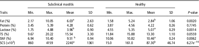
We evaluated the composition of buffalo milk through the estimation of SCS, fat, protein, lactose, TS and SNF (Table 2). Our results showed significant differences in fat, lactose, SNF and SCS between healthy animals and with subclinical mastitis. We found higher fat content and SCS in animals with subclinical mastitis, whereas the contents of SNF and lactose were higher in animals without mastitis. We observed no difference for protein and TS components.
Effect of subclinical mastitis in dairy buffaloes on the percentage of fat, protein, lactose, total solids (TS) and solids-not-fat (SNF) and somatic cell score (SCS)

Min. = minimum; Max. = maximum.
a,bValues within a row with different superscripts differ significantly at P <0.05.
Gene expression
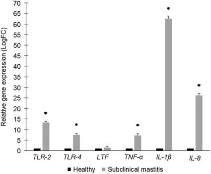
We studied the mRNA expression of TLR-2, TLR-4, TNF-α, IL-1β, IL-8 and LTF genes in the milk of buffaloes with and without subclinical mastitis (Figure 1). The TLR-2 gene showed expression levels 13.34 times higher in animals with mastitis compared to animals without mastitis. For the TLR-4 gene, mRNA expression was also higher (7.49 times) in buffaloes with mastitis. Expression of the TNF-α, IL-1β and IL-8 genes differed significantly between the groups with and without mastitis. The relative expression of the TNF-α, IL-1β and IL-8 genes was, respectively, 7.15, 62.49 and 26.20 times higher in animals with mastitis. We observed no significant difference between groups for the LTF gene.
Relative expression of the TLR-2 (toll-like receptor 2), TLR-4 (toll-like receptor 4), LTF (lactoferrin), TNF-α (tumor necrosis factor alpha), IL-1β (interleukin-1 beta) and IL-8 (interleukin-8) genes in the milk of buffalo with and without subclinical mastitis. Mean values with *differ significantly at P <0.05.

Association between the gene expression and milk components
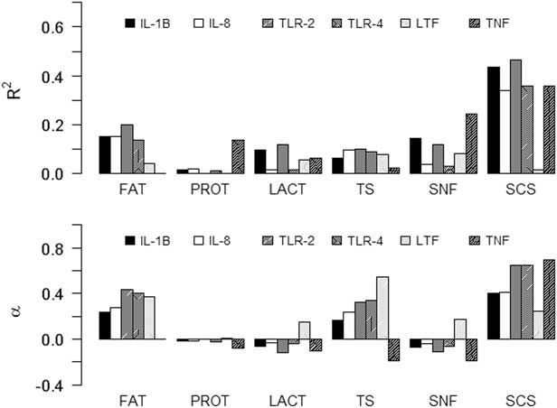
We used the coefficient of determination of linear regression (R 2) and linear regression coefficient (α) (Figure 2), to determine the relationship between the genes studied and the buffalo milk components. For SCS, the regressions were significant for the IL-1β, IL-8, TLR-2, TLR-4 and TNF-α genes, with P-values of 0.00112, 0.00543, 0.00095, 0.00409 and 0.00534, respectively. For fat, the regression was significant only with the TLR-2 gene (P = 0.04787), and for SNF only with the TNF-α gene (P = 0.02694). However, these associations were no longer significant after Bonferroni correction of these values for the number of components (0.05/6 = 0.0083). We observed the highest coefficients of determination between SCS and almost all genes, except for the LTF. Toll-like receptor-2 is the gene that most explains the variation in SCS. For every additional unit of ∆Ct for IL-1β, IL-8, TLR-2, TLR-4 and TNF-α genes, we expected to see an increase in SCS. The change in a unit of ∆Ct that would result in a higher increase in SCS would be obtained with the TNF-α gene.
Coefficient of determination of linear regression (R 2) and linear regression coefficient (α) between the target genes ∆Ct values and fat (FAT %), protein (PROT %), lactose (LACT %), total solids (TS %), solids-not-fat (SNF %) and somatic cell score (SCS, being [log2(SCS × 103 cells/ml per 100,000)] + 3) in the milk of buffalo with and without subclinical mastitis. IL-1β = interleukin-1 beta; IL-8 = interleukin-8; TLR-2 = toll-like receptors 2; TLR-4 = toll-like receptors 4; LTF = lactoferrin; TNF-α = tumor necrosis factor alpha.

Discussion
Effects of subclinical mastitis were observed in buffalo milk composition through the significant differences in fat, lactose, SNF, and SCS between the animals with and without mastitis. As reported by Harmon et al. (Reference Harmon1994) the diminished synthesis capability of mammary tissue promotes a decrease in lactose and fat in milk and is related to mastitis or elevated SCS. However, as in the present study, associations of increased SCS with increased fat and reduced lactose have been reported for cattle by Sarikaya et al. (Reference Sarikaya, Schlamberger, Meyer and Bruckmaier2006) who evaluated different milk fractions and different SCS levels. In accordance with these authors, the increased fat content was probably due to decreased lactose synthesis, which reduced milk volume. A significant change in SCS between the groups confirmed the difference between animals with and without mastitis considered for the analysis of expression of genes of the immune system.
Messenger RNA expression of the TLR-2 and TLR-4 genes were higher in buffalo cows with subclinical mastitis, resulting in the association of these genes with immune system alterations caused by mastitis in dairy buffaloes, as were also observed in cattle (Fonseca et al., Reference Fonseca, Cardoso, Higa, Giachetto, Brandão, Brito, Ferreira, Guimarães and Martins2015a). Similarly, a study by Goldammer et al. (Reference Goldammer, Zerbe, Molenaar, Schuberth, Brunner, Kata and Seyfert2004) in cattle, identified an increase in the gene expression of TLR-4 and TLR-2, which the gene products recognize different cell wall components of pathogens, and suggests that the expression of these receptors is regulated together at the beginning of the immune response in the mammary tissue. Toll-like receptors are the first-line host response to the presence of pathogens and are responsible for triggering the innate immune response in animals (Pasare and Medzhitov, Reference Pasare and Medzhitov2004).
After activation of TLRs, different molecules and defense mediators are released or activated (Oppenheim et al., Reference Oppenheim, Biragyn, Kwak and Yang2003), such as TNF-α, IL-1β, IL-8 and LTF, all of which were evaluated in this study. The TNF-α and IL-1β genes encode proinflammatory cytokines that trigger the process of inflammation. Both cytokines mediate endothelial activation to stimulate vasodilation and an increase in blood vessel permeability, promoting the recruitment of serum proteins and leukocytes to the site of infection (Medzhitov, Reference Medzhitov2007). In this context, the increase in mRNA expression of both genes in animals with mastitis is expected since it is associated with protection against and elimination of the invading pathogen in the mammary gland. For this and other reasons, TNF-α and IL-1β genes are considered key components of the innate immune system. As observed in cattle by Sarikaya et al. (Reference Sarikaya, Schlamberger, Meyer and Bruckmaier2006), Griesbeck-Zilch et al. (Reference Griesbeck-Zilch, Meyer, Kühn, Schwerin and Wellnitz2008) and Fonseca et al. (Reference Fonseca, Cardoso, Higa, Giachetto, Brandão, Brito, Ferreira, Guimarães and Martins2015a), higher mRNA expression of these cytokines was also seen in animals with mastitis.
The IL-8 gene encodes a chemokine (chemotactic cytokine) that attracts neutrophils to the site of infection. This cytokine is produced by a variety of cells, such as monocytes, T lymphocytes, macrophages and epithelial and endothelial cells (Oviedo-Boyso et al., Reference Oviedo-Boyso, Valdez-Alarcón, Cajero-Juárez, Ochoa-Zarzosa, López-Meza, Bravo-Patino and Baizabal-Aguirre2007), and its expression can be induced by proinflammatory stimuli such as TNF-α and IL-1β (Mukaida, Reference Mukaida2003). We may, therefore, suggest that the increased IL-8 mRNA expression in animals with mastitis is related to the increased mRNA expression of TNF-α and IL-1β genes, increasing the number of neutrophils and, consequently, of somatic cells. Increased mRNA expression of the IL-8 gene has also been reported for cattle (Zhu et al., Reference Zhu, Liu, Weng, Zhuge, Zhang, Ma, Qiu, Li, Zhang and Wang2012; Fu et al., Reference Fu, Zhou, Liu, Li, Liang, Liu, Song, Zhao, Fen, Li, Cao, Zhang, Zhang and Yang2013), suggesting that the mechanism of combating the pathogen seems to be similar in both species.
The LTF gene, the only gene for which no significant difference was observed between groups, encodes the glycoprotein lactoferrin that is part of the complement system of the innate immunity (Rainard and Riollet, Reference Rainard and Riollet2006). This protein has been shown to exert antimicrobial activity, as well as antitumor and anti-inflammatory properties (Anand et al., Reference Anand, Kanwar, Dubey, Vahishta, Sehgal, Verma and Kanwar2015). Thus, the expected result would be increased mRNA expression of this gene in the group with mastitis, which was not observed. According to Berlutti et al. (Reference Berlutti, Schippa, Morea, Sarli, Perfetto, Donnarumma and Valenti2006), LTF downregulates proinflammatory cytokines, that is, it prevents the release of inflammatory mediators and inhibits the production of cytokines (Kushibiki et al., Reference Kushibiki2011). However, the opposite effect seems to have occurred in our study. It is possible that the differential expression of the cytokines may have affected LTF mRNA expression, however, further studies are needed to confirm this result. The non-differential expression of LTF mRNA may also be the result of the changes caused by mastitis in mammary tissue, which could have affected epithelial cells that, together with leukocytes, are responsible for its production (Sordillo and Streicher, Reference Sordillo and Streicher2002).
In addition to the study of immune system-related genes, linear regression analysis was performed using the ∆Ct values obtained along with specific components (fat, protein, lactose, TS, SNF and SCS). To evaluate the association of the different genes with the milk components, the linear regression coefficient (α), which quantifies the expected change in the milk components with an increment of one unit in ∆Ct, was analyzed. The TLR-2 is the gene that most explains the variation in SCS, probably because its gene product is a cell membrane receptor which forms heterodimers with TLR-6 and TLR-1, possibly increasing the range of recognizable ligands (Yu et al., Reference Yu, Wang and Chen2010). Thus, the increase in SCS in the mammary gland would result in a higher synthesis of mRNA of this gene in milk.
The change in a unit of ∆Ct that would result in a higher SCS and, consequently, a higher inflammatory response would be obtained with the TNF-α gene. This finding is probably due to the cytotoxicity of this cytokine, in addition to its chemotactic effects. The possible absence of an association for fat, protein, lactose, TS, SNF and the target genes evaluated, indicates that these genes were adequate for the study of the immune response of the mammary gland (Zhu et al., Reference Zhu, Liu, Weng, Zhuge, Zhang, Ma, Qiu, Li, Zhang and Wang2012; Fu et al., Reference Fu, Zhou, Liu, Li, Liang, Liu, Song, Zhao, Fen, Li, Cao, Zhang, Zhang and Yang2013).
In conclusion, subclinical mastitis modifies the fat, lactose and SNF in milk and promotes changes in the expression of the TLR-2, TLR-4, TNF-α, IL-1β and IL-8 genes in dairy buffaloes. This study contributes to the characterization of the action mechanism of the innate immunity that occurs in subclinical mastitis in this species improving the knowledge about gene expression in milk cells associated with the immune response in buffalo with subclinical mastitis. Furthermore, the rapid elimination of pathogens may prevent changes in the quality or produced milk, since, the rapid and effective action of the studied genes, which participate in the non-specific defense mechanism, is the first line of defense against pathogenic organisms and could eliminate them rapidly before the adaptive immunity can be activated.
Acknowledgements
The authors thank the São Paulo Research Foundation (FAPESP, Brazil) for providing the financial support (Grants #2014/19321-4 and #2014/25309-7) to conduct the research project. F.T. received a PhD fellowship from FAPESP (2014/19321-4). N.B.S. was supported by a postdoctoral fellowship from Coordination for the Improvement of Higher Education Personnel (CAPES/PNPD). D.J.A.S. received a postdoctoral fellowship from FAPESP. M.I.T.F., E.G., L.G.A. and H.T. were supported by productivity research fellowships from National Council of Technological and Scientific Development (CNPq). The authors thank the Avian Reproduction, Department of Animal Reproduction, School of Veterinary Medicine and Animal Science of University of São Paulo (FMVZ – USP), São Paulo, State of São Paulo, Brazil. The authors thank the Laboratory of Biochemistry and Molecular biology, Department of Technology, College of Agriculture and Veterinary Sciences of São Paulo State University (FCAV – UNESP), Jaboticabal, State of São Paulo, Brazil.
Declaration of interest
The authors declare no conflicts of interest.
Ethics statement
Not relevant.
Software and data repository resources
Data were not deposited in an official repository.