


Cardiac output plays an important role in fetal circulation, as oxygen is delivered to organs through the blood flow from the heart. The function of the fetal heart is typically assessed by the combined cardiac output (CCO) of the left and right ventricles (Kiserud et al., Reference Kiserud, Ebbing, Kessler and Rasmussen2006). This is because blood flow from the left ventricle primarily goes to the upper part of the body, whereas blood flow from the right ventricle goes to the lower part of the body through the ductus arteriosus.
Placental circulation is crucial for evaluating fetal circulation as oxygen is supplied through the placenta. The blood flow in the umbilical artery was measured to assess the placental function. While this method provides a qualitative assessment of placental circulation, the umbilical venous flow volume (UVFV) allows for quantitative evaluation of the placental function (Acharya et al., Reference Acharya, Wilsgaard, Rosvold Berntsen, Maltau and Kiserud2005; Barbera et al., Reference Barbera, Galan, Ferrazzi, Rigano, Józwik, Battaglia and Pardi1999; Bellotti et al., Reference Bellotti, Pennati, De Gasperi, Battaglia and Ferrazzi2000; Flo et al., Reference Flo, Wilsgaard and Acharya2009, Reference Flo, Wilsgaard and Acharya2010; Ozawa et al., Reference Ozawa, Kanazawa, Mikami, Muromoto, Sugibayashi, Wada and Sago2023; Tchirikov et al., Reference Tchirikov, Rybakowski, Hüneke, Schoder and Schröder2002). UVFV has been measured in cases involving fetal growth restriction (FGR) or twin-twin transfusion syndrome (TTTS) to investigate pathophysiological conditions (Gungor et al., Reference Gungor, Glosemeyer, Huber, Hecher and Baschat2008; Maskatia et al., Reference Maskatia, Ruano, Shamshirsaz, Javadian, Kailin, Belfort, Altman and Ayres2016; Szwast et al., Reference Szwast, Tian, McCann, Donaghue, Bebbington, Johnson, Wilson and Rychik2007; Yamamoto et al., Reference Yamamoto, Nasr, Ortqvist, Bernard, Takahashi and Ville2007).
The normal reference ranges for CCO and UVFV have been evaluated in singleton fetuses (Acharya et al., Reference Acharya, Wilsgaard, Rosvold Berntsen, Maltau and Kiserud2005; Luewan et al., Reference Luewan, Srisupundit, Tongprasert, Traisrisilp, Jatavan and Tongsong2020; Mielke & Benda, Reference Mielke and Benda2001; Ozawa et al., Reference Ozawa, Kanazawa, Mikami, Muromoto, Sugibayashi, Wada and Sago2023; Rasanen et al., Reference Rasanen, Wood, Weiner, Ludomirski and Huhta1996). In uncomplicated fetuses, CCO and UVFV increase exponentially with advancing gestational age (Gerson et al., Reference Gerson, Wallace, Stiller, Paul, Weiner and Bolognese1987). However, the median weight-corrected CCO remains unchanged by gestational age (Gerson et al., Reference Gerson, Wallace, Stiller, Paul, Weiner and Bolognese1987; Kiserud et al., Reference Kiserud, Ebbing, Kessler and Rasmussen2006; Mielke & Benda, Reference Mielke and Benda2001; Rasanen et al., Reference Rasanen, Wood, Weiner, Ludomirski and Huhta1996), and the median weight-corrected UVFV slightly decreases with gestational age (Ozawa et al., Reference Ozawa, Kanazawa, Mikami, Muromoto, Sugibayashi, Wada and Sago2023). The proportion of UVFV to CCO represents the proportion of blood flow to the placenta (accounting for approximately one-third), but this proportion decreases to one-fifth after 32 weeks of gestation (Kiserud et al., Reference Kiserud, Ebbing, Kessler and Rasmussen2006).
However, the normal reference ranges for CCO and UVFV have not been reported for twin fetuses. It is not clear whether CCO and UVFV in twin fetuses differ from those in singleton fetuses (Almog et al. Reference Almog, Shehata, Aljabri, Levin, Shalom-Paz and Shrim2011). Moreover, the differences between monochorionic and dichorionic twins seem to be interesting because vascular anastomoses in the monochorionic placenta may influence CCO and UVFV in twins.
Measurement of CCO before 20 weeks of gestation is sometimes challenging because of the small diameter of the outflow tracts. In addition, the fetal position often does not allow for precise measurements of CCO in the third trimester. Therefore, this study aimed to establish normal reference ranges for CCO and UVFV in twin fetuses between 20 and 28 weeks of gestation and to evaluate the differences between monochorionic and dichorionic twins.
Methods
Study Design
This two-center prospective cohort study was conducted between January 2020 and July 2022 at Ramathibodi Hospital, Mahidol University, Thailand, and the National Center for Child Health and Development (NCCHD), Tokyo, Japan. The study was approved by the Institutional Review Board of Ramathibodi Hospital (COA. MURA2020/70), and the ethics committee of the NCCHD (No 2019-185). This study was conducted in accordance with the ethical principles of the Declaration of Helsinki.
Twin pregnancies that were managed at hospitals during the study period were included. The inclusion criterion was that we obtained agreement to perform ultrasound measurements at least once at 20 to 28 weeks of gestation. Written informed consent was obtained from all participants. We applied the following exclusion criteria: age <18 years or >45 years, first visit to hospitals at >16 weeks of gestation, monochorionic monoamniotic twin pregnancy, fetal structural or chromosomal abnormality, and severe hypertension or renal disease. We excluded complicated twin pregnancies, including FGR, TTTS, or twin anemia polycythemia sequence (TAPS), even if they developed later in the pregnancy. TTTS was defined as polyhydramnios (maximum vertical pocket >8 cm) in one twin and oligohydramnios (maximum vertical pocket <2 cm) in the other twin. TAPS was defined as a peak systolic velocity of the middle cerebral artery (MCA-PSV) >1.5, multiple of median (MoM) in one twin, and MCA-PSV <1.0 MoM in the other twin. Data on maternal age, height, weight, parity, conception method, and chorionicity were also collected. Pregnancy dating relies on crown-to-rump length (CRL) measurements of the larger twin during the first trimester or conception date in in vitro fertilization (IVF) pregnancies. In women presenting after the first trimester, a larger head circumference is used (Khalil et al., Reference Khalil, Rodgers, Baschat, Bhide, Gratacos, Hecher, Lewi, Salomon, Thilaganathan and Ville2016). Chorionicity and amnionicity were determined prenatally before 14 weeks of gestations using ultrasonographic findings including the number of gestational sacs and amniotic membranes, or ‘lambda’ sign for monochorionic twins and ‘T’ sign for dichorionic twins. Twin labeling was performed based on the location of the fetus and insertion of the umbilical cord.
Outcome Measurements
Ultrasound measurements were conducted by four certified maternal-fetal medicine specialists. Between 20 and 28 weeks of gestation, ultrasonographic examinations to measure UVFV and CCO were conducted using a Voluson E10 (GE Healthcare, Chicago, IL, US), Aplio i700 (Canon Medical Systems, Tochigi, Japan), or Arietta750 (Fujifilm, Tokyo, Japan) with a convex probe. The period was divided into three groups: 20–22 weeks of gestation, 23–25 weeks of gestation, and 26–28 weeks of gestation. The measurements were scheduled once during each period. Some cases were measured at all three time periods, while others were only measured at one or two time periods between 20 and 28 weeks of gestation, depending on the opportunity to measure UVFV and CCO.
Fetal weight was estimated and FGR was defined as an estimated fetal weight (EFW) below the 10th percentile in at least one twin. Selective FGR was excluded from the study and was defined as intertwin EFW discordance >25% (Khalil et al., Reference Khalil, Beune, Hecher, Wynia, Ganzevoort, Reed, Lewi, Oepkes, Gratacos, Thilaganathan and Gordijn2019). UVFV was measured using two different parameters: absolute UVFV (ml/min) and weight-corrected UVFV for EFW (UVFV/kg). The diameter and maximum flow velocity of the umbilical vein (UV) were measured according to a previous report (Tchirikov et al., Reference Tchirikov, Rybakowski, Hüneke, Schoder and Schröder2002). The diameter and flow velocity of the UV were measured at the point of the fetal abdomen between the insertion of the UV and branches of the portal vein. The diameter of the UV was measured from the inner wall to the inner wall across the lumen of the UV with appropriate magnification of the view using a perpendicular ultrasound beam. The diameter of the UV was calculated as the average of three measurements. The angle between the ultrasound beam and the vessel was kept as close as possible to 0° to measure the velocity of the UV flow, but the angle was allowed to fall within 30° after angle correction. The sample volume for Doppler interrogation was set slightly larger than the UV diameter. The maximum velocity of UV flow was calculated as the average of the two measurements. UVFV (ml/min) was calculated as follows: (UV diameter [cm]/2)2 × maximum velocity (cm/s) × 0.5 × 3.14 × 60).
CCO was measured using two different parameters: absolute CCO (ml/min) and weight-corrected CCO for EFW (CCO/kg). CCO was calculated by adding the left and right cardiac output (CO). CO (ml/min) was calculated as follows: (annulus diameter [cm]/2)2 × 3.14 × velocity time integral (VTI) × fetal heart rate (per minute). The inner diameter of the aortic valve annulus was measured during systole from the long-axis view of the left heart, while the inner diameter of the pulmonary valve annulus was obtained from the short-axis view of the fetal thorax during systole, with appropriate magnification of the view with a perpendicular ultrasound beam. The annulus diameter was calculated as the average of two measurements. The angle between the ultrasound beam and the vessel of the outflow tract was kept as close as possible to 0° to measure the velocity of the outflow, but the angle was allowed to fall within 30° after angle correction. Doppler tracings were recorded with the sample volume positioned just distal to the valve in the center of the vessel, with the sample volume covering the vessel’s width and the high-pass filter set at 100–200 Hz. At least five consecutive uniform Doppler velocity waveforms with the highest velocities and a narrow frequency band were recorded, and one cycle was analyzed. The VTI of the outflow was calculated as the average of two measurements.
Statistical Analysis
All statistical analyses were performed using STATA version 18.0 (Stata Corp., College Station, TX, USA), employing multilevel regression to explore the interplay between each ultrasound measurement (dependent variable) and gestational age (independent variable). Prior to the analysis, the normality of the data for each variable was evaluated. Furthermore, reference percentiles for UVFV and CCO in uncomplicated fetuses were established.
Results
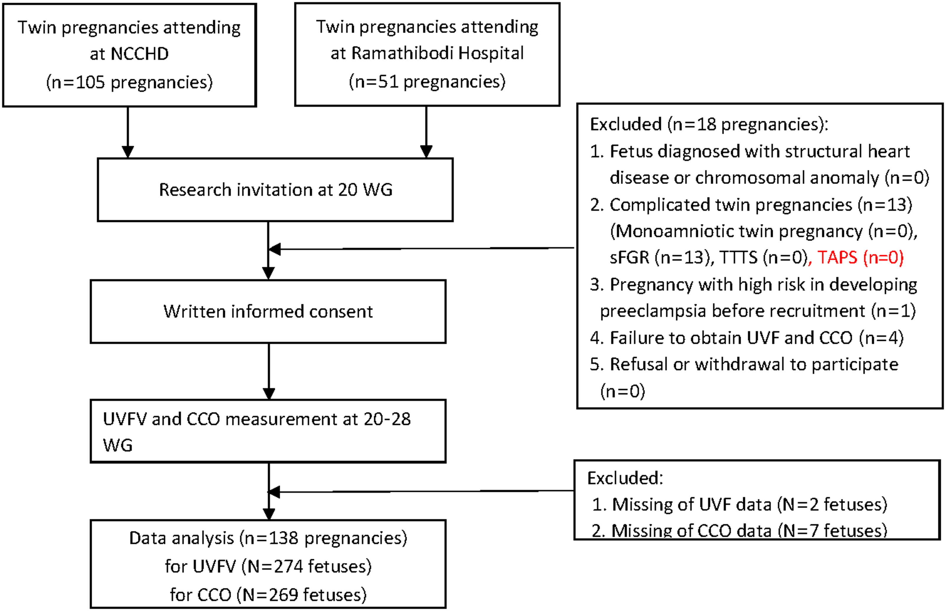
A total of 156 pregnant women were included in the study (Figure 1). After excluding 13 cases of selective FGR, one woman at high risk of preeclampsia, and four women in whom UVFV and CCO measurements could not be obtained due to fetal position, 138 women remained in the analysis. Although UVFV and CCO measurements could not be obtained from two and seven fetuses respectively, they were still analyzed from 274 and 269 fetuses respectively.
Study flow diagram.
Note: CCO, combined cardiac output; NCCHD, National Center for Child Health and Development; sFGR, selective fetal growth restriction; TAPS, twin anemia polycythemia sequence; TTTS, twin-twin transfusion syndrome; UVFV, umbilical venous flow volume.

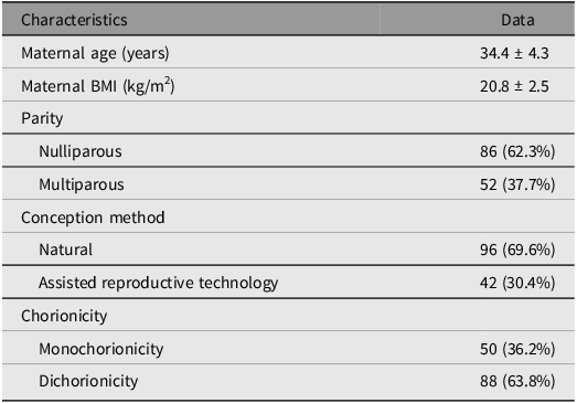
Table 1 presents the characteristics of 138 twin pregnancies. The average maternal age was 34.4 ± 4.3 years, and 62.3% of participants were nulliparous. This study included 50 monochorionic and 88 dichorionic twin pregnancies.
Characteristics of 138 twin pregnancies

Note: Data are given as the mean ± SD or n (%). BMI, body mass index.
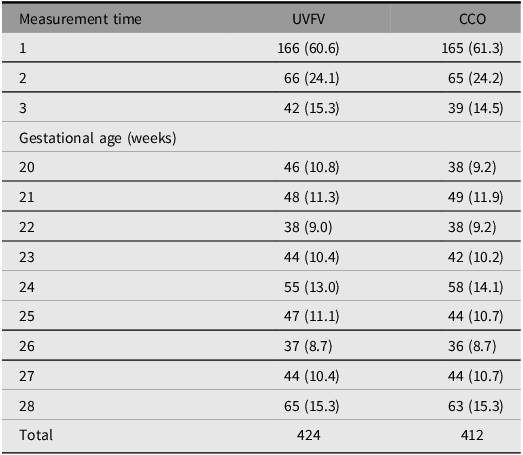
The measurement count per fetus and its distribution in relation to gestational age are shown in Table 2.
Number of fetuses measured according to measurement time and gestational age

Note: Data are given as n (%). UVFV, umbilical venous flow volume; CCO, combined cardiac output.
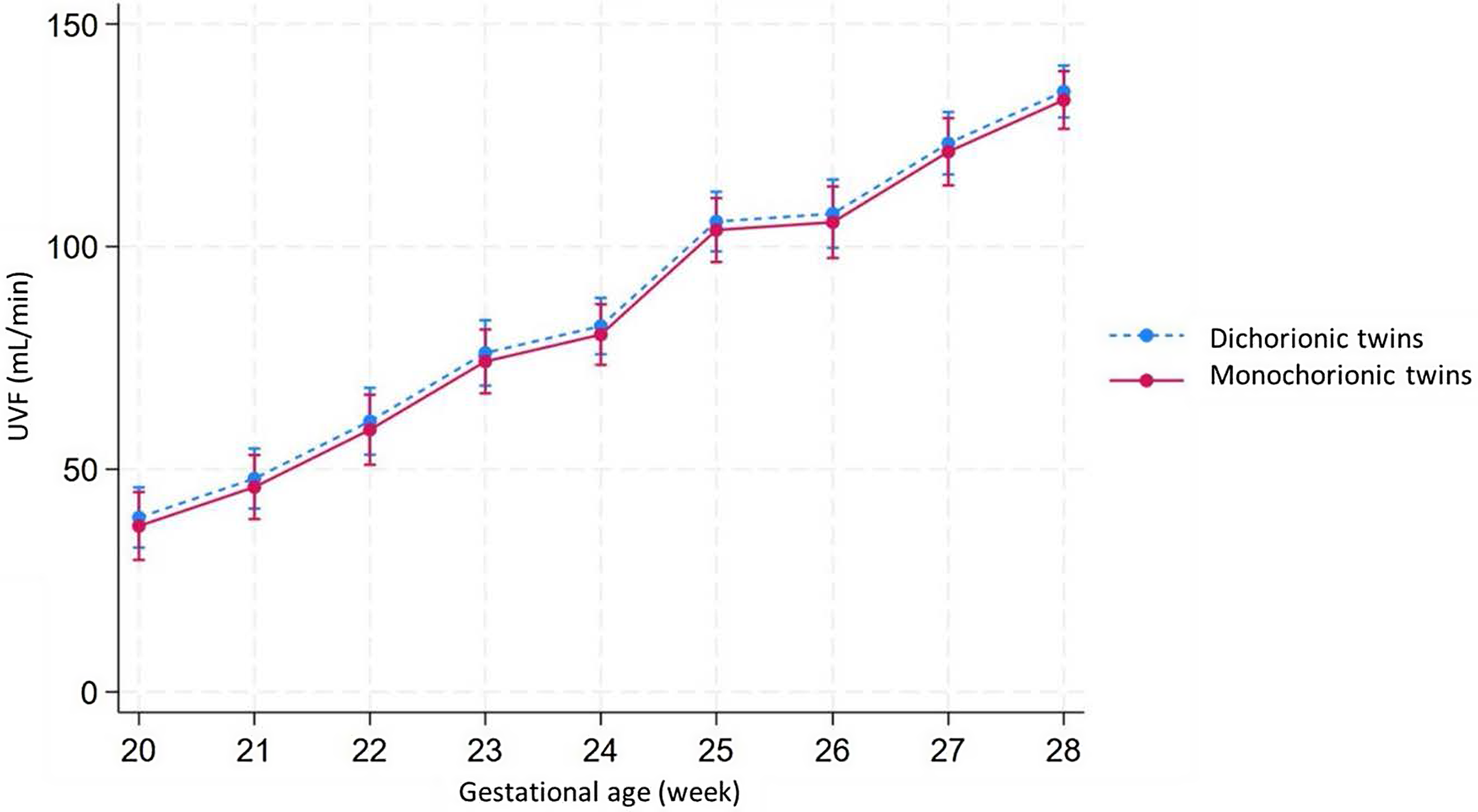
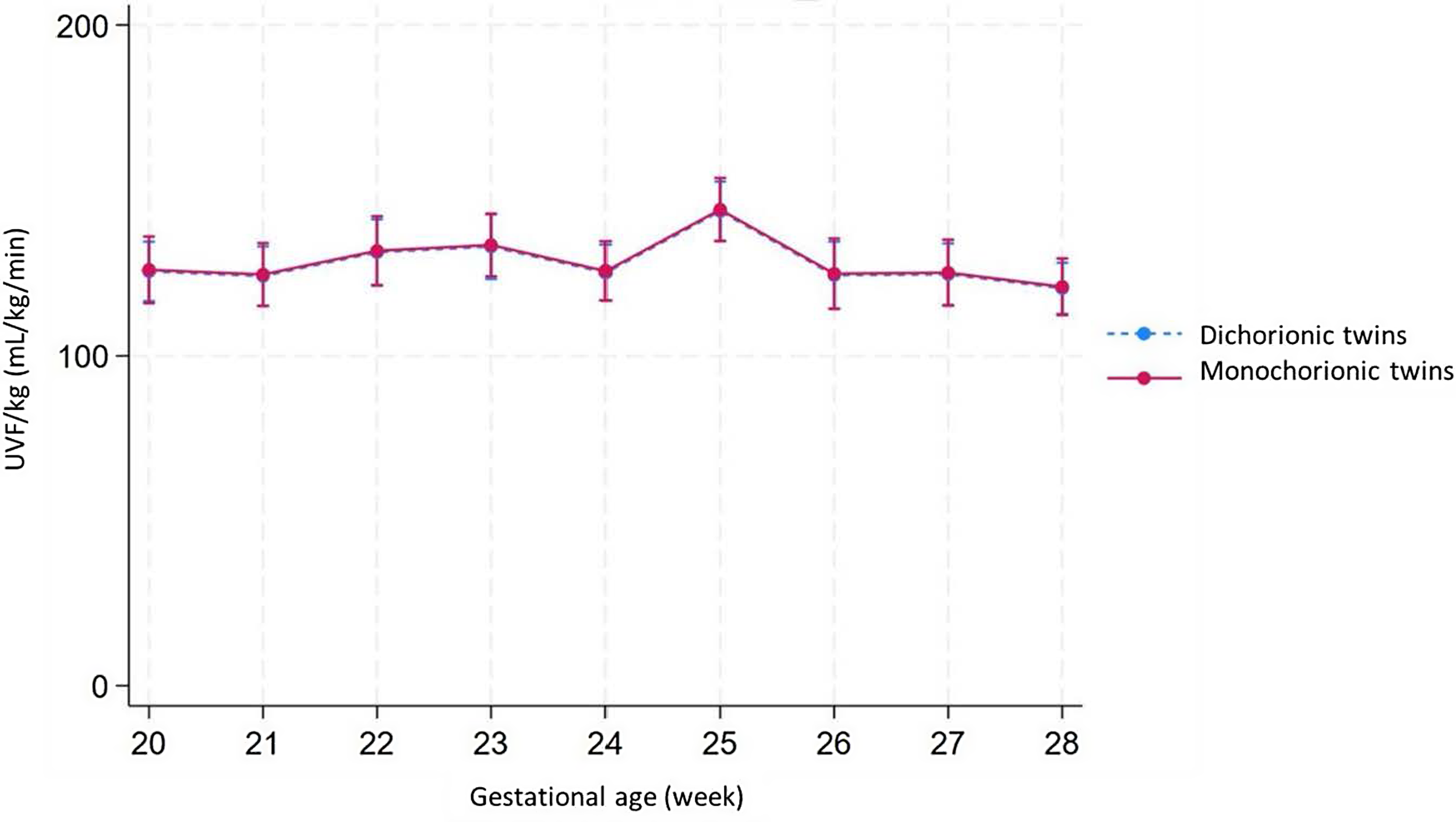
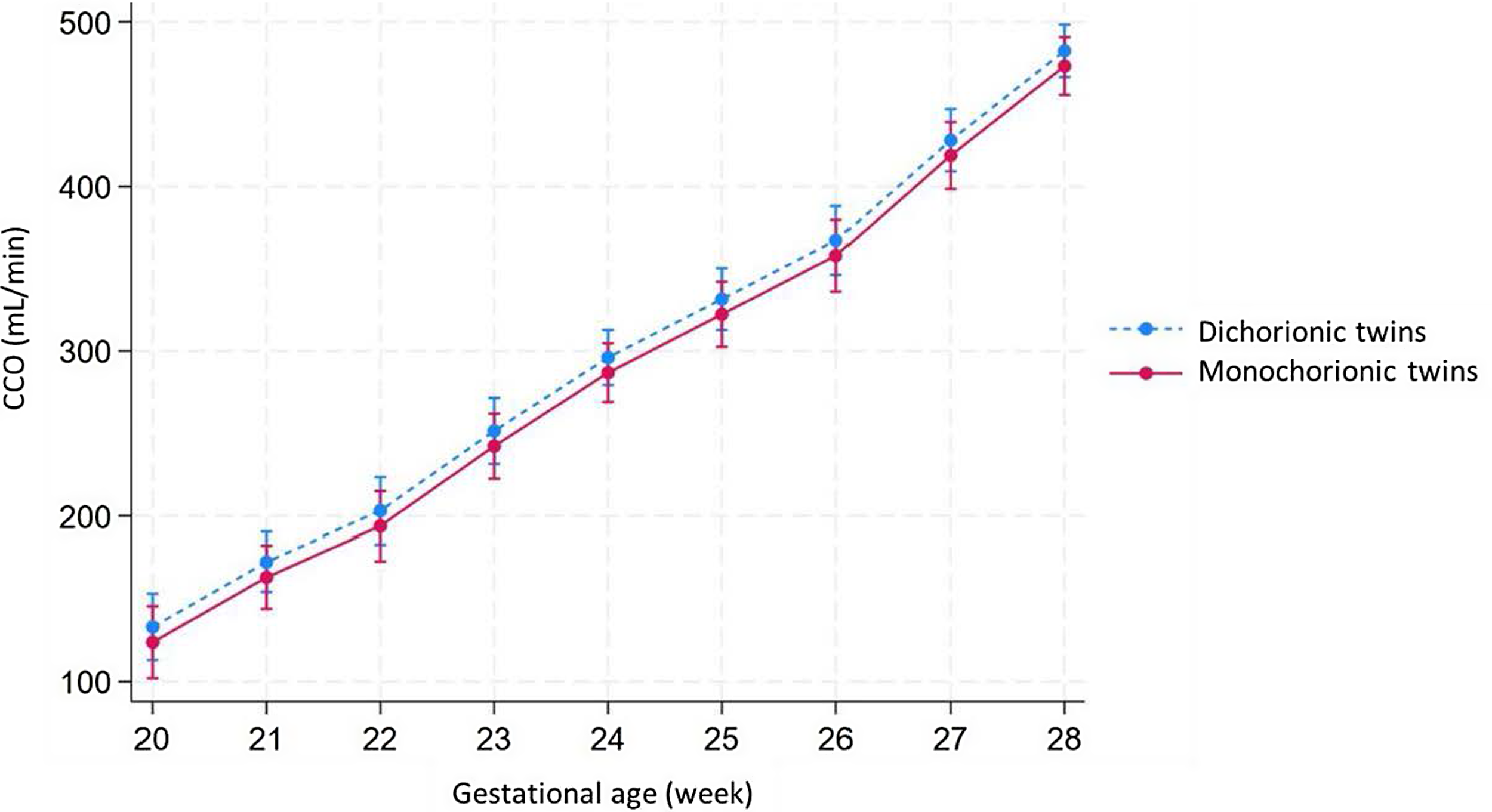
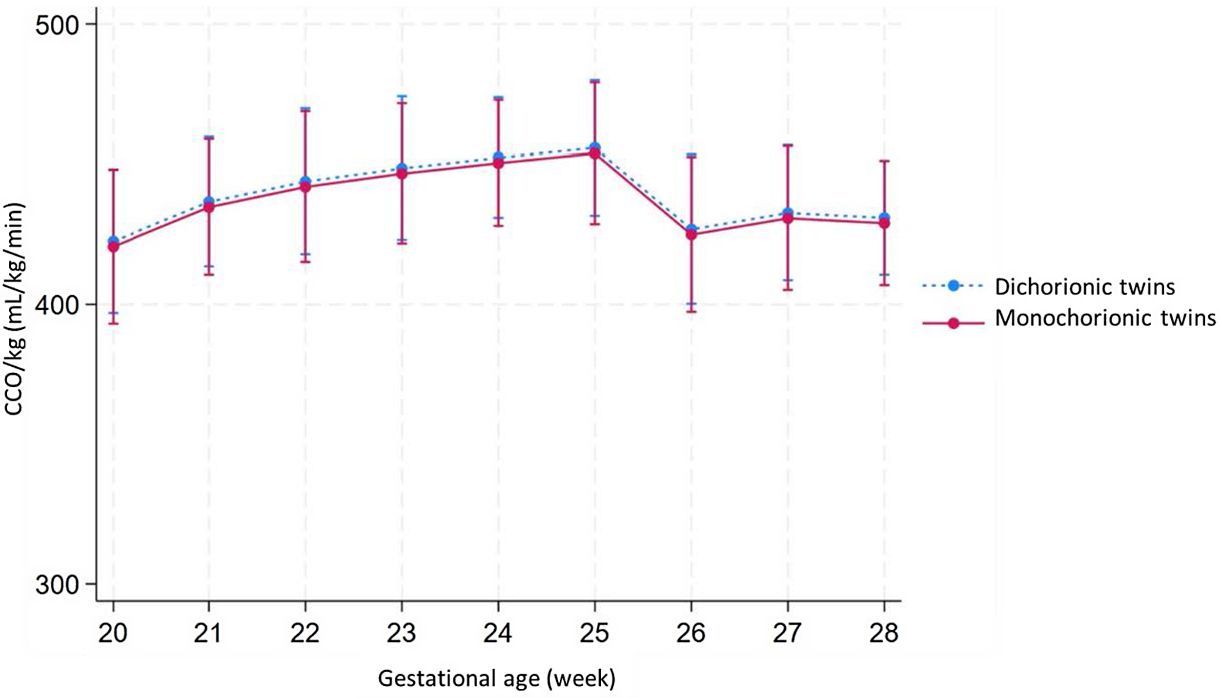
We employed linear regression mixed-effects models for UVFV and CCO predictions. UVFV and CCO levels increased as gestation progressed (Figure 2A and 2B). Weight-corrected UVFV (UVFV/kg) and CCO (CCO/kg) remained stable from 20 to 28 weeks of gestation (Figure 3A and 3B). UVFV and CCO did not differ between monochorionic and dichorionic twin fetuses.
The adjusted predictions of UVFV, along with 95% confidence intervals according to gestational age, in dichorionic and monochorionic twin fetuses.
Note: UVFV, umbilical venous flow volume.

The adjusted predictions of UVFV/kg, along with 95% confidence intervals according to gestational age, analyzed in dichorionic and monochorionic twin fetuses.
Note: UVFV, umbilical venous flow volume.

The adjusted predictions of CCO, along with 95% confidence intervals according to gestational age, analyzed in dichorionic and monochorionic twin fetuses.
Note: CCO, combined cardiac output.

The adjusted predictions of CCO/kg, along with 95% confidence intervals according to gestational age, analyzed in dichorionic and monochorionic twin fetuses.
Note: CCO, combined cardiac output.

The analyses of UVFV, UVFV/kg, CCO, CCO/kg, and the UVFV-to-CCO ratio in groups with one, two, and three measurements show similar trends and are available in the Supplementary material on the Cambridge Core website.
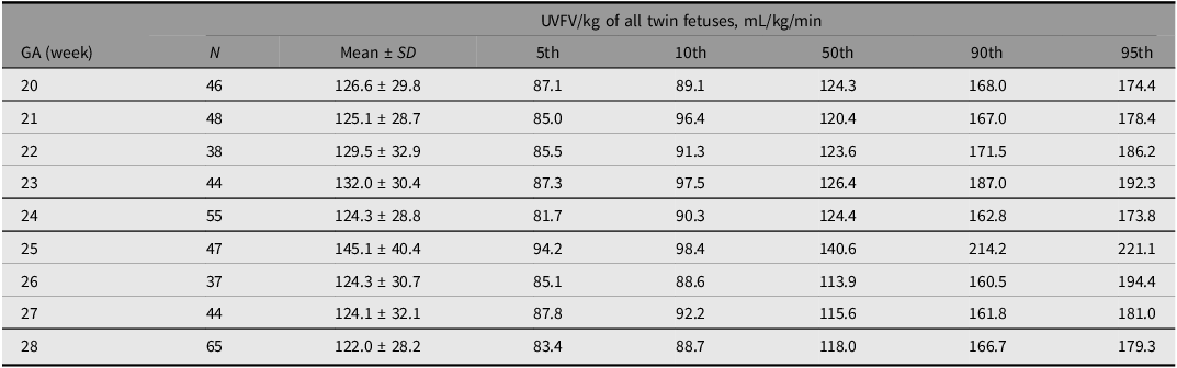
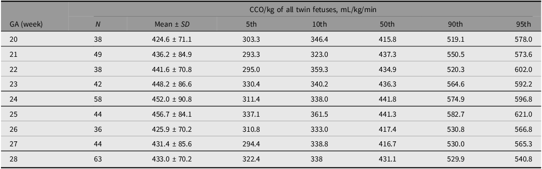
UVFV/kg and CCO/kg, along with the corresponding percentiles according to gestational age, are shown in Tables 3 and 4, respectively. The mean ± SD of UVFV/kg and CCO/kg were 127.8 ± 31.8 and 439.4 ± 80.1 mL/kg/min, respectively.
UVFV/kg with corresponding percentiles according to gestational age

Note: GA, gestational age; UVFV, umbilical venous flow volume.
The CCO/kg with corresponding percentiles according to gestational age

Note: GA, gestational age; CCO, combined cardiac output.
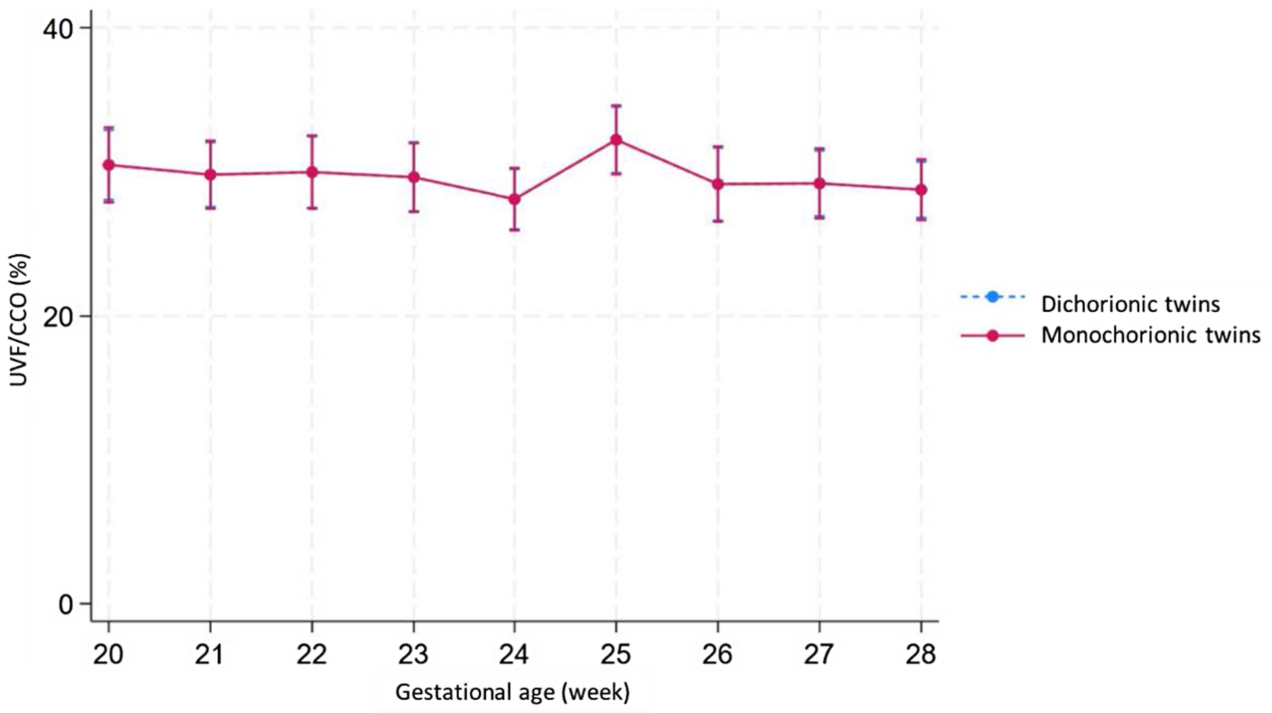
The UVFV-to-CCO ratio also remained stable from 20 to 28 weeks of gestation, ranging from 27.7% to 31.8%, as shown in Figure 4.
The proportion of UVFV to CCO as percentage, along with 95% confidence intervals according to gestational age, analyzed in dichorionic and monochorionic twin fetuses.
Note: CCO, combined cardiac output; UVFV, umbilical venous flow volume.

Discussion
We observed that UVFV and CCO levels increased with gestational age. Adjusted for fetal body weight, UVFV, and CCO remained stable from 20 to 28 weeks of gestation. We compared UVFV and CCO in dichorionic twin fetuses with those in monochorionic twin fetuses and found no significant differences between the two groups. The proportion of UVFV to CCO remained at approximately one-third and was stable throughout the gestational period. The values and ranges of UVFV/kg and mean CCO/kg in twins are similar to those in singletons.
UVFV and CCO increased with gestational age but were stable if adjusted by estimated fetal weight. Our study revealed that the mean UVFV/kg was 122–145 mL/kg/min, which was similar to previously reported singleton fetuses (Acharya et al., Reference Acharya, Wilsgaard, Rosvold Berntsen, Maltau and Kiserud2005; Bellotti et al., Reference Bellotti, Pennati, De Gasperi, Battaglia and Ferrazzi2000; Ozawa et al., Reference Ozawa, Kanazawa, Mikami, Muromoto, Sugibayashi, Wada and Sago2023), and the range of values was narrow from 20 to 28 weeks of gestation, consistent with previous studies in singleton pregnancies (Ozawa et al., Reference Ozawa, Kanazawa, Mikami, Muromoto, Sugibayashi, Wada and Sago2023)
UVFV/kg (mL/kg/min) with published 50th percentiles and 10th−90th percentiles according to gestational age in the present study and previous studies on singleton pregnancies

Note: GA, gestational age; UVFV, umbilical venous flow volume. The value is expressed as the 50th percentiles (10th-90th percentiles).
The mean CCO/kg in twin fetuses ranged from 425 to 457 mL/kg/min, which was similar to previously reported singleton fetuses (400–433 mL/kg/min; Kiserud et al., Reference Kiserud, Ebbing, Kessler and Rasmussen2006; Luewan et al., Reference Luewan, Srisupundit, Tongprasert, Traisrisilp, Jatavan and Tongsong2020; Mielke & Benda, Reference Mielke and Benda2001) (Table 6).
The mean CCO/kg (mL/kg/min) in the present study and previous studies on singleton pregnancies

Note: CCO, combined cardiac output.
The estimated fetal weight in twin pregnancies is lower than that in singletons after the late second trimester (Hiersch et al., Reference Hiersch, Okby, Freeman, Rosen, Nevo, Barrett and Melamed2020; Sekiguchi et al., Reference Sekiguchi, Mikami, Nakagawa, Ozaki, Tanigaki, Kobayashi, Miyasaka and Sago2019). However, because CCO and UVFV are dependent on fetal body weight in normal fetuses, the CCO/kg and UVFV/kg of twin fetuses were not different from those of singleton fetuses. The change in UVFV depending on the fetal growth pattern has been previously reported in singleton fetuses with fetal growth restriction (FGR; Boito et al., Reference Boito, Struijk, Ursem, Stijnen and Wladimiroff2002; Hamidi et al., Reference Hamidi, Driver, Steller, Peek, Monasta, Stampalija, Gumina, DeVore, Hobbins and Galan2023; Rigano et al., Reference Rigano, Bozzo, Ferrazzi, Bellotti, Battaglia and Galan2001) and fetal macrosomia (Ebbing et al., Reference Ebbing, Rasmussen and Kiserud2011; Rizzo et al., Reference Rizzo, Mappa, Bitsadze, Khizroeva, Makatsarya and D’Antonio2021; Rizzo et al., Reference Rizzo, Mappa, Bitsadze, Słodki, Khizroeva, Makatsariya and D’Antonio2020). Thus, further studies are needed to investigate whether UVFV may change selective FGR.
UVFV and CCO in dichorionic twin fetuses were not different from those in monochorionic twin fetuses. The placenta of a monochorionic twin pregnancy has anastomoses of vessels between fetuses, and blood flow passes through the anastomoses from one fetus to the other. This can affect UVFV and CCO in monochorionic twin fetuses. In TTTS, UVFV and CCO change due to pathogenic blood imbalance (Baschat et al., Reference Baschat, Gungor, Glosemeyer, Huber and Hecher2010; Gratacós et al., Reference Gratacós, Van Schoubroeck, Carreras, Devlieger, Roma, Cabero and Deprest2002; Gungor et al., Reference Gungor, Glosemeyer, Huber, Hecher and Baschat2008; Ishii et al., Reference Ishii, Chmait, Martínez, Nakata and Quintero2004; Maskatia et al., Reference Maskatia, Ruano, Shamshirsaz, Javadian, Kailin, Belfort, Altman and Ayres2016; Szwast et al., Reference Szwast, Tian, McCann, Donaghue, Bebbington, Johnson, Wilson and Rychik2007; Yamamoto et al., Reference Yamamoto, Nasr, Ortqvist, Bernard, Takahashi and Ville2007). Our study showed that UVFV and CCO do not change with chorionicity under normal physiological conditions.
The proportion of UVFV to CCO in twins remained at approximately one-third, similar to that in singletons. This proportion is regulated by the cardiac function and placental resistance (Ozawa et al., Reference Ozawa, Davey, Tian, Hornick, Mejaddam, McGovern, Flake and Rychik2021). If the proportion of UVFV to CCO decreases, then adequate oxygen and nutrition are not supplied. If it increases, the blood flow through the organs decreases. The proportion of UVFV to CCO often decreases in fetuses with severe FGR, suggesting placental circulation insufficiency. Similarly, it is important to assess placental circulation in twin pregnancies, such as cases of TTTS or selective FGR. Previous studies reported that the UVFV of donor and recipient twins in TTTS changed after fetoscopic laser surgery (Gratacós et al., Reference Gratacós, Van Schoubroeck, Carreras, Devlieger, Roma, Cabero and Deprest2002; Ishii et al., Reference Ishii, Chmait, Martínez, Nakata and Quintero2004; Yamamoto et al., Reference Yamamoto, Nasr, Ortqvist, Bernard, Takahashi and Ville2007), and the proportion of UVFV to CCO in fetuses with TTTS can vary before and after fetal treatment. In selective FGR, discordance in the placental area between twins can result in a difference in the proportion of UVFV to CCO.
The present study was associated with several limitations. First, the study was conducted at two centers in Asia, which may limit the applicability of the reference ranges to populations in other geographic locations. Second, the study excluded pregnancies with fetal structural heart disease or chromosomal anomaly, monoamniotic twin pregnancies, and those at high risk of developing preeclampsia before recruitment. This may limit the generalizability of the reference ranges for pregnancies with these conditions. Third, we did not adjust any correlations between the twins. Lastly, we measured UVFV and CCO during the mid-to-late second trimester, which may not capture the changes in these parameters that occur at other gestational ages. It is important to acknowledge that beyond 28 weeks’ gestation, twin fetuses have been observed to be smaller in size relative to singletons (Ghi et al., Reference Ghi, Prefumo, Fichera, Lanna, Periti, Persico, Viora and Rizzo2017; Kosińska et al., Reference Kosińska, Sierzputowska-Pieczara, Gadzinowski, Cygan and Szpecht2018; Sekiguchi et al., Reference Sekiguchi, Mikami, Nakagawa, Ozaki, Tanigaki, Kobayashi, Miyasaka and Sago2019; Shivkumar et al., Reference Shivkumar, Himes, Hutcheon and Platt2015; Stirrup et al., Reference Stirrup, Khalil, D’Antonio and Thilaganathan2015). Unfortunately, our study did not encompass this period, and consequently, we could not report the differential UVFV trend observed after 28 weeks of gestation. Consequently, the correlation between fetal growth patterns in twin pregnancies and UVFV/kg remains unexplained based on the findings of our study. Further research is needed to comprehensively investigate this relationship.
In conclusion, we documented the reference ranges for fetal UVFV and CCO in uncomplicated twin pregnancies. Our findings indicate that UVFV and CCO characteristics in twin fetuses were similar to those observed in singleton fetuses and did not differ by chorionicity.
Supplementary material
To view supplementary material for this article, please visit https://doi.org/10.1017/thg.2025.23.
Data availability
The data that support the findings of this study are not usually available since the Institutional Review Board of Ramathibodi Hospital and the Ethics Committee of the NCCHD do not permit secondary use of all published data. A new ethical review carried out by our institute is required for the secondary use of these data. The corresponding author will be happy to cooperate with any reasonable request.
Financial support
This research received no specific grants from any funding agency, commercial, or not-for-profit sectors.
Competing interests
None.
Ethical standards
We assert that all procedures contributing to this work comply with the ethical standards of the relevant national and institutional committees on human experimentation and the Helsinki Declaration of 1975, as revised in 2008.