Acute rheumatic fever is a late, non-suppurative, inflammatory complication of infection of the upper airways by the β-haemolytic Streptococcus of Lancefield group A. It is still a challenging disease, with high morbidity and mortality in developing countries.1 Acute rheumatic fever is currently diagnosed based on the major and minor criterions proposed by Jones, albeit that atypical cases with variable clinical presentations must also be taken into account.2
Carditis, the second most common manifestation, is the most severe clinical feature of acute rheumatic fever, and contributes to well recognized and long-standing sequels. Some patients with acute rheumatic fever have subclinical carditis, or silent valvitis detected by cross-sectional Doppler echocardiography, as they present with arthritis and/or chorea, with normal physical examination of the heart.2, 3 Thus, there has been much discussion on incorporating cross- sectional Doppler echocardiographic findings as part of the diagnostic criterions, as this technique is currently the most reliable noninvasive means of diagnosing valvar regurgitation, being more sensitive than cardiac auscultation, particularly when cardiac involvement is mild.4
On the other hand, due to the known frequency of physiologic regurgitation in the healthy population, it has remained moot as to whether or not cross-sectional Doppler echocardiography can accurately distinguish pathological from physiological regurgitation.5 Few studies using the technique to quantify mitral and aortic valvar regurgitation, however, have been performed better to define its specificity and predictive values.3, 6
Minich et al.6 used the technique to study a group of 68 infants and young adults with cardiac murmurs, 37 patients with rheumatic fever and 31 patients with innocent murmurs, who showed signs of mitral regurgitation. Pathological mitral regurgitation was defined by analysis of a specific set of quantitative and qualitative parameters. A specificity index of 94% and a positive predictive value of 93% were found to detect true mitral regurgitation. These authors concluded that the technique did permit organic but silent mitral regurgitation to be distinguished from physiological regurgitation. They suggested that the technique should be accepted as a secondary criterion in the diagnosis of rheumatic fever, but their findings, and their implications for therapy and secondary prophylaxis, remain the subject of debate.7
Considering the variable frequency of rheumatic fever in different populations, and the importance of detection and control of cardiac involvement, we conducted our prospective study aiming to use cross-sectional Doppler echocardiography to detect and quantify mitral and aortic valvar regurgitation in patients with acute rheumatic fever with or without manifestations of carditis as compared to a population of healthy controls.
Patients and methods
We evaluated prospectively 31 patients, 18 of whom were male, with a mean age of 8.1 years, with acute rheumatic fever diagnosed according to the revised criterions of Jones. We assessed different parameters using Doppler in 22 of the patients with clinical carditis, and 9 with subclinical carditis, these having arthritis and/or chorea without clinical carditis, comparing the findings to those obtained in 20 healthy controls with no structural heart disease, 11 being male, and the group having a mean age of 7.9 years. All patients and controls underwent clinical cardiac examination and cross-sectional Doppler echocardiographic assessment, both performed blindly. Criterions for inclusion were acute rheumatic fever in the acute phase and an age of less than 18 years. Criterions for exclusion were a previous history of chronic valvar disease. Echocardiographic evidence of chronic mitral or aortic valvar disease, such as restriction of mobility, also served as a criterion for exclusion.
Echocardiography was performed with a commercially available system (Philips SD 800, or Advanced Technology Laboratories HDI 3500) with a 2.0 or 3.5 megahertz transducer, a comprehensive examination being performed according to the recommendations of the American Society of Echocardiography, and including cross-sectional and M-mode imaging of the cardiac cavities and valves, as well as measurements of the velocity of flows and their spatial distribution by spectral Doppler and colour Doppler flow mapping.8 The images were recorded on 0.5 inch videotapes, and further reviewed for analysis and measurements in the Image Vue DCR 1.60 (Nova Microsonics, Mahwah, NJ) or in the echocardiographic imaging system used. In addition to the classic echocardiographic measurements, we also measured the thickness of the mitral valvar leaflets in diastole, and the aortic valvar leaflets in systole, as well as noting any failure of coaptation of the leaflets of the mitral valve. The normal thickness of the mitral valvar leaflets was considered to be below 2.5 milimetres during diastole; and for the aortic valve to be below 2.0 millimetres. The echocardiographer used the mitral valvar aspect to make subjective or qualitative impressions of the valve as being diseased or not. For quantification of mitral regurgitation, we measured the length of the jet in centimetres, and its area in the left atrium in square centimetres, and measured the so-called vena contracta, this being the narrowest part with the highest velocity of the jet as it crosses the regurgitant orifice, in centimetres, and the peak systolic velocity of the regurgitant jet in metres per second. For quantification of aortic regurgitation, we measured the width of the jet in the left ventricular outflow tract. We also analyzed the duration of mitral and aortic regurgitation in milliseconds, the presence of the region of convergence of flow for both regurgitations, and the variance in the image of the jet as seen in the left atrium. We did not presently analyze any instances of isolated tricuspid or pulmonary regurgitation. So as to characterize pathological valvar regurgitation, we used previously established criterions, but with some modifications to make them more rigid.6 Pathological mitral valvar regurgitation was defined according to the presence of:
- convergence of the region of flow, the vena contracta, and the jet in the receiving cavity, with variance;
- identification of a colour jet longer than 1.0 centimetre in the left atrium in at least two planes;
- an area of the jet more than or equal to 1.0 square centimetre;
- persistence of the jet throughout the entirety of systole, or at least for longer than 200 milliseconds as identified by continuous or pulsed wave Doppler; and
- peak systolic velocity of the regurgitant jet superior to 3.2 metres per second as revealed by continuous wave Doppler.
Patients who fulfilled more than 2 of these criterions were considered to present organic, that is pathological, mitral regurgitation.
Pathological aortic valvar regurgitation was defined if there was:
- a holodiastolic pattern and
- a jet wider that 0.1 centimetre in the left ventricular outflow tract.
All parents of patients and controls gave informed consent for their children to participate in the study, which had received prior approval from the hospital Investigational Review Board.
Statistic analysis was based on establishment of differences between the quantitative parameters among patients with subclinical carditis and controls, and likenesses among qualitative parameters in those with carditis and subclinical carditis. For analysis of quantitative parameters, we used Levine's test and the multiple comparison test. The Bonferroni procedure was applied when multiple comparisons were made. For analysis of qualitative parameters, we initially compared all parameter distributions using the Chi-square or exact Fischer's tests in the three groups individually. The groups that did not show difference were regrouped and compared to the other group. The level of significance was taken as p less than 0.05.
Results
We selected 31 patients for echocardiographic analysis of their mitral and aortic valves, comparing them with 20 controls.
Quantitative parameters
Mitral valve.
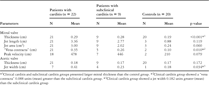
The leaflets of the mitral valves of patients with both clinical and subclinical carditis were thicker than those of the controls (p less than 0.001 – Table 1), with the values for the vena contracta across the mitral valve being statistically greater in those with clinical carditis when compared to those with subclinical carditis (p equal to 0.01 – Table 1).
Table 1. Quantitative parameters evaluated by cross-sectional Doppler echocardiography in 22 patients with clinical carditis, 9 with subclinical carditis, and 20 controls.

For additional quantitative variables, such as the length and area of the jet, and peak systolic velocity, we failed to find significant differences between those having clinical and subclinical carditis.
Aortic valve.
We found no differences in the thickness of the leaflets of the aortic valve between those having clinical and subclinical carditis as compared to their controls (p equal to 0.172). The mean values for the width of the aortic jet, however, were statistically greater in the patients with clinical carditis (p equal to 0.034 – Table 1).
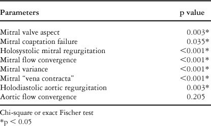
For qualitative analysis, we initially compared all distributions of different echocardiographic parameters for all three groups by the Chi-square or exact Fischer's tests. In all qualitative parameters, with the exception of convergence of aortic flow, we found statistical differences between the three groups (Table 2).
Table 2. Comparison of the three groups concerning qualitative parameters evaluated by cross-sectional Doppler echocardiography.

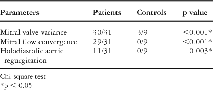
So as to compare those with clinical and subclinical carditis, we used a significance level α equal to 0.30 in order to reduce type II error. All patients with clinical and subclinical carditis presented mitral variance. In this way, we found statistically significant values in all variables except mitral variance, convergence of mitral flow, and aortic regurgitation, which presented p values of greater than 0.30. In other words, the valves of the patients with clinical and subclinical carditis were similar in terms of these last three variables, allowing them to be analyzed together. In this way, we observed a greater frequency of mitral variance (p less than 0.001), convergence of flow (p less than 0.001), and aortic regurgitation (p equal to 0.003), in those with rheumatic fever compared to their controls (Table 3).
Table 3. Presence of mitral variance, mitral flow convergence and holodiastolic aortic regurgitation in patients with rheumatic fever and their controls.

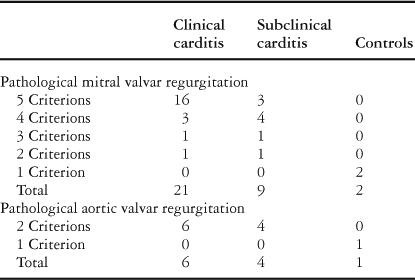
We show in Table 4 the frequency of distribution of fulfilled criterions, both quantitative and qual-itative parameters, for assessment of both valves using cross-sectional Doppler echocardiography in all groups. Both groups of patients, that is those with clinical and subclinical carditis, had a larger number of fulfilled criterions for mitral and aortic regurgitation than did the controls.
Table 4. Distribution of fulfilled quantitative and qualitative parameters for mitral and aortic assessment by cross-sectional Doppler echocardiography in patients with clinical carditis, subclinical carditis, and their controls.

Discussion
Quantification of mitral regurgitation remains a controversial topic. This is probably one of the reasons why the question of reliability has been raised regarding the capacity of cross-sectional Doppler echocardiography to distinguish accurately between pathological and physiological regurgitation.
Based on an analysis of a special set of criterions comprising both quantitative and qualitative parameters of regurgitant flow, Minich et al.6 observed a high specificity, and a positive predictive value, for these values when comparing patients with rheumatic fever and healthy individuals with physiological mitral regurgitation. According to them, such methodologies can accurately differentiate pathological from physiological regurgitation. In our analysis, we added still further modifications in order to achieve greater specificity.
We found significantly increased thickness of the leaflets of the mitral valve in patients with clinical and subclinical carditis when compared to their controls. Although no cut-off point has previously been described for this parameter, the thickness of the leaflets of the mitral valve is surely an important and specific finding, since it represents the structural abnormalities which characterize rheumatic carditis. Also, the fact that no statistical difference was observed in the thickness of the leaflets for the patients with either clinical or subclinical carditis when compared to their controls reinforces the already established concept that those with subclinical carditis belong within the spectrum of clinical carditis. The same could not be concluded for thickness of the leaflets of the aortic valve, due to the small number of patients with such findings.
Regarding additional parameters, such as the length and area of the mitral jet, and peak systolic velocity, we failed to observe differences between those with clinical and subclinical carditis, reinforcing again the likeness between the two conditions, despite higher absolute measures being found in those with clinical carditis. This might explain why a murmur is heard in patients with clinical carditis.
The so-called vena contracta of the regurgitant mitral valve, and the width of the aortic jet, are important parameters to quantify valvar regurgitation.8 These variables presented statistically higher values in our patients with clinical carditis as compared to those with subclinical carditis. We are not aware of other studies comparing these parameters in these groups, and it is possible that these findings are associated with the magnitude of the valvar lesion. We could not use the control group for comparison of these findings, since our normal subjects showed no evidence of regurgitation, not even minimal.
For the qualitative parameters, although the control group presented a reduced number of subjects with some abnormalities at cross-sectional Doppler interrogation, we opted first to compare all three groups in an attempt to identify parameters that could reinforce similarity between the groups with rheumatic fever and eventual differences with the control group.
Thus, we observed that the parameters for mitral variance, mitral flow convergence and aortic regurgitation were those that showed similarity between the patients with clinical and subclinical carditis. When grouped together, the patients then showed statistically higher frequency of these parameters than did their controls. The patients with clinical and subclinical carditis having mitral regurgitation could not be regrouped for comparison with their controls, since the latter had no such regurgitation. We should point out, nonetheless, the higher frequency of holosystolic mitral regurgitation and its specific characteristics in terms of convergence of flow, the vena contracta, and variance in the group with subclinical carditis as compared to the controls. The other qualitative variables did not show any difference among the three groups.
The criterions we have used to define pathological mitral regurgitation are slightly different from those used previously.6 It was also necessary for at least two of these criterions to be present in order for the regurgitant jet to be considered pathological. We think that these changes in the criterions may minimize the possibility of considering physiological regurgitation as pathological.
We treat carditis with prednisone given in doses from 1 to 2 milligram per kilogram per day divided into two doses in the first week and given as a single dose thereafter, tapering the dose over a period of 8 to 12 weeks. In the case of patients having carditis without sequels, we recommended intramuscular benzathine penicillin G every 3 weeks up to 25 years of age. In the other cases prophylaxis should be maintained indefinitely.
Use of steroids for subclinical carditis has not been well established, neither have standards been accepted for extended secondary prophylaxis in such cases. Some believe that the therapeutic approach to these patients should address the prevalence of acute rheumatic fever in one specific region.9 Little is known about the real impact of steroids in the course of silent chronic rheumatic disease, as well as their implications in avoiding progression of the disease. There are few studies characterizing the history of a prior episode of rheumatic carditis that required valvar surgery.10 Augestad et al.10 analyzed 296 patients undergoing mitral comissurotomy, and noted that more than half of the patients did not know they had had rheumatic fever, becoming aware of their rheumatic carditis only after mitral stenosis became evident. Chopra et al.,11 examining fresh left atrial appendages from 50 patients with the presumed diagnosis of chronic rheumatic disease, found Aschoff nodules and vast mononuclear infiltrates after mitral valvectomy, strongly suggesting the possibility of a silent course of subclinical carditis. According to Ozkutlu et al.,12 subclinical carditis should be considered as equivalent to a diagnosis of carditis. They observed that patients with subclinical carditis that received steroids showed statistically faster resolution of their valvar lesions when compared to those not receiving steroids. The diagnosis of acute rheumatic fever can be established by the presence of carditis or chorea by itself, dispensing the need for further Jones criterions. We should emphasize, nonetheless, that since cross- sectional Doppler echocardiography does not provide evidence of the aetiology of carditis, echocardiographic findings of subclinical carditis need to be considered in the context of additional signs and symptoms of acute rheumatic fever.
Thus, we conclude that the quantitative and qualitative parameters evaluated by cross-sectional Doppler echocardiography are able accurately to identify pathological mitral and aortic valvar regurgitation that was not evident on clinical auscultation, making this a specific and sensible approach for detecting subclinical carditis. Prospective studies evaluating and comparing patients with clinical and subclinical carditis to those with physiological mitral and/or aortic regurgitation, will be necessary in order to establish the hierarchy of the results of the technique as a diagnostic criterion for acute rheumatic fever.
Acknowledgement
Sources of funding: FAPESP – Fundação de Amparo à Pesquisa do Estado de São Paulo.