Introduction
A burgeoning, yet heterogeneous area of inquiry in major depressive disorder (MDD) relates to processing of emotions in faces. Individuals with MDD tend to exhibit decreased ability to recognize facial emotions (Persad & Polivy, Reference Persad and Polivy1993; Langenecker et al. Reference Langenecker, Bieliauskas, Rapport, Zubieta, Wilde and Berent2005, Reference Langenecker, Caveney, Giordani, Young, Nielson, Rapport, Biellauskas, Mordhorst, Marcus, Yodkovik, Kerber, Berent and Zubieta2007a; Csukly et al. Reference Csukly, Czobor, Szily, Takacs and Simon2009) and a negative response bias (Watkins et al. Reference Watkins, Vache, Verney, Muller and Mathews1996; Bouhuys et al. Reference Bouhuys, Geerts and Gordijn1999; Elliott et al. Reference Elliott, Rubinsztein, Sahakian and Dolan2002; Joormann & Gotlib, Reference Joormann and Gotlib2006; Wright et al. Reference Wright, Langenecker, Deldin, Rapport, Nielson, Kade, Own, Akil, Young and Zubieta2009) compared with healthy controls (HCs). These deficits may be greatest among those with the most severe depressive symptomatology (Gur et al. Reference Gur, Erwin, Gur, Zwil, Heimberg and Kraemer1992), yet also may represent a trait risk for MDD observed outside of symptomatic episodes (LeMoult et al. Reference LeMoult, Joormann, Sherdell, Wright and Gotlib2009). Using a challenging facial emotion perception (FEP) paradigm, our group observed that young women with MDD are less accurate at detecting emotions, particularly fear, than both HC women and depressed men, whereas young depressed men performed similarly to control men (Wright & Langenecker, Reference Wright, Langenecker, Hernandez and Alonso2008; Wright et al. Reference Wright, Langenecker, Deldin, Rapport, Nielson, Kade, Own, Akil, Young and Zubieta2009). In a recent meta-analysis, subjects with MDD performed worse than controls in all emotions, trending toward worse skills in men with MDD (and bipolar disorder, see Kohler et al. Reference Kohler, Hoffman, Eastman, Healey and Moberg2011).
There are also reports of functional magnetic resonance imaging (fMRI) activation abnormalities in persons diagnosed with MDD during viewing of emotional faces and related emotional stimuli (Sheline et al. Reference Sheline, Barch, Donnelly, Ollinger, Snyder and Mintun2001; Fu et al. Reference Fu, Williams, Cleare, Brammer, Walsh, Kim, Andrew, Pich, Williams, Reed, Mitterschiffthaler, Suckling and Bullmore2004; Frodl et al. Reference Frodl, Scheuerecker, Albrecht, Kleemann, Muller-Schunk, Koutsouleris, Moller, Bruckmann, Wiesmann and Meisenzahl2009). Neuroimaging studies in adults with MDD typically report heightened limbic (Sheline et al. Reference Sheline, Barch, Donnelly, Ollinger, Snyder and Mintun2001; Surguladze et al. Reference Surguladze, Brammer, Keedwell, Giampietro, Young, Travis, Williams and Phillips2005) and basal ganglia responses (Fu et al. Reference Fu, Williams, Cleare, Brammer, Walsh, Kim, Andrew, Pich, Williams, Reed, Mitterschiffthaler, Suckling and Bullmore2004; Surguladze et al. Reference Surguladze, Brammer, Keedwell, Giampietro, Young, Travis, Williams and Phillips2005) with mixed activation differences in frontal regions (Lawrence et al. Reference Lawrence, Williams, Surguladze, Giampietro, Brammer, Andrew, Frangou, Ecker and Phillips2004; Lee et al. Reference Lee, Seok, Lee, Cho, Yoon, Lee, Chae, Choi and Ham2008; Demenescu et al. Reference Demenescu, Renken, Kortekaas, van Tol, Marsman, van Buchem, van der Wee, Veltman, den Boer and Aleman2011). In MDD, there is some indication of a shift in laterality of frontal activation for emotional stimuli, from a left-greater-than-right pattern observed in healthy adults to right-greater-than-left or greater bilateral frontal activation. Specifically, in healthy individuals, left laterality in emotion labeling/identification has been associated with inferior and medial frontal activation in meta-analysis (Fusar-Poli et al. Reference Fusar-Poli, Placentino, Carletti, Landi, Allen, Surguladze, Benedetti, Abbamonte, Gasparotti, Barale, Perez, McGuire and Politi2009), although others have reported greater activation in the left middle frontal gyrus associated with the regulation of emotion (Mak et al. Reference Mak, Hu, Zhang, Xiao and Lee2009). A few limited studies with electroencephalography (EEG) and fMRI suggest that a right-greater-than-left or more bilateral activation pattern shift is present in MDD, but this finding has not been frequently tested (Henriques & Davidson, Reference Henriques and Davidson1991; Grimm et al. Reference Grimm, Beck, Schuepbach, Hell, Boesiger, Bermpohl, Niehaus, Boeker and Northoff2008; Langenecker et al. Reference Langenecker, Lee, Bieliauskas, Grant and Adams2009) nor is it clear whether this shift might be related to FEP accuracy.
The large majority of neuroimaging studies investigating FEP in MDD have utilized experimental tasks with oblique emotion-processing paradigms, such as passive viewing of stimuli (Lee et al. Reference Lee, Seok, Lee, Cho, Yoon, Lee, Chae, Choi and Ham2008), presenting masked faces theoretically outside of conscious awareness (Dannlowski et al. Reference Dannlowski, Ohrmann, Bauer, Kugel, Arolt, Heindel and Kersting2007, Reference Dannlowski, Ohrmann, Bauer, Deckert, Hohoff, Kugel, Arolt, Heindel, Kersting, Baune and Suslow2008), or gender/age discrimination tasks (Canli et al. Reference Canli, Cooney, Goldin, Shah, Sivers, Thomason, Whitfield-Gabrieli, Gabrieli and Gotlib2005; Costafreda et al. Reference Costafreda, Khanna, Mourao-Miranda and Fu2009; Demenescu et al. Reference Demenescu, Renken, Kortekaas, van Tol, Marsman, van Buchem, van der Wee, Veltman, den Boer and Aleman2011; Thomas et al. Reference Thomas, Elliott, McKie, Arnone, Downey, Juhasz, Deakin and Anderson2011). One limitation of oblique paradigms is that implicit emotion processing may still occur and vary across individuals, which limits the performance differences that might be observed as compared with explicit paradigms. A few studies have utilized tasks requiring explicit emotion judgments, although these tasks have required binary emotional classification decisions about facial expressions (e.g. emotional versus neutral; Almeida et al. Reference Almeida, Versace, Hassel, Kupfer and Phillips2010), or matching emotions presented in different faces (Frodl et al. Reference Frodl, Scheuerecker, Albrecht, Kleemann, Muller-Schunk, Koutsouleris, Moller, Bruckmann, Wiesmann and Meisenzahl2009). Often, explicit studies have not capitalized upon challenging depressed subjects to the point of dysfunctional performance, which then precludes the analysis of the functional correlates of poor performance. As a result, although oblique or simple explicit matching paradigms are an excellent way to understand perturbations of emotion-processing circuitry, they do not provide an exportable behavioral paradigm for ecologically valid translation to clinical settings. Moreover, FEP paradigms with low levels of challenge have precluded the direct investigation of the role that FEP deficits play in the neural responses to facial emotions and in risk for and expression of disease. In contrast, simple explicit and oblique paradigms do not have a potential for disease by performance confounds, which difficult explicit paradigms must address. It is important to evaluate the strengths and weaknesses of the different approaches, and to consider whether the goal is to focus more on internal or external validity.
The current study addressed some of the limitations of the knowledge base by investigating disrupted explicit FEP in MDD. First, to address the relationship between performance and activation directly, the experimental paradigm involved an explicit emotion classification task previously demonstrated to elicit performance deficits in individuals with MDD (Langenecker et al. Reference Langenecker, Bieliauskas, Rapport, Zubieta, Wilde and Berent2005, Reference Langenecker, Caveney, Giordani, Young, Nielson, Rapport, Biellauskas, Mordhorst, Marcus, Yodkovik, Kerber, Berent and Zubieta2007a). Second, in light of our recent findings suggesting female-specific vulnerability to FEP decrements in MDD (Wright et al. Reference Wright, Langenecker, Deldin, Rapport, Nielson, Kade, Own, Akil, Young and Zubieta2009), the current sample was comprised exclusively of women. It was hypothesized that depressed women would exhibit hyperactivity in frontal, basal ganglia and limbic regions (Sheline et al. Reference Sheline, Barch, Donnelly, Ollinger, Snyder and Mintun2001; Fu et al. Reference Fu, Williams, Cleare, Brammer, Walsh, Kim, Andrew, Pich, Williams, Reed, Mitterschiffthaler, Suckling and Bullmore2004; Lawrence et al. Reference Lawrence, Williams, Surguladze, Giampietro, Brammer, Andrew, Frangou, Ecker and Phillips2004; Surguladze et al. Reference Surguladze, Brammer, Keedwell, Giampietro, Young, Travis, Williams and Phillips2005; Lee et al. Reference Lee, Seok, Lee, Cho, Yoon, Lee, Chae, Choi and Ham2008). In addition, it was expected that these areas of disrupted activation in MDD would be related to FEP performance, irrespective of the effects of medications. Finally, we hypothesized shifted laterality of activation in MDD (right greater than left), and we explored the relationship of laterality to FEP performance.
Method
Participants
A total of 24 women with MDD and 22 female HCs participated. The groups did not differ with regard to age (MDD: mean = 37.8, s.d. = 14.5 years; HCs: mean = 31.7, s.d. = 14.4 years; t 44 = − 1.42, p = 0.16), education (MDD: mean = 16.3, s.d. = 2.3 years; HCs: mean = 16.2, s.d. = 2.4; t 44 = − 0.09, p = 0.93), or Shipley intelligence quotient (MDD: mean = 113.1, s.d. = 9.3; HCs: mean = 115.5, s.d. = 7.1; t 42 = 0.954, p = 0.35). Informed consent procedures were completed according to the University of Michigan Institutional Review Board guidelines and consistent with the Declaration of Helsinki. Exclusion criteria for participants included: a diagnosis of schizophrenia, bipolar disorder, brain injury, neurological condition, substance abuse in the last 2 years or other such condition that would affect cognitive functioning. Non-depressed HC participants had no personal history of any psychiatric illness.
MDD diagnosis was from Structured Clinical Interview for DSM-IV (First et al. Reference First, Spitzer, Gibbon and Williams1996) criteria by a licensed psychologist with mean age of onset of 24.4 (sd = 13.7) years. Severity of depressive symptomatology was evaluated with the 17-item Hamilton Depression Rating Scale (HAMD; Hamilton, Reference Hamilton1960; MDD: mean = 15.8, s.d. = 7.2) and Beck Depression Inventory-II (mean = 21.2, s.d. = 10.3). Of the MDD participants, 10 were unmedicated. Of the medicated group, 11 were taking antidepressants only (e.g. selective serotonin reuptake inhibitors, monoamine oxidase inhibitors, tricyclic antidepressants, and buproprion), and three were taking antidepressants plus/or medications with reported cognitive side effects (one prescribed Seroquel, Neurontin and Lamictal, one prescribed Ativan, Cymbalta and Trazodone, and one prescribed Xanax PRN, Ambien, Celexa and Wellbutrin). The study was a cross-sectional, naturalistic design; therefore, it was not possible to control for dose, severity or effects of expectation (placebo) on medicated volunteers.
Affect classification paradigm
The Facial Emotion Perception Test (FEPT; Rapport et al. Reference Rapport, Friedman, Tzelepis and Van Voorhis2002; Langenecker et al. Reference Langenecker, Bieliauskas, Rapport, Zubieta, Wilde and Berent2005, Reference Langenecker, Caveney, Giordani, Young, Nielson, Rapport, Biellauskas, Mordhorst, Marcus, Yodkovik, Kerber, Berent and Zubieta2007a) was used to assess the accuracy and speed of identification of facial expressions. There are parallel computer (facial stimuli from Ekman & Friesen, Reference Ekman and Friesen1976) and imaging (MacBrain, http://www.macbrain.org/; color photographs of faces from the NimStim stimuli; Tottenham et al. Reference Tottenham, Tanaka, Leon, McCarry, Nurse, Hare, Marcus, Westerlund, Casey and Nelson2009) versions. Participants were required to categorize faces into one of four emotion categories (happy, sad, angry or fearful). Each presentation (total of 21 blocks, 147 stimuli) began with an orienting cross in the center of the screen (500 ms), followed by a facial emotion stimulus (300 ms), then a visual mask to prevent visual afterburn (100 ms), followed by a response period (2600 ms). As a control task, blocks requiring participants to identify and categorize pictured animals (eight blocks, 56 total stimuli) were interspersed amongst facial emotion recognition blocks. These animal blocks were used as a method of controlling for activation related to visual processing and praxis, and for response selection and execution. The task consisted of five 3.5-min runs with counterbalanced animal and face blocks, each run ending with a rest block. Each presentation in the imaging experiment was a novel presentation of an emotional stimulus for an actor/actress, although individual actors/actresses did repeat.
Prior to entering the fMRI scanner, all participants completed a computer-only version of the FEPT. The computer test utilized the Ekman faces (Ekman & Friesen, Reference Ekman and Friesen1976) and was presented on a standard computer monitor. The practice version was one 6-min run and consisted of 12 animal trials and 54 face trials and had parameters for presentation and response times identical to the fMRI version.
Performance on the FEPT task was captured by accuracy and reaction time for correct responses for each emotional expression, in addition to animal classification.
Scanning procedures
MRI acquisition
Whole brain imaging was performed using a GE Signa 3-T scanner (release VH3) (General Electric, USA). fMRI series consisted of 30 contiguous oblique-axial 4 mm sections acquired using a forward–reverse spiral sequence, which provides excellent fMRI sensitivity. The image matrix was 64 × 64 over a 24 cm field of view for a 3.75 × 3.75 × 4 mm voxel. The 30-slice volume was acquired serially at 1750 ms temporal resolution for a total of 590 time points for FEPT. A total of 106 high-resolution fast SPGR IR axial anatomic images [echo time = 3.4 ms, repetition time = 10.5 ms, 27° flip angle, number of excitations (NEX) = 1, slice thickness = 1.5 mm, field of view = 24 cm, matrix size = 256 × 256] were obtained for each participant for co-registration purposes.
MRI processing
Pre-processing of all fMRI data was conducted in SPM2 (http://www.fil.ion.ucl.ac.uk/spm), similar to our previously reported work (Langenecker et al. Reference Langenecker, Kennedy, Guidotti, Briceno, Own, Hooven, Young, Akil, Noll and Zubieta2007b, Reference Langenecker, Weisenbach, Giordani, Briceno, Guidotti-Breting, Schallmo, Leon, Noll, Zubieta, Schteingart and Starkman2012). This process included realignment, slice timing correction, co-registration of anatomical and functional images, normalization to Montreal Neurological Institute space, and smoothing with a full width at half maximum filter of 5 mm. Contrast images were created using the subtraction method, such that the blood oxygen level-dependent (BOLD) signal for animal processing blocks was subtracted from that of faces processing blocks, in order to examine the BOLD response for the stimuli specifically related to facial emotion processing (faces minus animals for all imaging contrasts). First-level individual and second-level group models were completed in SPM5. Coordinates for activation foci were transformed to the Talairach system.
Statistical analyses
Prior to analyses, data were screened for assumptions associated with the statistical models employed according to guidelines recommended by Tabachnick & Fidell (Reference Tabachnick and Fidell2007). Analyses of the neuroimaging data began with one-sample t tests of the ‘faces minus animals’ contrast for the HC and MDD groups individually. Next, to examine whether this sample resembled those observed in prior research (i.e. a pattern of poorer performance among women with MDD), FEPT performance within the scanner was compared between MDD and HC groups using one-tailed t tests and effect sizes in Cohen's d. Analysis of covariance (ANCOVA) was then performed with group as the independent variable, and overall faces accuracy and age as covariates, as prior research has shown that these are pertinent features for which to control in understanding group differences (Gunning-Dixon et al. Reference Gunning-Dixon, Gur, Perkins, Schroeder, Turner, Turetsky, Chan, Loughead, Alsop, Maldjian and Gur2003; Wright et al. Reference Wright, Langenecker, Deldin, Rapport, Nielson, Kade, Own, Akil, Young and Zubieta2009). Age was moderately correlated with accuracy (r = − 0.45, p = 0.002). Finally, to evaluate the role of performance in task activation, a separate regression model with only MDD subjects was conducted, with task activation as the criterion and overall faces accuracy as the predictor. The index analysis between the MDD and HC groups was false discovery rate (FDR) corrected at p < 0.05 at whole brain resolution and extent threshold of 160 mm3, and that threshold was used for all analyses. For the animals-only contrast, HC and MDD groups differed only in lateral, posterior globus pallidus bilaterally (MDD > HC, x = +20/ − 20, y = −19, z = −8/ − 2, Z′s 3.79/3.82), indicating that any between-group differences in activation for the ‘faces minus animals’ contrast outside these small regions are more likely to be related to social/emotional facial processing involved in the faces condition.
Results
Individual group tests for facial emotion processing
Individual group maps for the ‘faces minus animals’ contrast for the MDD group alone and the HC group alone are illustrated in Fig. 1 (a, b) (MDD participants in blue, HCs in green). The areas of overlapping activation for both groups (purple) included the bilateral inferior frontal gyrus (IFG) and middle frontal gyrus, insula, inferior parietal lobule, precuneus, posterior putamen and the medial dorsal nucleus of the thalamus. The HC group exhibited significant activation that was not present in the MDD group in primarily posterior subcortical areas, such as the bilateral parahippocampal gyrus, right caudate, right pulvinar and left pons/raphe. The MDD group exhibited activation that was not present in the HC group in the right precentral and postcentral gyrus, left anterior cingulate, as well as the bilateral superior temporal gyrus, left parahippocampal gyrus/amygdala, substantia nigra and left cerebellum. Independent activation foci for HC and MDD groups are included in Supplementary Tables S1 and S2 for the interested reader.
Lateral activation in the healthy control (HC) and major depressive disorder (MDD) groups, including evidence for laterality shifts by extent and degree of activation. (a, b) Significant areas of activation in the MDD and HC groups alone are shown in blue and green, respectively. Areas of overlapping activation are displayed in purple. Values are x axis coordinates in the Talairach system. (c) Increased right signal in MDD subjects relative to the left in the ventral and dorsal inferior frontal gyrus (IFG) clusters (* p < 0.05), with no significant difference between left and right signals in HCs. (d) Percentage of significant suprathreshold voxels in each group including the ventro-lateral prefrontal cortex (VLPFC), corresponding to Brodmann areas 9, 10, 11, 44, 45, 46 and 47.

Laterality tests by group – height of activation
Fig. 1 (a, b) illustrates the evidence of left frontal laterality in the HC group, and an absence of this effect in MDD participants. Thus, two sets of post hoc analyses tested whether the reduced left laterality of activation in the MDD patients was significant and meaningful in magnitude. First, the HC left inferior frontal cluster shown in Fig. 1 was used as a center of mass for a 5 mm region of interest (ROI) from which to extract a mean BOLD signal for each subject using MarsBaR (Brett et al. Reference Brett, Anton, Valabreque and Poline2002). This cluster in the ventral IFG (VIFG: −45, 30, −1) is similar in coordinates to activation foci in healthy subjects reported for angry and fearful facial expressions in a meta-analysis of facial emotion processing by Fusar-Poli et al. (Reference Fusar-Poli, Placentino, Carletti, Landi, Allen, Surguladze, Benedetti, Abbamonte, Gasparotti, Barale, Perez, McGuire and Politi2009) (−46, 33, 4 for fear, and −44, 26, 2 for anger). A right-hemisphere homologue cluster was created by inverting the x coordinate (x = 45). In addition, a right-hemisphere cluster in the dorsal aspect of the IFG (DIFG) of significant activation in the MDD subjects (DIFG: 40, 26, 17) that was not observed in the HC group was used to create a similar sphere with a 5 mm radius ROI and matching bilateral homologue. Mean BOLD activation was extracted for each subject for these two ROIs and contralateral homologue ROIs and then subjected to a 2 (group) × 2 (hemisphere) × 2 [location (dorsal, ventral)] analysis of variance. In the IFG, there was an interaction between group and hemisphere (F 1,44 = 5.11, p = 0.03), with greater right than left activation in the MDD group (t 23 = − 2.12, p = 0.045, d = − 0.38), and non-significantly greater left than right IFG activation in the HC group (p = 0.30). There were no other significant effects or interactions (displayed in Fig. 1 c, labeled left and right IFG). The mean extracted BOLD signals from these four MarsBaR ROIs were used to compute a laterality index (R > L) using the formula (RVIFG + RDIFG)/(LVIFG + LDIFG) for each subject and this index was used in subsequent post hoc analyses (see Matsuo et al. Reference Matsuo, Chen and Tseng2012, for a description of some approaches to laterality indexing).
Laterality tests by group – extent of activation
As a second, novel post hoc strategy for evaluating shifted inferior frontal laterality of MDD, χ2 analysis tested the number of significant voxels activated in the MDD group relative to the HC group, primarily within the IFG and middle frontal gyrus [Brodmann areas 9, 10, 11, 44, 45, 46, 47 using WFU Pickatlas (Maldjian et al. Reference Maldjian, Laurienti, Kraft and Burdette2003); dilation 2 mm]. The HC group exhibited a strong left laterality for activation during identification of emotions in faces, with a ratio of 2.8:1 (left to right) activated clusters. In contrast, the MDD group exhibited two effects of interest. There were substantially more activated frontal voxels in both the left ventro-lateral prefrontal cortex (VLPFC) [χ2(1, n = 46) = 2238.45, p < 0.0001] and the right VLPFC [χ2(1, n = 46) = 13440.01, p < 0.0001] for the MDD group as compared with the HC group. More specifically, the pattern of left-specific frontal activation in the HC group for this contrast was absent in the MDD subjects. In the MDD group, the ratio was 0.93:1 (left to right) activated clusters, revealing an inverted frontal laterality pattern in the MDD group for identification of emotions in faces. The percentage of significant voxels activated for each group in the entire middle and inferior frontal Brodmann area masks are presented in Fig. 1 d (labeled left and right PFC).
Between-group differences in fMRI activation for facial emotion processing
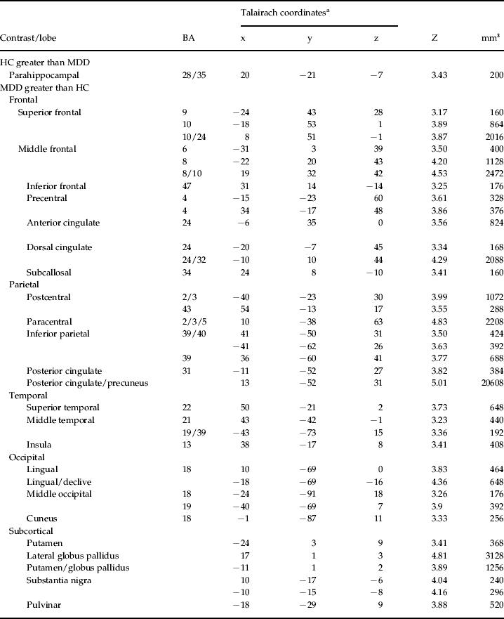
Group comparisons were conducted using ANCOVA in SPM5. Results are reported in Table 1 and displayed in Fig. 2 (a–d), with HC > MDD in yellow and MDD > HC in cyan (p < 0.05, FDR, whole brain-corrected). The HC group exhibited greater activation relative to the MDD group in a right parahippocampal cluster in the ‘faces minus animals’ contrast. In contrast, the MDD group had greater activation relative to the HC group in a large number of cortical and subcortical areas bilaterally, including the bilateral superior frontal, middle frontal and precentral gyri, anterior, dorsal, and posterior cingulate, lingual gyrus, inferior parietal lobule, superior and middle temporal gyrus, middle occipital gyrus, cuneus, putamen, pulvinar and substantia nigra. To investigate group effects within the amygdala (Sheline et al. Reference Sheline, Barch, Donnelly, Ollinger, Snyder and Mintun2001; Dannlowski et al. Reference Dannlowski, Ohrmann, Bauer, Kugel, Arolt, Heindel and Kersting2007), a MarsBaR Pickatlas ROI was created, with a dilation of 1.5 and a threshold of p < 0.05, uncorrected. The MDD group exhibited greater bilateral amygdala activation than HCs (left: t = 3.41, p = 0.0007, k = 29; right: t = 2.15, p = 0.019, k = 10).
Greater activation in patients with major depressive disorder (MDD) for facial emotion identification compared with healthy control (HC) subjects. The figure illustrates areas of significantly greater activation in HC subjects relative to MDD subjects in yellow. Clusters with greater activation in MDD relative to HC subjects are shown in cyan. Values are z axis coordinates in the Talairach system.
Differences between HC and MDD groups in facial emotion processing
HC, Healthy control; MDD, major depressive disorder; BA, Brodmann area.
a x, y, z = Talairach coordinates of significant effects.
Explicit emotion identification in MDD participants and evaluation of medication effects
Prior to investigating relationships between performance and activation, one-tailed t tests and effect sizes in Cohen's d were used to examine the groups on FEPT performance. For overall accuracy, MDD participants performed more poorly than did HCs (t 44 = 1.86, p = 0.04, d = 0.75). Of note, although power for this comparison is low due to sample size, the pattern of effect sizes observed closely parallels prior studies that compared FEP in depressed and non-depressed adults (Langenecker et al. Reference Langenecker, Caveney, Giordani, Young, Nielson, Rapport, Biellauskas, Mordhorst, Marcus, Yodkovik, Kerber, Berent and Zubieta2007). Moderate effect sizes indicating lower accuracy in MDD patients versus HCs were observed for identifying happy (t 44 = 2.07, p = 0.02, d = 0.90) and fearful faces (t 44 = 1.57, p = 0.06, d = 0.55). Performance did not differ between the medicated and unmedicated MDD groups on any FEPT variables. Supplementary Table S3 displays accuracy and reaction time data and statistical results for within-scanner performance. Thus, performance–activation analyses could proceed on the supported premise that this sample performed similarly to those in prior studies.
Correlates of intact and disrupted emotion perception performance in MDD participants
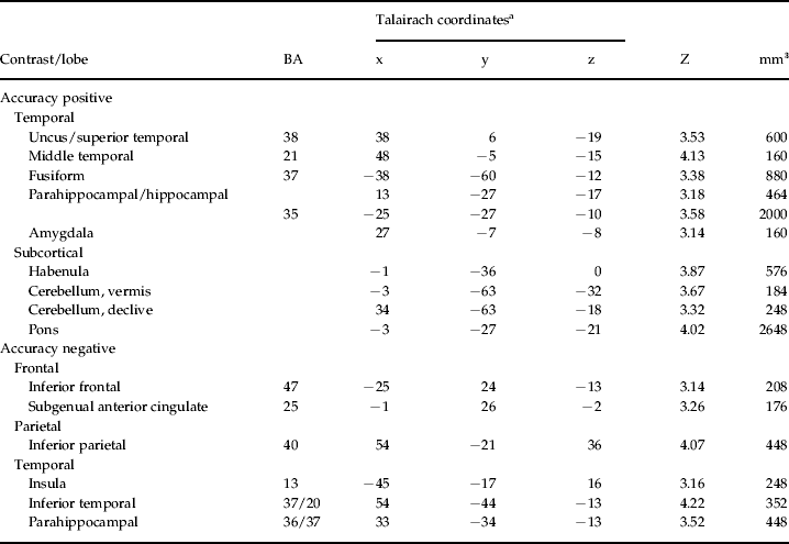
To investigate the role of performance on activation within the MDD group, a regression analysis was conducted with overall faces accuracy regressed upon FEP activation in the MDD group only. Results from this analysis are reported in Table 2, with some foci illustrated in Fig. 3. A number of foci were positively related to task performance, including the right amygdala, bilateral hippocampal and parahippocampal foci, and the left fusiform gyrus, in addition to the cerebellum and habenula. In contrast, regions inversely associated with accuracy included the left IFG and insula, right inferior parietal and parahippocampal foci, plus the bilateral subgenual anterior cingulate gyrus (sgAC).
(a, b, c) Regions of positive and negative association in activation for major depressive disorder (MDD) subjects during facial emotion identification. The figure illustrates areas of activation that are positively associated with facial emotion accuracy classification in green, as well as areas of activation negatively correlated with activation in red. Values are z axis coordinates in the Talairach system. (d) Activation for the subgenual anterior cingulate is displayed in the scatterplot relative to performance accuracy in MDD in red. Activation for healthy controls (HCs) was extracted for comparative purposes and is displayed in white.
Regions positively and negatively associated with facial emotion identification accuracy within the MDD group
MDD, Major depressive disorder; BA, Brodmann area.
a x, y, z = Talairach coordinates of significant effects.
Relationship of shifted laterality extent of activation with emotion perception performance
To investigate the relationship between laterality and FEPT performance, a right:left ratio was created from the MarsBaR ROIs extracted in the ‘height’ of activation, post hoc analysis (Fig. 1). The sum of the two right hemisphere IFG clusters was divided by the sum of the two left hemisphere IFG clusters. Correlations were conducted examining the relationship of ratio scores with task performance. Right-to-left ratio scores were positively associated with accuracy in the MDD group and showed a substantial effect (r = 0.56, p = 0.004, Fisher's exact Z = 0.63), whereas this relationship showed a small and non-significant effect size in the HC group (r = 0.21, p = 0.34, Fisher's exact Z = 0.21). The HC group showed a trend inverse relationship between performance and right IFG activation (r = − 0.41, p = 0.06), but not in left IFG activation (r = − 0.08, p = 0.74). In the MDD group, an opposite pattern was observed: a positive relationship between performance and left IFG activation (r = 0.31, p = 0.14) and the same direction but not as strong for right IFG activation (r = 0.15, p = 0.47; Supplementary Fig. S1). The Fisher's test of significance for one-tailed comparisons of correlations between MDD patients and HCs for comparisons of the correlations of left IFG, right IFG, and right-to-left ratio scores exceeded the p < 0.05 threshold difference score of 0.263 in all three sets of correlations. These effects were similar when computing a partial correlation correcting for depression severity (HAMD), including the correlation of right-to-left IFG ratio score (r = 0.57, p = 0.005) and sgAC activation (r = − 0.41, p = 0.05) with FEPT accuracy.
This laterality–performance relationship was explored further to determine whether it was primarily driven by activation or deactivation on the right or left in the MDD group. Among the 19 MDD participants who showed increased right hemisphere activation (i.e. right + /left+ or right + /left–), there was a strong relationship between the laterality ratio and FEPT performance (r 2 = 0.39, p = 0.004); in contrast, among participants who did not show right hemisphere activation, the laterality ratio was unrelated to FEPT performance (r 2 = 0.08, p = 0.65). Taken together, these results indicate that less activation of left relative to right (i.e. left < right IFG, or deactivation of the left IFG combined with right IFG activation) is associated with poor performance in women with MDD.
Evaluation of possible medication effects related to hyperactivation within the MDD group
To determine whether aberrant activation in the MDD group was associated with psychotropic medication status, extracted (MarsBaR) activation values from the ‘faces minus animals’ contrast in which group differences were observed (see Table 1) were evaluated. There were no significant effects of medication status using an uncorrected threshold of p < 0.05.
Discussion
During facial emotion processing, women with MDD showed hyperactivation in a large bilateral network of cortical and subcortical regions. The hyperactivity observed in women with MDD was characterized by shifted inferior frontal laterality compared with HCs. Whereas non-depressed healthy women exhibited a bilateral frontal network in response to the challenge of identifying facial emotions, women with MDD exhibited right-greater-than-left hyperactivation in the IFG, combined with extensive bilateral IFG activation. Furthermore, right-greater-than-left IFG laterality was associated with preserved performance in women with MDD. In contrast, left-greater than-right IFG and increased subgenual cingulate activation were associated with poor performance in women with MDD. Overall, these findings help to illustrate neural correlates of the relative impairments in facial emotion identification in women with MDD, whereby some effects were associated with diagnosis and others directly linked to diminished performance.
This is the first fMRI study to demonstrate that women with MDD show a shifted frontal laterality of activation during classification of emotional faces relative to female HCs. To our knowledge, the only related study demonstrating shifted laterality in response to emotion processing in women with MDD was conducted with EEG (Henriques & Davidson, Reference Henriques and Davidson1991). The present findings support this prior work and further demonstrate that the laterality shift has two components, characterized by a greater magnitude of activation in the right compared with the left inferior gyrus, in addition to greater bilateral spatial extent of activation in response to the challenge of processing facial emotion.
Although the pattern of shifted laterality in women for FEP during fMRI has not previously been reported, there is some support for right hyperactivity in individuals with MDD during tasks involving viewing emotional words and pictures (Rotenberg, Reference Rotenberg2004; Langenecker et al. Reference Langenecker, Lee, Bieliauskas, Grant and Adams2009). Evidence for shifted laterality has also been demonstrated in other types of emotion-processing paradigms. For example, Johnstone et al. (Reference Johnstone, van Reekum, Urry, Kalin and Davidson2007) reported a bilateral pattern of IFG activation among MDD patients during an affect regulation task. IFG activation among HCs was left-lateralized; however, this effect was not quantified. Furthermore, Mathersul et al. (Reference Mathersul, Williams, Hopkinson and Kemp2008) reported left-lateralized frontal α-EEG activity in healthy young adults, accompanied by symmetrical frontal activity among adults with subclinical MDD. In contrast, two studies (Davidson et al. Reference Davidson, Irwin, Anderle and Kalin2003; Grimm et al. Reference Grimm, Beck, Schuepbach, Hell, Boesiger, Bermpohl, Niehaus, Boeker and Northoff2008) reported reduced left lateral prefrontal activation combined with equivalent right hemisphere activation among adults with MDD compared with HCs while viewing emotionally salient pictorial stimuli. These prior fMRI studies used generally qualitative interpretations of laterality shifts, unlike the quantitative approach utilized by EEG studies (Henriques & Davidson, Reference Henriques and Davidson1991; Mathersul et al. Reference Mathersul, Williams, Hopkinson and Kemp2008) and the present study. In a complementary study, Almeida et al. (Reference Almeida, Versace, Mechelli, Hassel, Quevedo, Kupfer and Phillips2009) demonstrated reduced top-down control in effective connectivity from the orbital medial frontal seed to the amygdala in MDD and bipolar disorder patients relative to HCs for happy stimuli, and at the trend level for sad stimuli. Deficient medial and orbital frontal ‘automatic’ top-down control of emotional responses has been carefully considered, although the relationship with lateral more ‘effortful’ control regions remains tenuous (Egner et al. Reference Egner, Etkin, Gale and Hirsch2008; Phillips et al. Reference Phillips, Ladouceur and Drevets2008; Price & Drevets, Reference Price and Drevets2012).
In women with MDD, poor emotion identification performance was associated with lower left relative to right IFG activation, suggesting a link between shifted laterality and emotion-processing skill. Reduced activation of the left, but not right IFG, was associated with poor performance. Intriguingly, a different pattern of results was observed in non-depressed women, such that those with greater right IFG activation performed worse on the task, and no relationship was observed between performance and the left IFG or right:left IFG ratio. A speculative interpretation of these findings is that in a subset of women with MDD, the left IFG may not effectively engage during emotion processing, and successful recruitment of the right IFG in this context may serve a compensatory function. In non-depressed women, the meaning of increased right IFG activation is unclear. It might reflect an effort to compensate for less efficient emotion-processing skill, to regulate emotional response to stimuli that interfere with performance, or reflect unexpressed illness in the presence of risk.
To our knowledge, the present study is also the first to show that increased activation in the sgAC is related to poor FEP performance in women with MDD. The cluster bridges an important overlapping region between anterior Brodmann area 25 and ventral Brodmann area 24. This is similar to the rostral/subgenual cingulate region that is positively associated with treatment response after deep brain stimulation in treatment-resistant MDD (Lozano et al. Reference Lozano, Mayberg, Giacobbe, Hamani, Craddock and Kennedy2008). A recent study (van Wingen et al. Reference van Wingen, van Eijndhoven, Tendolkar, Buitelaar, Verkes and Fernandez2011) reported that dorso-rostral cingulate and dorsal cingulate regions were more active in current MDD than recovered MDD for emotion matching, with an inverse pattern for emotion labeling. The active MDD group also performed more poorly in labeling (but not matching) emotions relative to the recovered MDD group, making interpretation of the results difficult in light of the relationships observed in the current study. Hyperactivity of the sgAC in response to facial expressions has been observed among individuals with MDD (Gotlib et al. Reference Canli, Cooney, Goldin, Shah, Sivers, Thomason, Whitfield-Gabrieli, Gabrieli and Gotlib2005) and has been linked to greater disease severity (Keedwell et al. Reference Keedwell, Drapier, Surguladze, Giampietro, Brammer and Phillips2009), regulation in emotional but not cognitive interference tasks (Egner et al. Reference Egner, Etkin, Gale and Hirsch2008; Etkin et al. Reference Etkin, Egner and Kalisch2011) as well as prediction of positive response to pharmacological treatment (Keedwell et al. Reference Keedwell, Drapier, Surguladze, Giampietro, Brammer and Phillips2010; Pizzagalli, 2011).
The general finding of hyperactivation in MDD in response to emotion processing is consistent with several prior reports (Frodl et al. Reference Frodl, Scheuerecker, Albrecht, Kleemann, Muller-Schunk, Koutsouleris, Moller, Bruckmann, Wiesmann and Meisenzahl2009; Keedwell et al. Reference Keedwell, Drapier, Surguladze, Giampietro, Brammer and Phillips2009; Demenescu et al. Reference Demenescu, Renken, Kortekaas, van Tol, Marsman, van Buchem, van der Wee, Veltman, den Boer and Aleman2011), but not uniformly so (Lawrence et al. Reference Lawrence, Williams, Surguladze, Giampietro, Brammer, Andrew, Frangou, Ecker and Phillips2004; Lee et al. Reference Lee, Seok, Lee, Cho, Yoon, Lee, Chae, Choi and Ham2008). Hyperactivation findings suggest that individuals who are depressed may require substantially greater cortical and subcortical circuitry across a broad network to perform emotional–cognitive tasks. The present study suggests that there exists a subset of depressed women with facial emotion identification deficits who may: (1) engage relatively inefficient neural systems to support emotion processing; (2) use additional neural resources in an attempt to compensate for a weaker system; (3) engage in self-referential processing irrelevant to the task at hand; or (4) exhibit hyperactivation as a result of attempts at emotion regulation. In contrast, activation of the right parahippocampal gyrus, important in emotion processing and regulation in healthy adults (Phillips et al. Reference Phillips, Drevets, Rauch and Lane2003), was reduced in the MDD group compared with the HC group. One prior study also reported less hippocampal activation in individuals with MDD as compared with HCs when viewing sad faces (Lee et al. Reference Lee, Seok, Lee, Cho, Yoon, Lee, Chae, Choi and Ham2008). This decreased activation may pertain directly to why women with MDD have difficulty in accurately identifying emotions in faces, especially given the extensive cluster in the hippocampus being positively associated with performance in MDD.
There are several limitations of the present study. First, there was some variability in the severity of depressive symptoms within this sample, although the modal participant was in the moderately depressed range. Second, participants varied with regard to medication status; however, there were no activation differences based upon psychotropic medication status. Medication status likewise did not affect performance on the task. Thus, the network of hyperactive regions observed in the depressed group appears specific to disease process, rather than to medication artifacts, consistent with a recent meta-analysis of antidepressant effects on emotion processing in MDD (Delaveau et al. Reference Delaveau, Jabourian, Lemogne, Guionnet, Bergouignan and Fossati2011). Due to the relatively modest sample size and the lack of placebo-controlled medication trial, we can only state that medications do not appear to play a meaningful role in the results for this experiment; we cannot comment on the broader medication effect literature. Also due to sample size and power constraints we did not investigate whether individual emotion (e.g. happiness) × disease interactions are driving the hyperactivation in MDD (e.g. Eugene et al. Reference Eugene, Joormann, Cooney, Atlas and Gotlib2010). Future studies with focus on event-related designs can better elaborate potential emotion × disease interactions. In addition, the use of ROI strategies for post hoc analyses of laterality is inherently explanatory in nature, and we must rely on replication in a separate sample to substantiate the pattern of shifted laterality in women with MDD. Finally, the present study focused exclusively on women, which may be viewed as both a limitation and a strength. This study was designed to be performed in women with MDD to minimize disease heterogeneity and capitalize on our previous findings, but this may also limit generalizability. Future research could test whether these phenomena are only present in females, as would be suggested by previous non-imaging studies from our group (Wright & Langenecker, Reference Wright, Langenecker, Hernandez and Alonso2008; Wright et al. Reference Wright, Langenecker, Deldin, Rapport, Nielson, Kade, Own, Akil, Young and Zubieta2009) or are also present in similar males diagnosed with MDD.
In summary, the present study provides novel links between activation abnormalities in MDD and performance on an explicit facial emotion identification task with known performance decrements in MDD (Langenecker et al. Reference Langenecker, Bieliauskas, Rapport, Zubieta, Wilde and Berent2005). The study also extends existing findings of disease-specific abnormalities in emotion-processing networks in MDD. Facial emotion-processing decrements in women with MDD were associated with a unique pattern of disrupted laterality in brain activation and enhanced subgenual cingulate activation. The role of this laterality shift in the genesis and maintenance of MDD remains unclear, but is an exciting lead toward better definition of homogeneous subtypes in MDD, with potential downstream benefits for treatment tailoring and evaluation of the efficacy of existing treatments.
Supplementary material
For supplementary material accompanying this paper visit http://dx.doi.org/10.1017/S0033291712002176.
Acknowledgements
This project was supported by: a KL2 Career Development Award (NIMH RR024987, S.A.L.); a K23 Award (NIMH074459, S.A.L.); the Psychiatry Research Committee (S.A.L.); a National Alliance for Research in Schizophrenia and Depression Young Investigator Award (S.A.L.), a General Clinical Research Center pilot grant to M.N.S. and S.A.L. (for some control fMRI scans, from no. MO1 RR00042); Rachel Upjohn Clinical Scholars Awards (S.A.L., S.L.W.); some fMRI scans from the University of Michigan functional MRI laboratory (S.A.L., S.L.W.); as well as the Phil F. Jenkins Foundation (J.K.Z.). Allison M. Kade, Michael L. Brinkman, Leslie Guidotti-Breting, Benjamin D. Long, Lawrence S. Own, Thomas M. Hooven and Karandeep D. Singh are thanked for their assistance in data collection and analysis for this work. The staff and faculty at the University of Michigan fMRI laboratory are gratefully acknowledged for their assistance and support in completing this work.
Declaration of Interest
None.