Age-related macular degeneration (AMD) is the leading cause of blindness in the developed world, causing 50% of blindness in Europe, the USA and Australia (Ref. Reference Resnikoff1). It is a disease that is prevalent with increasing age, and affects up to 64% of the population over the age of 80 years (Refs Reference de Jong2, Reference Friedman3). Most patients with AMD have the ‘dry’ form of AMD, which causes gradual deterioration of central vision as a result of atrophy of the retina and retinal pigment epithelium (RPE), and is at present untreatable. However, a significant fraction of patients develop ‘wet’ AMD, which causes sudden visual loss due to neovascularisation. This results in structural damage of the macula and consequent loss of central vision. Wet AMD can be treated with intraocular injections of monoclonal antibody fragments that inhibit vascular endothelial growth factor (VEGF), which can lead to an improvement in vision in up to 30% of patients (Refs Reference Brown4, Reference Rosenfeld5). Occasionally, laser treatment or surgery may help. The dry form is more prevalent, although the relative proportion of each form of AMD is variable and depends on how the disease is classified and which population is studied. However, regardless of subtype, vision improves in few patients; there is no restorative treatment for dry AMD, or any form of adequate prevention for either form of AMD.
Oxidative damage is one of the key disease-inducing processes in the human body. Although AMD is recognised as a multifactorial disease, oxidative stress may have a pivotal role in its development. In this article, we provide a brief description of AMD and the oxidative processes in the body, followed by evidence of oxidative damage in relation to the pathogenesis of AMD, and how these may be translated into clinical practice.
Anatomical changes to the retina in AMD
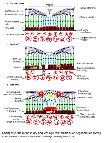
The structure of the normal retina is illustrated in Figure 1a. With progressive age, the retina develops characteristic anatomical features. Localised deposits form under the retina, usually between the RPE and Bruch's membrane. These are called ‘drusen’ and usually appear either as small, discrete yellow dots, called ‘hard drusen’, or larger more confluent and less discrete yellow spots, called ‘soft drusen’. Drusen can also collect more internally to the RPE and within the photoreceptor cell layer. These deposits are called ‘reticular drusen’ and appear as an interlacing network of small yellow round or oval spots (Ref. Reference Schmitz-Valckenberg6). Both soft and reticular drusen are associated with increased progression of AMD (Refs Reference Klein7, Reference Knudtson8). Histologically, these are composed of a multitude of proteins (e.g. complement proteins, immunoglobulins, acute-phase proteins, amyloid-β) and lipids (e.g. phospholipids, cholesterol, apolipoproteins) (Ref. Reference Lotery and Trump9).
Changes in the retina in dry and wet age-related macular degeneration (AMD). (a) Structure of the normal retina. The outermost layer of the retina consists of a monolayer of retinal pigment epithelial (RPE) cells, containing granules (melanosomes) with melanin. Microvilli project from the inner surface of RPE cells, which interdigitate with the outer segments of photoreceptors (rods, green; cones, blue). Multiple photoreceptors usually communicate with each bipolar cell (not shown), which in turn synapse with ganglion cells. The long axons of ganglion cells constitute the nerve fibre layer, which conducts nerve impulses to the optic nerve and then onto the visual cortex in the brain. A network of capillaries is found in the bipolar cell layer, and further capillaries are found within the ganglion cell and nerve fibre layers. Numerous other supporting cells are found in the retina, including amacrine, horizontal and Muller cells (not shown). The outer layer of the retina, the RPE layer, sits on Bruch's membrane. This is composed of collagen and elastin, and acts as a barrier between the RPE cells and the external choroid. The inner layer of the choroid is called the choriocapillaris and is composed of fenestrated capillaries. Larger vessels constitute the outer choriodal layer (not shown). The macula demonstrates characteristic anatomical features compared with the rest of the retina. The bipolar and ganglion cells are displaced to allow maximal light exposure to the photoreceptors, and there are no blood vessels within the retinal layers. The centre of the macula, the fovea, is concave in cross-section and consists of cone photoreceptors only. Furthermore, the RPE cells are taller. (b) Specific anatomical changes are seen in dry AMD. Drusen (shown in yellow) accumulates between RPE cells and Bruch's membrane. Reticular drusen (also shown in yellow) is a specific form of drusen that accumulates between the RPE layer and photoreceptors. Both atrophy and hypertrophy of RPE cells occur, along with choriocapillaris atrophy and thickening of Bruch's membrane. (c) Wet AMD is characterised by the presence of choroidal neovascular membrane (CNV). This forms within the choroid and enters the sub-RPE space through a break in Bruch's membrane. The CNV then proliferates, which can lead to further growth between the retina and RPE layers, leakage of fluid (shown in yellow) and haemorrhage (shown in red). This disruption of retinal layers can cause image distortion. These changes lead to thickening and elevation of the retina, which can be detected clinically.

AMD is also associated with alterations in the architecture of the retina and choroid. The cells of the RPE, retina and choroid can all diminish in size and number, eventually becoming atrophic. RPE cells can also become hypertrophic, which is possibly a reactive change. Bruch's membrane becomes thicker and more impermeable. The photoreceptor cells at the macula are inadequately supported because of these changes and become atrophic, possibly leading to long-term impairment of central vision. In addition, as a result of neovascularisation (see below), photoreceptor cells can lose their normal orientation and become disorganised. This results in visual symptoms such as distortion and metamorphopsia (change in image size).
Neovascularisation, a late sign of AMD, originates either in the choroid [choroidal neovascularisation (CNV)] or within the retina [retinal angiomatous proliferation (RAP)]. In CNV, endothelial cells begin to proliferate within the choroid, and a defect in Bruch's membrane allows these blood vessels to pass into the sub-RPE or subretinal space. In RAP, these endothelial vessels proliferate within the retina itself. In both cases, these vessels are fragile and tend to leak or bleed easily. Oedema tends to form either in the subretinal space or within the retinal tissue (intraretinal fluid). Haemorrhage that occurs in the sub-RPE or subretinal space can break through the retina to sit in the potential space between the retina and vitreous (subhyaloid space), or can break through into the vitreous. New vessels tend to proliferate for several months, eventually undergoing fibrosis, which can potentially lead to irreversible vision loss. Figure 1b and c depict the typical changes, as seen in cross-section, in dry and wet AMD.
Clinical features of AMD
Early AMD can be asymptomatic, despite the presence of drusen and changes in the RPE. Patients with dry AMD tend to exhibit a gradual deterioration in the ability to read small print, which sometimes progresses to distortion and central scotomas (blind spots). These changes are very gradual and can develop and progress over many years. Drusen may increase in size and number and this is accompanied by hypo- or hyperpigmentation that results from changes in the RPE. As the choriocapillaris layer becomes atrophic, the larger choroidal vessels become visible through areas of RPE atrophy. Eventually, the neurosensory retina becomes atrophic. Large areas of RPE and retinal atrophy are termed ‘geographic atrophy’ (GA) (Refs Reference Klein7, Reference Knudtson8). Widespread, extensive GA of the neurosensory retina and RPE at the macula, associated with severe central visual impairment, represents end-stage dry AMD.
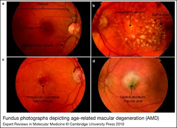
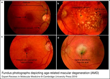
In wet AMD, the development of neovascularisation typically presents suddenly, with a dramatic worsening of central vision, distortion and development of a central scotoma. Clinically, fluid, hard exudates and haemorrhage may be present, which stem from the abnormal neovascularisation. Eventually, the abnormal blood vessels will either regress or fibrose, leaving a scar. End-stage wet AMD signifies the establishment of fibrous tissue at the macula, known as a ‘disciform’ scar (because of its circular or ‘disc-like’ shape). The majority of severe visual loss occurring in AMD is associated with the wet form (neovascularisation). The overall prevalence of GA or CNV is about 3% among all patients with AMD (Ref. Reference Augood10). Figure 2 depicts the various forms of AMD, as observed clinically.
Fundus photographs depicting age-related macular degeneration (AMD). (a) Fundus of a normal right eye. The optic disc is a pink–yellow area that represents the beginning of the optic nerve. It serves as an anatomical landmark during clinical examination. Radiating out from the optic disc in four quadrants are the retinal blood vessels. Veins are darker and slightly thicker than arteries. Note the ‘arcades’ created by the vessels on the left of the disc as they arc round the macula. The macula, encompassed by the arcades, lies temporal to the optic disc. The fovea is the centre of the macula and appears darker because of increased xanthophyll pigment levels and taller retinal pigment epithelial (RPE) cells. (b) Severe dry AMD. There is extensive soft drusen, which is visible as yellowish clumps. This patient has geographic atrophy as a result of extensive RPE cell and choriocapillaris atrophy. The sharply demarcated pale area with visible large choroidal vessels is characteristic. As a result this patient may have significantly reduced central vision. (c) Acute wet AMD. This patient developed sudden deterioration of central vision, as a result of subretinal or intraretinal haemorrhage from the choroidal neovascular membrane (CNV). (d) End-stage wet AMD — macular scar. Untreated wet AMD will eventually result in a permanent disciform (disc-shaped) macular scar. Central vision will inevitably be poor.
Oxidation and the body
A full description of the oxidative processes in the body and their relation to ageing is beyond the scope of this article; however, readers are directed to a review by Muller for further information (Ref. Reference Muller11). Reactive oxygen species (ROS) are highly reactive atoms, ions or molecules that contain oxygen. These are either free radicals or peroxides. Free radicals have an unpaired electron in the outer shell. Oxygen-containing free radicals include hydroxyl (OH•), hydroperoxyl (HO2•), superoxide anion (O−2•) and singlet oxygen (1O2). Peroxides, by definition, contain a single oxygen–oxygen bond and include hydrogen peroxide (H2O2). ROS within the body are formed as normal byproducts of the body's normal metabolic processes, including glycolysis and the Krebs cycle. The superoxide anion is produced by all cells in the mitochondria during the electron transport stage of cellular respiration, and is converted to H2O2 by the enzyme superoxide dismutase (SOD) (Ref. Reference Muller11). ROS are also formed by the immune system as a defence against invading pathogens, and by environmental factors such as smoking, pollution and radiation. The majority of ROS in the body are produced by mitochondria through the electron transport chain, which converts almost 4% of all utilised oxygen into ROS (Ref. Reference Chance, Sies and Boveris12).
The two main targets of oxidative damage are mitochondria and lipids. Damage from ROS results in point mutations and deletions to mitochondrial DNA (mtDNA) (Ref. Reference Golden and Melov13). Oxidative damage also leads to earlier senescence (loss of ability of cells to divide) (Refs Reference Hayflick and Moorhead14, Reference Yuan, Kaneko and Matsuo15), and this may be related to damage and shortening of telomeric DNA (Refs Reference Adelfalk16, Reference Rubio, Davalos and Campisi17). Furthermore, mtDNA is highly susceptible to oxidative damage, as it is preferentially damaged over nuclear DNA in the presence of ROS (Ref. Reference Ballinger18). This could be because mtDNA is closer to the site of creation of ROS, is not protected by histones, and is more prone to errors in replication owing to a less-effective repair system than that for nuclear DNA (Ref. Reference Cai19). Mitochondrial damage also leads to the release of lethal enzymes, such as caspase and cytochrome c, which can lead to apoptosis (Refs Reference Susin20, Reference Yang21). ROS damage lipids by oxidation in a process called lipid peroxidation. Polyunsaturated fatty acids (PUFAs) form a significant part of cell membranes. They are most vulnerable because they carry highly reactive hydrogen atoms (Ref. Reference Lundberg22). These oxidised lipids, in turn, are toxic and can cause further damage by combining with other molecules (especially proteins), leading to modification of function.
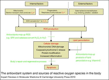
Uncontrolled ROS will cause severe damage to cells. The body has therefore evolved a protective system of antioxidants, which neutralise the damaging effects of ROS. This system includes a wide range of enzymes that are measurable in the circulation, such as SOD, glutathione peroxidase (GPX), catalase, metallothionein, peroxiredoxin and thioredoxin, as well as non-enzymatic compounds, such as vitamin C, vitamin E and the carotenoids (Ref. Reference Muller11). Normally, these antioxidants help to eliminate free ROS to prevent cellular damage. Despite this, some inevitable cellular damage occurs owing to the highly reactive state of ROS. This is usually kept in check by the body's own repair and regeneration processes. However, if ROS levels exceed antioxidation capacity, as a result of either excess ROS production or reduced antioxidant capability, then oxidative damage may ensue. Unfortunately, this is what tends to happen with increasing age, because these steps are key drivers of the ageing process. Furthermore, the body's repair and regeneration capabilities also decrease with age (Ref. Reference Muller11). This can lead to progressive dysfunction due to abnormal production of proteins and lipids that are essential for cellular activity (Ref. Reference Harman23). In particular, mtDNA damage increases with age as a result of accumulation of mutations (Ref. Reference Golden and Melov13). Figure 3 summarises the main oxidative processes that occur in the body.
The antioxidant system and sources of reactive oxygen species in the body. Mitochondria are the main source of reactive oxygen species (ROS) in the body. The highly reactive superoxide anion (O2•−) is generated within the mitochondrial inner membrane via the electron transport chain, an essential component of cellular respiration. Its function is to produce energy, after-glycolysis and the Krebs cycle. ROS are also produced by phagocytosis via the NADPH system. This process, called the ‘respiratory burst’, leads to O2•− production, required for digestion of internalised particles and pathogens. This is an important component of the immune system, enabling neutrophils and macrophages to clear invading microorganisms. These ROS in turn generate further toxic products by oxidisation of polyunsaturated fatty acids, especially in cellular membranes. ROS can exert a range of toxic effects, including protein modifications, mitochondrial DNA damage and release of the apoptosis-associated enzymes caspase and cytochrome c (ROS and their effects shown in red). Increased inflammation as a result of cellular damage may worsen the situation. Antioxidants (shown in green) help to prevent cellular damage. Most O2•− is converted, or ‘dismutated’ to H2O2 by superoxide dismutase (SOD). H2O2 itself is also toxic, but this in turn is usually converted to H2O by glutathione peroxidase (GPX) and catalase. A range of other antioxidants (e.g. thioredoxin, metallothionein, peroxiredoxin, vitamin C, vitamin E and carotenoids) also help to mop up ROS. Any ROS that are not eliminated could still produce some cellular damage. However the body's repair and regenerative facilities come into play to maintain normal function (also in green).

The retina and oxidative stress
The retina is exposed to a constant source of oxidative stress from incoming light, leading to photo-oxidation, which in turn generates ROS within the retina. Male albino rats raised for 12 months in dark conditions had a reduced age-related susceptibility to damage from intense photo-oxidative stress, in the form of intense light, compared with rats raised in dim cyclic light conditions (Ref. Reference Organisciak24). Intense light also increases H2O2 levels in the outer retina of albino mice (Ref. Reference Yamashita25). Blue wavelength [ultraviolet-B (UV-B)] light in particular is considered the more harmful constituent of visible light. In-vitro-cultured RPE cells exposed to blue light develop increased levels of intracellular ROS and stress-related proteins (including osteopontin, heat shock protein 27 and cathepsin), show increased formation of advanced glycation end products and have greater mitochondrial damage (demonstrated by structural changes and reduced membrane potential). These changes are accompanied by a compensatory increase in antioxidant levels (Refs Reference van Kuijk and Buck26, Reference Roehlecke27). Furthermore, high levels of easily oxidised polyunsaturated fats are present in the retina, as measured by gas chromatography in vitro (Ref. Reference Yamashita25). Such lipid peroxidation products are in turn toxic to the retina (see below). Even the phagocytic process of the RPE itself generates ROS through the nicotinamide adenine dinucleotide phosphate (NADPH) oxidase system. In vitro studies of cultured human RPE cells incubated with probe-labelled bovine outer segments demonstrate increased production of intracellular H2O2 accompanying increased phagocytosis (Ref. Reference Miceli, Liles and Newsome28).
Oxidative damage induces RPE apoptosis through loss of mitochondrial membrane potential, cytochrome c production and caspase 3 activation, as seen in vitro in human cultured RPE cells exposed to t-butylhydroperoxide (tBHP) (Ref. Reference Jiang29). Human cultured RPE cells also exhibit premature senescence when exposed in vitro to oxidative stress from tBHP or H2O2, as recognised by increased senescence-associated β-galactosidase activity, altered gene expression and consequent alteration in cellular function (Refs Reference Glotin30, Reference Yu31). Because of their intimate relationship with RPE cells, photoreceptor cells are susceptible to secondary damage once RPE cells are affected, but can also be directly damaged by oxidative stress. Cultured photoreceptor cells exposed to sodium nitroprusside in vitro demonstrate increased activation of protease enzymes such as caplains and caspases, with consequent apoptosis (Ref. Reference Sanvicens32). ROS damage also increases the expression of connective tissue growth factor, plasminogen activator inhibitor-1, collagen type IV and fibronectin in vitro in cultured human RPE cells. This can lead to accumulation of the extracellular matrix in the RPE layer and might also be a factor in the thickening of Bruch's membrane seen in AMD (Ref. Reference Fuchshofer33). Oxidative stress also appears to impair retinal vascular endothelial function. Normally, retinal vessels undergo vasoconstriction in the presence of high oxygen levels; a randomised placebo-controlled clinical trial on 21 healthy volunteers demonstrated a reduction in this response during oxidative stress upon administration of intravenous lipopolysaccharide (Ref. Reference Pemp34). The choriocapillaris could also be a source of ROS. Protoporphrin IX, a photosensitive precursor of haemoglobin normally found in erythrocytes and plasma, produces ROS when experimentally exposed to light, as detected by electron spin resonance spectrometry (Ref. Reference Gottsch35).
A disorder of the immune system is considered to be the major underlying pathological feature of AMD. Several interactions exist between components of the immune system and the oxidative process within the normal retina. Mononuclear phagocytes increase intracellular ROS levels, activate caspase, decrease mitochondrial membrane potential and induce apoptosis in mouse cultured RPE cells. These changes are more exaggerated in Sod2-knockout mice RPE cells and after mononuclear phagocyte stimulation with interferon gamma (Ref. Reference Yang36). The oxidative process may activate the complement cascade, which can cause tissue damage, as demonstrated by the following in vitro experiments. Cultured human and mouse RPE cells normally express high levels of complement factor H (CFH), a regulator of the alternate complement cascade. Prolonged phagocytosis of the outer segments reduces intracellular CFH protein production by RPE cells (Ref. Reference Chen, Forrester and Xu37). Cultured RPE cells exposed to oxidative stress induced by H2O2 demonstrate increased surface deposition of C3 and reduced surface expression of the complement inhibitors DAF, CD55 and CD59 (Ref. Reference Thurman38). Mice immunised in vivo with carboxyethylpyrrole (CEP), a product of lipid peroxidation, fix complement C3 in Bruch's membrane, and develop features of AMD (Ref. Reference Hollyfield39). SERPINA3K, a serine protease inhibitor, is an endogenous regulator of inflammation. It has been shown to significantly decrease ROS production and increase antioxidant levels in vitro in cultured retinal cells exposed to hypoxia (Ref. Reference Zhang40).
VEGF is upregulated in AMD (Ref. Reference Kliffen41), and as mentioned previously, anti-VEGF intravitreal injections are now the mainstay of treatment of wet AMD (Refs Reference Brown4, Reference Rosenfeld5). Cultured RPE cells exposed in vitro to oxidative stress from tBHP demonstrate upregulation of VEGF (Ref. Reference Kannan42). Anti-VEGF treatment could exert some therapeutic benefit by reducing oxidative stress. Bevacizumab, one of the anti-VEGF treatments available clinically, reduces ROS production, lipid peroxidation and apoptosis in choroid and retinal tissue when injected in the eyes of rabbits in vivo (Ref. Reference Sari43).
Protective mechanisms in the normal retina
Because the retina is highly susceptible to oxidative damage, it has developed a range of protective mechanisms. These include carotenoids, antioxidant enzymes and other substances. The term ‘carotenoid’ encompasses a range of diet-derived substances, which indude carotenes (α-carotene, β-carotene and lycopene) and xanthophylls (zeaxanthin, lutein and β-cryptoxanthin) (Ref. Reference Young and Lowe44). Lutein and zeaxanthin are found in the retina and might be protective through reduction of lipid peroxidation (Refs Reference Packer45, Reference Chucair46). Pretreatment of cultured rat photoreceptors with lutein and zeaxanthin protects from oxidative damage due to paraquat and H2O2 exposure (Ref. Reference Nakajima47). Zeaxanthin also protects cultured rat retinal ganglion cells from oxidative damage induced by in vitro H2O2 exposure or serum deprivation (Ref. Reference Lim48). Furthermore, proteomic studies utilising high-performance liquid chromatography (HPLC) and mass spectrometry demonstrate the presence of several carotenoid oxidation products in extracts of human donor retina, demonstrating their ability to mop up ROS (Refs Reference Lakshminarayana49, Reference Khachik, Bernstein and Garland50).
The retina and RPE may have higher levels of antioxidant activity compared with other ocular tissues. In vitro measurement of antioxidant enzyme activity in human eyes by direct spectrophotometry shows that the retina has high levels of the copper and zinc-containing isoform of SOD (CuZn-SOD), but relatively low levels of the other SOD isoforms (Ref. Reference Behndig51). However, there seems to be no correlation between RPE SOD levels and the presence of AMD or ageing (Ref. Reference Liles, Newsome and Oliver52). Human donor RPE cells have six times more catalase activity than other ocular tissues (Ref. Reference Liles, Newsome and Oliver52).
Antioxidant levels increase as a protective mechanism during conditions of oxidative stress (Refs Reference Sreekumar53, Reference Kutty54, Reference Melo55). In vitro exposure of cultured RPE cells to H2O2 demonstrates increased expression of the gene coding for the antioxidant enzyme methionine sulfoxide reductase within the cytosol and mitochondria. This enzyme is also found in sub-RPE drusen from the macula of donors with AMD (Ref. Reference Sreekumar53). Another antioxidant, haem oxygenase-1 (HO-1), is barely detectable normally, but mRNA levels for this enzyme increase dramatically in vitro in rat retinas exposed to intense visible light. However, there does not appear to be an increase in HO-2 levels in similar conditions, suggesting that the two isoforms have different functions (Ref. Reference Kutty54). Methamphetamine, a psychostimulant that induces oxidative stress, increases SOD levels in the retina as well as in plasma when given to rats in vivo (Ref. Reference Melo55). Other substances with antioxidant properties found in the retina indude melanin and melantonin. In rat eyes, melanin appears to protect retinal and choroidal cells from the phototoxic effects of light (Ref. Reference Peters56). Melatonin (N-acetyl-5-methoxytryptamine), a hormone produced by the pineal gland and retina, might also have antioxidant effects because cultured RPE cells are protected from oxidative damage by the prolonged addition of melatonin (Ref. Reference Liang57).
Changes in oxidative stress in the retina with age
Advancing age increases the susceptibility of the retina to oxidative stress. With age, RPE cells accumulate granules containing a fluorescent pigment called lipofuscin. This is composed of undigested end products from the phagocytic process of photoreceptor cell outer segment discs (Ref. Reference Ryan58). Autophagy also results in accumulation of lipofuscin granules in photoreceptor inner segments (Ref. Reference Iwasaki and Inomata59). When purified from extragranular debris, lipofuscin granules contain toxic substances, including retinoids (products of the visual cycle), and modified protein and lipid products (Ref. Reference Ng60). In vitro, RPE cells loaded with lipofuscin exhibit a significant reduction in phagocytic capacity, therefore compounding the problem (Ref. Reference Nilsson61). When exposed to light in vitro, lipofuscin can form ROS, which in turn can damage the RPE. In vitro illumination of lipofuscin with visible light results in extragranular lipid peroxidation, enzyme inactivation and protein oxidation, which are reduced upon the addition of antioxidants (Ref. Reference Wassell62). Using liquid chromatography, mass spectrometry and immuno-analysis, more than 180 proteins in lipofuscin have been identified, many of which are modified, possibly as a result of oxidative influence (Refs Reference Ng63, Reference Schutt64). Lipofuscin can also reduce intracellular antioxidant capacity; isolated lipofuscin granules fed to human cultured RPE cells in vitro can reduce the activity of the antioxidant enzymes SOD and catalase by up to 60% (Ref. Reference Shamsi and Boulton65).
One key component of lipofuscin is N-retinylidene-N-retinylethanolamine (A2E), a byproduct of the retinoid visual cycle. Oxidation of A2E can lead to harmful ROS, which may damage DNA. A2E self-generates singlet oxygen, which in turn reacts with A2E to form A2E epoxide. This damages DNA in cultured RPE cells, as detected by the presence of ‘comet tails’ on single-cell gel electrophoresis, indicating DNA strand breaks (Ref. Reference Sparrow66). Cell death can follow. Human cultured RPE cells loaded with A2E exposed in vitro to photo-oxidative stress in the form of blue light show increased apoptosis compared with RPE cells devoid of A2E (Ref. Reference Sparrow, Nakanishi and Parish67). A rise in cytoplasmic caspase, which is associated with cell apoptosis, appears to accompany this process (Ref. Reference Sparrow and Cai68). There is also evidence that the retinoids within lipofuscin can activate the complement cascade. Oxidative products of A2E in lipofuscin can activate complement in vitro, which in turn can damage local tissue if left unchecked (Ref. Reference Zhou69). Protection against the harmful effects of lipofuscin may be one of the roles of melanin. The presence of calf melanin in cultured RPE cells in vitro is protective against photo-oxidation from blue light (Ref. Reference Wang, Dillon and Gaillard70). Furthermore, pigmented rabbit and bovine cultured RPE cells fed photoreceptor outer segments over 4 weeks accumulate significantly less lipofuscin compared with nonpigmented RPE cells. This suggests that melanin may also reduce lipofuscin build-up in RPE cells (Ref. Reference Nilsson61).
In vitro studies suggest that the antioxidant capacity of the RPE and retina decreases with age: the rat retina shows decreased SOD, GPX and catalase activity at 4 months compared with 1 month (Ref. Reference Castorina71). Catalase levels appear to diminish in RPE cells of donor eyes with increasing age (although in this study, SOD levels appear unchanged) (Ref. Reference Liles, Newsome and Oliver52). Furthermore, cumulative light exposure of RPE cells with age may reduce the effectiveness of local antioxidants. Porcine RPE cells exposed to high-intensity light demonstrate degradation of melanosomes, reduced melanin levels, with increased ROS production (Ref. Reference Zareba72). Cultured RPE cells fed melanosomes in vitro from older human donors, exposed to blue light, sustain greater cellular damage with prolonged exposure to blue light than RPE cells fed melanosomes from younger human donors (Ref. Reference Rozanowski73).
The macula and oxidation
There are several reasons why the macula may be particularly susceptible to oxidative damage. The optics of the eye are designed to focus incoming light rays on the macula, therefore maximising photo-oxidative stress, especially from UV-B light. The highest metabolic activity within the retina occurs at the macula. This can be demonstrated indirectly in several ways. The multifocal electroretinogram is an objective, noninvasive method of recording retinal function at several areas throughout the retina simultaneously, after exposure to a light stimulus. This shows greatest response at the macula (Ref. Reference Feigl, Stewart and Brown74). Blood flow within the choroid is greatest in the macular region (Ref. Reference Nork75). Furthermore, lipofuscin levels are also relatively high at the macula (with a dip at the fovea due to its anatomical structure – see Fig. 1a), suggesting high levels of phagocytosis (Ref. Reference Wing, Blanchard and Weiter76). Finally, in keeping with increased oxidative stress at the macula, concentrations of the antioxidants lutein and zeaxanthin are also highest in the macula, and specifically in the outer segments of photoreceptor cells, as measured by HPLC in vitro in the adult human retina (Refs Reference Bone77, Reference Rapp, Maple and Choi78).
Oxidation and AMD
Certain factors that increase oxidative stress may also be associated with an increased risk of AMD. Cigarette smoke is highly damaging to RPE cells in vitro. Cultured human RPE cells exposed to cigarette smoke extract or components of cigarette smoke (including hydroquinone, acrolein and cadmium) exhibit ROS production and reduction in antioxidant capacity, leading to impairment of mitochondrial function and eventually apoptosis of RPE cells. These detrimental processes can be reduced experimentally with the addition of antioxidants (Refs Reference Bertram79, Reference Jia80, Reference Kalariya81). This in vitro effect may explain the strong association of smoking with an increased risk of AMD seen epidemiologically, as demonstrated by three large population-based cross-sectional studies (Refs Reference Klein82, Reference Smith, Mitchell and Leeder83, Reference Vingerling84).
The Beaver Dam Study (1993) in the USA examined 4771 patients aged between 43 and 89 years, and found that the relative odds for the presence of wet AMD in smokers was 3.29 and 2.50 (in males and females, respectively) compared with nonsmokers. However, there was no relationship between the presence of GA and smoking (Ref. Reference Klein82). The Blue Mountain Eye Study (1996) in Australia looked at 3654 patients aged 49 years and over. This study found an odds ratio (OR) of 3.20 for the presence of wet AMD in smokers and 4.54 for GA, compared with nonsmokers (Ref. Reference Smith, Mitchell and Leeder83). Yet another large study, the Rotterdam Study (1996), examined 6174 patients from the Netherlands aged 55 years and over, and found, in subjects younger than 85 years, an OR of 6.6 for the presence of wet AMD in smokers compared with non-smokers. However, there was no association with GA. There was also a dose-dependent relationship, with a significant increased risk (relative risk 6.5) of wet AMD in those smoking in excess of 10 pack years (Ref. Reference Vingerling84).
The association between increased sunlight exposure and AMD has been studied extensively; however, both retrospective and prospective epidemiological studies have been inconclusive. The following studies have all used a questionnaire-based system for ascertaining sunlight exposure, which may introduce subject variability. A cohort of 2764 patients from the Beaver Dam Study, aged 43–86 at baseline were followed up prospectively over 10 years. An association of increased incidence of early AMD (including drusen and pigmentary changes) and increased exposure to the summer sun was found. However, no association was found with sun exposure and progression of AMD, or with the incidence of more advanced AMD (Ref. Reference Tomany85). A retrospective study of 769 watermen over 50 years old working on Chesapeake Bay, Maryland in the USA found no relationship between light exposure (either UV-A or UV-B) and presence of AMD (Ref. Reference West86). The researchers subsequently reported a subgroup analysis on the same population, and found that patients with advanced AMD (extensive GA or wet AMD, n = 8) had significantly higher blue and visible light exposure in the previous 20 years compared with age-matched controls (Wilcoxon sign rank test, P = 0.027). However, the small numbers of patients with advanced AMD should be noted; furthermore, there was no association when considering lifetime sunlight exposure (Ref. Reference Taylor87). The Beaver Dam Study found, in 4771 subjects aged 43–84 years, that the presence of ‘late maculopathy’ (defined as GA or wet AMD, n = 77) was significantly associated with the amount of leisure time spent outdoors in summer (OR 2.19). However, there was no association between estimated UV exposure and AMD (Ref. Reference Cruickshanks, Klein and Klein88). Furthermore, in Newcastle, Australia, 409 patients with AMD actually had significantly less sun exposure than 286 controls (P>0.0001) (Ref. Reference Darzins, Mitchell and Heller89). More recently, 4753 patients in the European Eye Study aged 65 years and over showed no association between adult lifetime blue light exposure and early or wet AMD. However, a subgroup analysis showed that patients with wet AMD and low blood antioxidant levels had increased blue light exposure, suggesting that antioxidant supplements could be beneficial (Ref. Reference Fletcher90).
Drusen is the earliest manifestation of AMD and may also contribute to oxidative damage. For example, amyloid-β, which is found in drusen, increases ROS formation and reduces antioxidant capacity in RPE cells (Ref. Reference Bruban91). However, drusen formation itself may be infuenced by the oxidative process. Liquid chromatography and mass spectrometry analysis of drusen from donor human retinas demonstrates oxidative protein modifications, including CEP protein adducts (see below), as well as crosslinking of tissue metalloproteinase inhibitor-3 and vitronectin (Ref. Reference Crabb92).
As a result of oxidative damage at the macula, local alterations in cellular function can occur. To offset this, compensatory local antioxidant activity within the retina appears to increase in AMD. SOD levels measured in vitro are higher in histological specimens in the cytoplasm and lysosomes of RPE cells in eyes with AMD compared with levels in disease-free eyes. However, this particular experiment demonstrated no change in GPX levels (Ref. Reference Frank, Amin and Puklin93). Furthermore, antioxidant activity seems to increase with AMD severity (Ref. Reference Decanini94). This suggests that there is a localised compensatory response to increased ROS activity with progressive AMD. Nevertheless, ROS activity may exceed antioxidant capacity within the macula, with subsequent progression of AMD.
Systemic markers of oxidation in AMD
It is possible that development and progression of AMD is influenced by a systemic imbalance between ROS and the antioxidant system. In vitro, RPE cells are more likely to be damaged in the presence of a more oxidative extracellular redox environment (Ref. Reference Jiang29). In vivo, plasma or serum levels of both ROS and antioxidants in patients with AMD have been investigated, with varying results.
Systemic ROS levels and AMD
There are no consistent associations of systemic ROS levels in AMD. Recently, significantly higher levels of serum protein carbonyl (a marker of protein oxidation), 8-hydroxy-29-deoxyguanosine (a marker of oxidative damage to DNA) and total serum oxidation state were found in 47 patients with wet AMD compared with 25 healthy controls (Ref. Reference Totan95). Nitric oxide (NO) is the most abundant free radical in the body; however, levels of circulating NO are not consistent in AMD. A relatively small study (n = 66) found elevated plasma levels of NO with AMD compared with controls (Ref. Reference Evereklioglu96). However, in a smaller study (n = 30), the opposite was seen, implying possible damage to endothelial cells and reduced endothelial production of NO as a result (Ref. Reference Totan97). Another study found no difference between AMD subgroups (Ref. Reference Tsai98). Homocysteine is an amino acid that rapidly oxidises to form ROS, but again with inconsistent associations with AMD. In a large cohort of over 2000 patients from the Blue Mountain Study population, higher systemic homocysteine levels were found in patients with AMD than in controls (Ref. Reference Rochtchina99). Numerous other smaller studies have confirmed this association (Refs Reference Seddon100, Reference Coral101, Reference Ates102, Reference Nowak103, Reference Vine104, Reference Kamburoglu105). Other studies show increased homocysteine levels in wet AMD compared with dry AMD (Refs Reference Kamburoglu105, Reference Axer-Siegel106). However, in the third National Health and Nutrition Examination Survey study of more than 3000 patients, no association was found with serum homocysteine (Ref. Reference Heuberger107). This was also the case in another study drawn from the Blue Mountain population (with 630 patients) (Ref. Reference Wu108).
Systemic antioxidant levels and AMD
In contrast to the above, there is possibly greater evidence suggesting an association between systemic levels of certain antioxidants and AMD. Of the individual antioxidants, carotenoids have been studied the most extensively, perhaps because of the ease with which dietary supplementation is possible. The Eye Disease Case-Control Study Group looked at the serum levels of carotenoids in a large number of patients (n = 1036), stratifying levels into low, medium and high groups. They found that patients with medium and high levels of carotenoids had up to a 50% reduced risk of wet AMD (Ref. 109). In 722 Japanese patients, reduced total carotene and total carotenoid serum levels, as well as individual levels of α-tocopherol and β-cryptoxanthin levels, were associated with late AMD. However, there was no association with early AMD (Ref. Reference Michikawa110). A smaller study on 334 subjects from the Beaver Dam population found similar levels of individual carotenoids in both patients and controls; only very low levels of lycopene were associated with AMD (Ref. Reference Mares-Perlman111). Further smaller studies have reported variable results. A French study (n = 55) did not find any difference in the total serum carotenoid level, or in the other carotenoids between AMD patients and controls (again with the exception of reduced serum lycopene levels in the AMD group) (Ref. Reference Cardinault112). In a study of Italian patients (n=94), lower levels of circulating vitamin C, vitamin E, β-cryptoxanthin and total carotenoids were found in patients with late AMD (GA or wet AMD) than in those with early AMD. However, there was no difference in total antioxidant capacity and individual carotenoid levels between the two groups. Furthermore, there was no difference in all the measured values between patients with AMD and controls (except again, for lower lycopene levels in the AMD group)(Ref. Reference Simonelli113). Another study found increased serum total antioxidant capacity in patients with AMD compared with controls (Ref. Reference Totan95).
There is less consistency of evidence depiciting the association between systemic levels of antioxidant enzymes and AMD. Several studies have found that systemic antioxidant enzyme levels, including SOD GPX (Refs Reference Evereklioglu96, Reference Prashar114), catalase (Refs Reference Evereklioglu96, Reference Yildirim115), glutathione reductase (Ref. Reference Cohen116) and paroxonase (Refs Reference Ates102, Reference Baskol117), are lower in AMD patients compared with controls. Lower antioxidant levels have also been reported in patients with late AMD compared with early AMD (Ref. Reference Evereklioglu96). Some studies have, however, report no difference in systemic antioxidant levels and AMD (Refs Reference Cohen116, Reference De, Paz, Zhang and Fridovich118, Reference Delcourt119). This may be associated with the Furthermore, several studies actually report an increase in systemic antioxidant levels in AMD. A higher level of plasma GPX was associated with a ninefold increase in prevalence of advanced AMD (Ref. Reference Delcourt119).
Other antioxidants include copper (found in RPE cells) and melatonin. The serum levels of ceruloplasmin (a globulin that binds copper, found in RPE cells) are higher in patients with AMD (Ref. Reference Newsome120). Daytime serum levels of melatonin are significantly increased in patients with AMD than in controls (Ref. Reference Schmid-Kubista121). This may be associated with the higher systemic levels of products of lipid peroxidation found in AMD (discussed below). Overall, the evidence suggests that lower systemic antioxidant levels, particularly total carotenoid levels, as well as lycopene individually, are associated with AMD, especially the late form. However the hypothesis that a systemic imbalance of ROS and antioxidant levels is a predisposing factor in AMD, is far from confirmed.
Lipid peroxidation and AMD
The retina is abundant in fatty acids (FAs), including palmitic, stearic, oleic, arachidonic and docosahexaenoic acids (Ref. Reference van Kuijk and Buck26). Under conditions of oxidative stress, PUFAs are particularly susceptible to oxidation (peroxidation). The oxidation of phospholipids in RPE and photoreceptor cells from donor eyes increases with age and in AMD (Ref. Reference Suzuki122). These oxidative products can induce apoptosis of RPE cells (Ref. Reference Choudhary123). One omega-3 long-chain PUFA particularly susceptible to oxidative damage is docosahexaenoate (DHA), which is found at high concentrations in photoreceptor cells, where it comprises 80% of the total amount of PUFAs in the outer segment discs (Ref. Reference Fliesler and Anderson124). Interestingly, the concentration of FAs is lower at the macula than in the peripheral retina, perhaps because of increased lipid peroxidation in the macular area (Ref. Reference van Kuijk and Buck26).
CEP is produced by the oxidation of DHA-containing phospholipids, which can subsequently form bonds (adducts) with proteins, leading to functional modification (Ref. Reference Gu125). CEP-adducted proteins tend to accumulate in the outer retina, in rod outer segments and in the RPE, as shown in vitro in mouse and human donor retina (Ref. Reference Gu126). Several studies suggest that CEP-adducted proteins have a key role in the pathogenesis of AMD. Mice immunised with CEP-adducted albumin accumulate complement C3 in Bruch's membrane, develop drusenoid changes under the RPE and also develop RPE atrophic-like changes (Ref. Reference Hollyfield39). CEP-adducted proteins are elevated in Bruch's membrane and drusen in donor eyes with AMD (Ref. Reference Crabb92). New vessel growth can be stimulated by CEP adducts that exacerbate laser-induced CNV (Ref. Reference Ebrahem127). Plasma anti-CEP antibodies are elevated in patients with AMD (Ref. Reference Gu126). Proteomic analysis also demonstrates increased plasma levels of CEP adducts and CEP autoantibodies in AMD, especially in association with the ARMS2 and HTRA1 risk alleles (Ref. Reference Gu128).
CEP is likely to be only one of several products of lipid peroxidation associated with AMD (Ref. Reference Gu125). For example, levels of oxidised LDL as well as susceptibility of LDL to oxidation are both increased in the plasma of AMD patients (Refs Reference Ikeda129, Reference Javadzadeh130). 4-Hydroxy-2-nonenal (HNE) is a highly toxic product of lipid peroxidation. However, levels of HNE-adducted proteins found at the macula in donor eyes with AMD are similar to levels in the periphery. Furthermore, HNE levels do not increase with AMD severity. This suggests that the retina has sufficient antioxidation capacity to remove this product (Ref. Reference Ethen131). Malondialdehyde (MDA), a product of lipid peroxidation, is used as a marker of oxidative stress in studies of oxidation. Several studies have shown that circulating MDA levels are increased in AMD, compared with controls (Refs Reference Totan95, Reference Evereklioglu96, Reference Totan97, Reference Yildirim115, Reference Baskol117).
Genetic studies of oxidation and AMD
Antioxidant enzyme genes
Current studies in the literature examining the association between genes encoding antioxidant enzymes and AMD in human populations are not confirmatory; however, antioxidant gene knockout mice do develop retinal features reminiscent of AMD. The most studied antioxidant genes are those encoding SOD proteins. The SOD1 protein is the most abundant and is located in the cytoplasm (associated with copper or zinc, gene position 21q22.1); SOD2 is found in the mitochondria (associated with manganese, gene position 6q25.3); SOD3 is found in the extracellular space (gene position 4p15.3–p15.1) (Refs Reference Johnson and Giulivi132, 133). An association with a polymorphism in the SOD2 gene with AMD was found in a Japanese population (Ref. Reference Kimura134). However, two other groups found no association in Japanese populations (Refs Reference Gotoh135, Reference Kondo136), and futhermore, no association was observed in a Northern Irish population (Ref. Reference Esfandiary137). Interestingly, it has also been suggested that a SOD gene polymorphism could actually be protective in AMD (Ref. Reference Gotoh135). Other antioxidant genes have been studied in human populations, again with variable outcomes. One report suggests that there is no association between polymorphisms in the GPX or catalase gene and AMD (Ref. Reference Esfandiary137). However, polymorphisms in the paroxonase gene (which protects LDL from oxidation) have been associated with AMD (Ref. Reference Ikeda129).
Despite the above results, animal models of AMD have been created by manipulation of antioxidant genes. Sod1-knockout mice are more likely to develop CNV in the presence of VEGF (Ref. Reference Dong138). Sod1-knockout mice also demonstrate necrotic swelling in the inner and outer nuclear layer of the retina, as well as mitochondrial degeneration associated with increased drusen, thickened Bruch's membrane and CNV (Refs Reference Imamura139, Reference Hashizume140). RPE cells from Sod2 heterozygous knockout mice have lower levels of SOD2 correlating with disruption of mitochondrial transmembrane potential, release of cytochrome c and resulting apoptosis (Ref. Reference Kasahara141). However, overexpression of SOD in transgenic mice can be detrimental, suggesting that a careful balance of SOD levels in the body is usually essential (Ref. Reference Lu142). The lack of translation from animal studies to human populations suggests that AMD pathogenesis in humans is likely to be a far more complex, multifactorial disease.
ARMS2
Because mitochondria have a key role in oxidative damage (as described above), it is possible that genes affecting mitochondrial function alter the susceptibility to oxidative damage. The ARMS2/LOC387715 gene located on chromosome 10q6 has been associated with AMD and might have a role in mitochondrial function (Ref. Reference Jakobsdottir143). A single-nucleotide polymorphism (SNP) (G→T in exon 1, with A69S amino acid change) at this locus has been associated with AMD (Ref. Reference Rivera144). The same group later reported an association of several further SNPs with AMD as well as a deletion–insertion (del443ins54) in the ARMS2 gene (Refs Reference Fritsche145). The gene encodes a 12 kDa protein; however, its function is not known. ARMS2/LOC387715 mRNA has been detected in vitro in the human retina, localising to the mitochondrial outer membrane in mammalian cells and in particular the mitochondria-rich ellipsoid region of photoreceptors (Refs Reference Fritsche145, Reference Kanda, Chen and Othman223). However, more recently, ARMS2 protein was found in the cytosol in cultured RPE cells (Ref. Reference Wang146). As mentioned previously, patients with AMD are more likely to have elevated systemic markers of lipid peroxidation (plasma CEP adduct or CEP antibody levels) if also carrying the G→T SNP (Ref. Reference Gu128). Interestingly, the same SNP almost doubled the risk of AMD in patients who smoked (OR, 2.7 versus 1.2) in heterozygotes (GT) and almost quadrupled the risk (OR, 8.2 versus 2.1) in homozygotes (TT), compared with nonsmokers, when taking into account CFH status (Ref. Reference Schmidt147). It may be that SNPs in the ARMS2 gene increase susceptibility to oxidative damage, possibly in association with smoking. However, this is speculative and further studies are indicated.
Mitochondrial genes
Despite the integral role of mitochondria in oxidative damage, there are few studies documenting the association of genes within the mitochondria and AMD. Varying outcomes are reported for different mitochondrial haplogroups. Haplogroup H appears to be protective and is associated with reduced AMD prevalence. Haplogroup J is associated with a higher prevalence of large soft drusen, and haplogroup U with higher prevalence of retinal pigment abnormalities (Ref. Reference Jones148). Patients with variations in the mitochondrial T2 haplogroup and complex I genes are up to 2.5 times more likely to have advanced AMD (Ref. Reference SanGiovanni149). More studies would be useful to investigate the incidence of genetic mutations in mtDNA and AMD.
Complement genes
As mentioned above, there may be an interaction between the immune system and the oxidative process. The immune system has a key role in the pathogenesis of AMD (Refs Reference Lotery and Trump9, Reference Swaroop150). Several genes encoding components of the complement cascade have been associated with AMD. Three landmark publications in 2005 documented the association of the Y402H sequence variation in the complement factor H (CFH) gene and susceptibility to AMD (this gene encodes a regulator of the alternate complement pathway) (Refs Reference Klein151, Reference Haines152, Reference Edwards153). Additional strong genetic associations with AMD have also been found with mutations in other complement-related genes, including complement C3 (Ref. Reference Yates154), complement factor B (CFB) (Ref. Reference Gold155), complement factor I (Refs Reference Ennis156, Reference Fagerness157) and a deletion in CFHR1 and CFHR3 (Refs Reference Hageman158, Reference Hughes159). Recently, an association with AMD and the SERPING1 gene was reported (this encodes a C1 inhibitor, which is a regulator of the classic complement pathway) (Ref. Reference Ennis160). Whether these genetic variants increase the risk of AMD by increasing oxidative stress as well as through inflammatory processes remains to be seen.
Clinical implications
Treatment aimed at reducing oxidative damage can therefore be approached in two different ways, either by reducing the source of oxidative damage or by increasing protection against oxidative damage, using antioxidants. The following measures can reduce the risk of AMD in patients by lowering oxidative stress.
Reducing the source of oxidative damage
As described above, the association between sunlight exposure and AMD is not confirmatory. Appropriately filtering sunglasses can reduce A2E and ROS production in RPE and photoreceptor cells in vitro (Ref. Reference Meyers, Ostrovsky and Bonner161). However, there is no evidence that sunglasses can reduce the risk of developing AMD (Ref. Reference Tomany85). Smoking, by contrast, is strongly associated with AMD. Stopping smoking is associated with a reduced risk of developing AMD, and individuals who have not smoked for more than 20 years have a risk of AMD that is equivalent to that of nonsmokers (Ref. Reference Khan162).
Antioxidant supplementation
In vitro, lipofuscin content in cultured RPE cells is significantly reduced by the addition of antioxidants such as α-tocopherol, lycopene, zeaxanthin and lutein (Refs Reference Nilsson61, Reference Sundelin and Nilsson163). Oral supplementation of antioxidants is a relatively simple intervention, and such products are widely available. However, there does not seem to be sufficient evidence to suggest that oral antioxidants can prevent the onset of AMD. A meta-analysis of three prospective interventional randomised controlled trials and nine prospective questionnaire-based studies looking at incidence of AMD over 5–18 years was recently performed. The authors concluded that ‘vitamin A, vitamin C, vitamin E, zinc, lutein, zeaxanthin, α carotene, β carotene, β cryptoxanthin, and lycopene have little or no effect in the primary prevention of early AMD’ (Ref. Reference Chong164).
Alternatively, there may be a place for oral antioxidants in reducing the progression of pre-existing mild AMD. The Age Related Eye Disease Study (AREDS) was a 6 year study observing the effects of high-dose antioxidants, including vitamins C and E and β-carotene, along with zinc, in over 3000 subjects. This demonstrated an odds-risk reduction of one-third in the highest risk group of developing progression to severe AMD (as observed on fundus images). The highest risk group was defined as those with extensive intermediate-size drusen (greatest linear diameter 63–124 μm), at least one large druse (>124 μm diameter), noncentral GA in one or both eyes, or advanced AMD or vision loss because of AMD in one eye (Ref. 165). Another study found that the AREDS formulation appeared to protect patients from retinal vascular endothelial dysfunction produced by oxidative stress (Ref. Reference Pemp34). In an ongoing study, a group from Vanderbilt University, USA is investigating whether the original AREDS vitamins change the plasma redox state in 140 patients over 5 years (Ref. 166). Use of β-carotene, however, is contraindicated in smokers because of an association with lung cancer (Ref. Reference Omenn167); AREDS ‘smokers’ formulations' are available as alternatives. Nevertheless, the benefits of taking such a formulation are likely to be outweighed by the harm resulting from continued smoking.
Potential future treatments
There is a wide spectrum of compounds under investigation that may provide antioxidant benefit. A brief description of the clinical and non-clinical studies of potential therapeutic agents in the literature follows.
Dietary antioxidants
The AREDS2 study is an ongoing 5-year prospective study based in the USA, ending in 2013, that is designed to look at the effects of oral supplementation of xanthophylls (lutein and zeaxanthin) and omega-3 FAs (DHA and eicosapentaenoic acid – EPA) on the risk of progression of pre-existing early AMD to late AMD. A total of 550 patients will be randomised into one of three groups: one taking 10 mg lutein and 2 mg zeaxanthin, another taking 350 mg DHA and 650 mg EPA, and the third taking a combination of both (Ref. 168).
Other oral supplements may be beneficial in providing antioxidant protection. The antioxidant enzyme SOD is available in oral form, called ‘Glisodin’ (derived from SOD found in melons). A French team has completed a 2 year study (no results are available at the time of writing) investigating whether this supplement can reduce the progression of AMD in 46 patients who have unilateral wet AMD in the fellow eye (Ref. 169). Epigallocatechin gallate, a component of green tea, appears to protect the retina against oxidative damage when injected into rats' eyes (Ref. Reference Zhang and Osborne170). The National Eye Institute, USA has completed a study examining the effects of oral lutein plus green tea extract, on macular pigment density (a reflection of lutein and zeaxanthin concentrations), as measured by heterochromatic flicker photometry, in 40 patients over 4 months (no results are available at the time of writing) (Ref. 171). However, this combination does not appear to alter oxidative stress systemically in adequately nourished older adults (Ref. Reference Li172).
Several other potential supplements appear to produce antioxidant benefits in vitro, but have yet to be studied in well-structured clinical trials. Bilberry extract appears to reduce the formation of reactive products from the oxidation of A2E in cultured RPE cells (Ref. Reference Sparrow66). Curcumin, a naturally occurring compound found in the spice turmeric, appears to have an antioxidant effect through several mechanisms, including inhibition of the nuclear factor kappa B (NF-κB) pathway (see below) (Ref. Reference Singh and Aggarwal173), upregulation of antioxidant gene expression (Ref. Reference Scapagnini174) and downregulation of gene expression of inflammatory mediators (Ref. Reference Abe, Hashimoto and Horie175). Cells of rats given a curcumin-supplemented diet for 2 weeks appear to sustain less photo-oxidative damage from light exposure. Furthermore, cultured RPE cells undergo lower rates of apoptosis when pretreated with curcumin (Ref. Reference Mandal176). Lipoic acid, an essential cofactor for mitochondrial enzymes, is found in most foods and appears to reduce the oxidative damage to RPE cells in vitro (Ref. Reference Jia80). This suggests that lipoic acid supplements may be beneficial, although no longitudinal trials have been carried out in patients with AMD. The extract from Ginkgo biloba has been used as a traditional Chinese medicine for many years and it might have antioxidant properties (Ref. Reference Dubey177). However, the addition of Ginkgo biloba experimentally does not appear to reduce the formation of toxic products from the photo-oxidation of A2E in cultured RPE cells (Refs Reference Lakshminarayana49, Reference Sparrow66). Resveratrol, which is found in red wine, reduces oxidative damage and proliferation of RPE cells in vitro (Ref. Reference King, Kent and Bomser178). Its beneficial action may be mediated via inhibition of NF-κB or its anti-inflammatory properties (Ref. Reference Kumar and Sharma179). Quercetin, a flavinoid, also appears to protect cultured RPE cells against oxidative stress (Ref. Reference Kook180). It should be noted that some supplements, such as St John's Wort, an over-the-counter antidepressant medication, could actually be harmful. One of its components, hypericin, increases lipid peroxidation and decreases antioxidant levels in human cultured RPE cells exposed to photo-oxidative stress (Ref. Reference Wielgus181).
A new antioxidant compound of interest is OT-551 (Othera Pharmaceuticals), which has the novelty of being administrable in topical eye drop form. A Phase II clinical study has recently been reported, evaluating the benefit of treating GA. Ten patients with bilateral GA were treated with OT-551 eye drops three times a day in one eye, with the other eye used as a control. After 2 years, there was greater loss of vision in the untreated eye (–11.3±7.6 letters) compared with the treated eye (+0.2±13.3 letters) (P = 0.0259), and it was well tolerated. This raises the possibility that antioxidant eye drops could be a possible means of preserving vision in patients with GA. However, the authors express caution in interpreting these results, because there was no difference in all secondary outcomes between the study and control eye (Ref. Reference Wong182). A larger Phase III study would provide further answers.
Genetic manipulation
Genetic manipulation is increasingly being considered as a treatment option for many diseases. Cellular genes can be manipulated to decrease production of harmful ROS, to increase antioxidant capacity or to boost cellular repair. There are a numerous pathways through which genetic manipulation can reduce the production of ROS. NF-κB is a protein complex found in the nucleus, which controls DNA transcription (Ref. Reference Wan and Lenardo183). In particular, it regulates the expression of genes involved in apoptosis (Ref. Reference Dutta184), inflammation and the immune response (Ref. Reference Hatada, Krappmann and Scheidereit185). Abnormalities in its regulation may be an underlying factor in several diseases, including rheumatoid arthritis and inflammatory bowel disease (Ref. Reference Mankan186). Thioredoxin is a ubiquitous protein with antioxidant properties (Ref. Reference Nordberg and Arner187), and might act by inhibiting NF-κB, in addition to other pathways (Ref. Reference Berndt, Lillig and Holmgren188). Thioredoxin protects photoreceptor cells from photo-oxidative stress, both in vitro and in vivo in mice (Refs Reference Wang189, Reference Tanito190). However, by reducing apoptosis, it may also be implicated in cancer pathogenesis. Indeed, thioredoxin system inhibitors are being evaluated as cancer therapy agents (Ref. Reference Arnér and Holmgren191). The Shc group of proteins act as adaptors (accessories) in many signal transduction pathways. One Shc protein, p66Shc, might have a key role in oxidative stress damage. P66Shc-deficient mice demonstrate increased resistance to oxidative stress, with a 30% increase in life span (Ref. Reference Migliaccio192). Short-interfering ribonucleic acid (siRNA) directed against p66Shc in cultured RPE cells results in reduced apoptosis from oxidative stress. This results in reduced levels of ROS and increased levels of antioxidant enzymes. Animal eyes injected with siRNA to knock down p66Shc have reduced loss of retinal function on electroretinograms when exposed to oxidative stress (Ref. Reference Wu193). However, impaired expression of p66Shc can also lead to loss of apoptosis, uncontrolled proliferation and cancerous growth (Ref. Reference Capitani194). One of the primary sources of ROS is NADPH enzyme system, which is involved in glycolysis within the cytoplasm and in the electron transport chain within mitochondria. siRNA against a subunit of NAPDH (p22phox) delivered in the subretinal space in a mouse eye through an adeno-associated virus vector inhibits CNV formation by downregulation of NADPH (Ref. Reference Li195).
Increasing cellular protection by the introduction of genes encoding antioxidant enzymes is an attractive therapeutic model. Positive results of antioxidant gene therapy have been reported in mice. ROS were generated in the eyes of mice by inducing retinal ischaemia (by increasing intraocular pressure for 2 h) and then allowing immediate reperfusion; 48 h preceding this, one eye was injected intravitreally with plasmids containing the Sod2 and Cat (catalase) genes. The fellow eye was used as a control. The pretreated eyes demonstrated significant reduction in ROS production and endothelial cell apoptosis within the retina (Ref. Reference Chen196). In another study, RPE cells in vitro transfected with adenovirus carrying the catalase gene were protected from damage from administration of H2O2. In vivo, mice eyes injected with the same vector and exposed to photo-oxidative stress demonstrate increased catalase activity in RPE cells, which also appear to confer protection to neighbouring photoreceptors (Ref. Reference Rex197). Furthermore, direct upregulation of GPX expression in RPE cells appears to protect them from oxidative damage; additionally, this protection is conferred in transgenic mice overexpressing GPX4 (an isomer of GPX). However, this same study also found that RPE cells overexpressing SOD1 or SOD2 might sustain increased oxidative damage, suggesting that a level of homeostasis is required (Ref. Reference Lu142).
Other genes can be upregulated to increase protection against oxidative damage. Upregulation of phase 2 genes, which encode enzymes involved in drug and toxin metabolism, might have an antioxidant protective effect (Ref. Reference Gao and Talalay198). Sulforaphane, found in Ginkgo biloba, broccoli and a wide range of vegetables, may exert its beneficial effects in this way (Refs Reference del199, Reference Liu200, Reference Talalay201, Reference Talalay, Dinkova-Kostova and Holtzclaw202). Pharmacological induction of phase 2 genes is under investigation for cancer therapy (Refs Reference Talalay201, Reference Talalay, Dinkova-Kostova and Holtzclaw202) and could offer a novel approach for the prevention of AMD. Furthermore, transgenic mice modified to express increased levels of glial-cell-line-derived neurotrophic factor (GDNF) sustain less retinal apoptosis when exposed to various models of oxidative damage, suggesting that gene therapy with GDNF might be protective (Ref. Reference Dong203).
Finally, genes can be manipulated to increase repair of damage sustained from oxidative stress. BCL2 is a gene that has important functions, including repair of mtDNA and promotion of cell survival. When this gene is transfected in vitro in human RPE cells, there is up to 50% less mitochondrial dysfunction after exposure to oxidative stress from H2O2 (Ref. Reference Godley204).
Other potential avenues of treatment
There are many other compounds with antioxidant properties that could form the basis of future therapies in AMD.
Nanotechnology, or the use of particles with diameters less than 1 μm, is an emerging science. Applications include delivery of gene therapy by particles small enough to pass through nuclear membranes. Currently, there are limited reports of the use of nanotechnology in reducing oxidative stress within the retina. Engineered cerium oxide nanoparticles (nanoceria particles) can act as scavengers of ROS, and reduce ROS production in vitro in rat retinas (and reduce visual loss in vivo), under conditions of photo-oxidative stress (Ref. Reference Chen205). Flupirtine, a neuroprotectant and antioxidant, decreases lipid peroxidation and protects photoreceptor cells from oxidative damage when injected intraocularly in rats (Ref. Reference Fawcett and Osborne206). The terpenoids, or isoprenoids, are polymers of isoprene units (five-carbon hydrocarbon) and include many organic compounds such as carotene and vitamins A, K and E. Derivatives of one such terpinoid, 2-cyano-3,12-dioxooleana-1,9-dien-28-oic acid (CDDO), appear to have an antioxidant effect on cultured RPE and photoreceptor cells, by inhibiting the phosphatase and tensin homologue (PTEN) protein, and by reducing apoptosis (Ref. Reference Pitha-Rowe207). However, other papers report that CDDO derivatives increase ROS production and apoptosis, and as a result are being evaluated as a potential treatment for cancer (Ref. Reference Petronelli, Pannitteri and Testa208). Anti-CEP adduct therapy may also be effective. CEP induces VEGF-independent new vessel growth (see above), which may be reduced by anti-CEP antibodies (Ref. Reference Ebrahem127).
The endocannabinoid system (ECS) consists of a group of lipids and receptors that have a wide range of roles in the body (Ref. Reference Onaivi209). Post-mortem eyes of patients with AMD have increased levels of endocannabinoids in the choroid and (less so) in the retina (Ref. Reference Matias210). Cultured human RPE cells express cannabinoid receptors, and these are upregulated under conditions of oxidative stress. Furthermore, cannabinoid receptor agonists protect RPE cells from oxidative damage (Ref. Reference Wei, Wang and Wang211). The potential clinical benefits of modulating the ECS have not been explored in relation to the eye, but trials have studied the potential benefits in reducing cardiovascular risk, in neurodegenerative diseases, in pain relief and in obesity; however, psychiatric side effects have limited progress (Refs Reference Onaivi209, Reference Butler and Korbonits212). Erythropoietin (EPO) is a hormone produced by the kidneys in response to hypoxia, with a primary role in the regulation of red blood cell levels (Ref. Reference Lacombe and Mayeux213). EPO also has an antioxidant effect (Ref. Reference Katavetin214) and is antiapoptotic (Ref. Reference Chung215). A receptor to EPO has been found in the retina (Ref. Reference Shah, Tsang and Mahajan216). Cultured RPE cells are protected from oxidative damage when treated with EPO (Ref. Reference Wang217), and subretinal EPO injections in mice appear to be neuroprotective (Ref. Reference Rex218). Furthermore, patients with wet AMD have higher serum EPO levels (Ref. Reference Yodoi219).
Caloric restriction is the only proven means of reducing the effects of ageing, as shown in a variety of species, including rodents, dogs and monkeys. Reduced oxidative stress is one proposed mechanism by which caloric restriction works; others include reduced metabolic rate, altered gene expression and enhanced DHA repair (Ref. Reference Mattison220). Ageing rats exhibit retinal changes, including reduced retinal cell density, increased protein insolubility and reduced levels of the antioxidants GSH and ascorbic acid. Rats fed a gradually increasing calorie-restricted diet demonstrate a reduced decline of these parameters with age. The authors of this study propose that the increase in protein insolubility is due to oxidative modification, which is reduced by caloric reduction (Ref. Reference Li, Sun and Wang221). However, rhesus monkeys on caloric restriction (reduction of 30%) for up to 19 years did not demonstrate any significant difference in prevalence of drusenoid maculopathy compared with control monkeys (on a normal diet) (Ref. Reference Gouras222). No human studies are evident in the literature as yet.
Many avenues of research have been explored to find suitable antioxidant therapeutic agents. The most practical and readily available intervention, diet supplementation, is relatively cheap and simple to implement; ongoing clinical studies will serve to increase our understanding of which supplements might be most beneficial. Perhaps the most promising therapeutic option is gene manipulation, which might offer more effective and longer-lasting protection from the effects of oxidative damage.
Conclusion
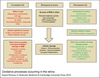
We have highlighted the important role of oxidative damage in the pathogenesis of AMD and the potential benefits of antioxidants. This is summarised in Figure 4. However, oxidative damage is likely to be only one of several pathways involved in the pathogenesis of a multifactorial disease such as AMD. Further studies are required to investigate the possible roles of the potential therapies as listed above, especially because some might have oncogenic potential. Nevertheless, future treatments based on protecting against antioxidant damage are likely to be part of a multifaceted approach, encompassing a deeper understanding of the disease processes underlying AMD.
Oxidative processes occurring in the retina. Reactive oxygen species (ROS) are normally produced in the retina and retinal pigment epithelial (RPE) as a result of normal cellular metabolic activity. In addition, incoming light itself is a potent generator of oxidative stress, as is RPE phagocytosis of outer segments. ROS may also be produced in the choriocapillaris from the effects of light. ROS can cause retinal cellular damage directly, or indirectly by peroxidation of polyunsaturated fatty acids, found in especially high levels in retinal cell membranes. These products of lipid peroxidation are in turn toxic. Furthermore, ROS may predispose to increased inflammation, for example by activating the complement cascade, which in turn may cause further cellular damage. Normally, the adverse effects of ROS are kept under control by antioxidants in the retina, including antioxidant enzymes [e.g. superoxide dismutase (SOD), catalase, haem oxygenase (HO)], carotenoids, melanin and others such as the hormone melatonin. Any cellular damage from ROS not mopped up by the antioxidant system is repaired by the retina. Increasing age may increase the potential for oxidative damage in the retina, with a reduction in antioxidant enzyme levels, a reduction in the retina's capacity for repair and increased build-up of a toxic fluorescent substance called lipofuscin in RPE and photoreceptor cells. The macula in particular is highly susceptible to oxidative damage because of the increased levels of incoming light centred on the macula and the consequent high level of metabolic activity. Other factors that may increase oxidative stress and are associated with age-related macular degeneration (AMD) include smoking, exposure to sunlight, drusen formation, systemic antioxidant levels and genetic variations. The risk of oxidative damage may be therapeutically decreased by oral antioxidant supplementation and by reducing the exposure to risk factors such as smoking and excess sunlight. Future therapeutic options include genetic manipulation and pharmacological modification.

Acknowledgements and funding
This work was supported by grants from the Frimley Park Hospital NHS Trust, the TFC Frost Charitable Trust and the Gift of Sight Appeal. We thank the peer reviewers for their comments.