Introduction
Small cell neuroendocrine carcinoma most commonly occurs in the lungs. It accounts for 20 per cent of all lung carcinomas. Extrapulmonary small cell neuroendocrine carcinoma represents 4 per cent of all small cell neuroendocrine carcinoma. Head and neck small cell neuroendocrine carcinoma was first reported in 1965.Reference Ibrahim, Briggs and Corbishley1 Small cell neuroendocrine carcinoma in the nasal cavity and paranasal sinuses is very rare. It has been suggested that the occurrence of small cell neuroendocrine carcinoma of the nasal and paranasal cavity is associated with the existence of accessory salivary glands.Reference Koss, Spiro and Hajdu2
Neuroendocrine tumours are divided into three main groups: carcinoid tumour, which is a well differentiated neuroendocrine carcinoma; atypical carcinoid tumour, which is a moderately differentiated neuroendocrine carcinoma; and small cell neuroendocrine carcinoma, which is a poorly differentiated neuroendocrine carcinoma.Reference Babin, Rouleau, Vedrine, Toussaint, de Raucourt and Malard3
Small cell neuroendocrine carcinoma of the nasal cavity has very familiar histology to small cell tumour of the lung, but it differs clinically, with a propensity for local recurrence rather than early metastatic spread.Reference Rejowski, Campanella and Block4 The treatment for this tumour is complete surgical excision followed by chemotherapy and radiotherapy.
We report a rare case of small cell neuroendocrine carcinoma of the nasal cavity and paranasal sinuses with intracranial extension, which was successfully treated with surgery alone, and there was no evidence of recurrence five years post-operation.
Case report
A 55-year-old Malay gentleman presented with a 6-month history of bilateral spontaneous, intermittent epistaxis, nasal blockage and nasal bridge swelling. There were no associated neurological or eye-related symptoms, and both neurological and eye examinations were normal.
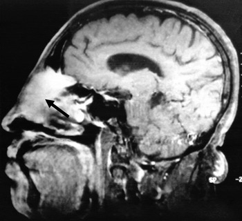
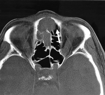
Anterior rhinoscopy revealed nasal masses filling both the nasal cavities, hindering further endoscopic examination. Computed tomography and magnetic resonance imaging (MRI) of the paranasal sinuses revealed a well-defined enhancing mass arising from the nasal septum and occupying most of the nasal cavities. The tumour had destroyed the bony structures and had encroached into the cribriform plate, bilateral medial canthi, and anterior ethmoidal and frontal sinuses (Figures 1 and 2). Computed tomography imaging showed no evidence of distant metastasis. Punch biopsy of the nasal tumour enabled a diagnosis of small cell neuroendocrine carcinoma.
Sagittal computed tomography image of the nasal cavity and paranasal sinuses, which shows a well-defined enhancing mass (arrow) occupying most of the nasal cavity, destroying bone and encroaching into the cribriform plate, and anterior ethmoidal and frontal sinuses.

Axial computed tomography image of the nasal cavity and paranasal sinuses showing the tumour (arrow) occupying the nasal cavity and paranasal sinuses.

A multidisciplinary approach was adopted. The cranial aspect was tackled using bifrontal craniotomy. En bloc resection of the tumour was followed by rhinectomy for removal of the tumour involving the nasal passages. Intra-operatively, a frozen section pathological assessment confirmed negative excision margins. The cranial defect was covered intracranially with vascularised frontal pericranium, and the nasal defect was covered with a free rectus abdominis flap.
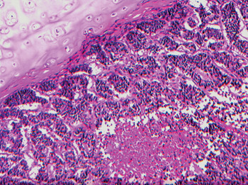
Histopathological examination of the excised tumour revealed small, dark, round cells arranged in a nesting pattern separated by varying amounts of loose eosinophilic, fibroconnective tissue stroma (Figure 3). The tumour cells contained regular, small hyperchromatic nuclei with scanty cytoplasm. In the more cellular areas, the tumour cells exhibited little nuclear pleomorphism with nuclear moulding, and increased mitotic activity. Tumour necrosis and vascular invasion were detected. Mild dysplasia of the adjacent squamous epithelium was noted, but the overlying skin was spared. The cells expressed immunoreactivity for neuron-specific enolase, with focal positivity for MNF 116 (pan-cytokeratin), but they were negative for chromogranin.
Photomicrograph of the tumour, which was composed of nests of uniform, small dark cells with scanty cytoplasm, separated by fibrous septa; areas of tumour necrosis can also be observed. (H&E; ×200)

As the histopathological examination revealed clear surgical margins around the tumour, the patient was given neither radiotherapy nor chemotherapy. Six months after resection, the patient remained clinically well and disease-free, as confirmed by magnetic resonance imaging, and he remained disease-free for the five years of follow up.
Discussion
Small cell neuroendocrine carcinoma involving the nasal cavity and paranasal sinuses is very rare. Less than 250 cases of head and neck small cell neuroendocrine carcinoma have so far been reported in the literature. These included 48 cases of small cell neuroendocrine carcinoma in the nasal and paranasal cavities over a 40-year period.Reference Mineta, Miura, Takebayashi, Araki, Ueda and Harada5
Histologically, small cell carcinoma of the nose and the lungs are very familiar. They are vascular and compact, with intermediate sized cells, and are arranged in sheets, nests or cords. The main difference between small cell neuroendocrine carcinoma of the nasal cavity and that of other sites is its propensity for local recurrence rather than metastatic spread to distant organs.
Radiological examination is important in order to determine the extent of local tumour invasion and distant metastasis. Computed tomography or MRI imaging of the nasal cavity and paranasal sinuses are more useful than conventional radiography when assessing the extent of local invasion of the tumour and are better for the planning of further treatment.
• We report a case of small cell neuroendocrine carcinoma of the nasal cavity and paranasal sinuses
• This is a very rare, locally aggressive tumour, with a high rate of recurrence
• Early aggressive surgical excision, with or without radiotherapy or chemotherapy, can improve outcome and prognosis
• Regular, close follow up is needed to detect any local or distant recurrence
The treatment of small cell neuroendocrine carcinoma has changed considerably over the years. Current therapeutic methods include surgery, radiotherapy and chemotherapy. In the 1980s, the favoured treatment was surgery followed by radiotherapy; this approach has been a resurgence in recent years.Reference Smith, Som, Fahmy, Lawson, Sacks and Brandwein6 This type of tumour should be treated with complete local tumour excision. However, if the surgical margins are suspicious, this should be followed by radiotherapy and adjuvant chemotherapy. In the case reported here, a single treatment modality was adopted as the surgical margins were clear. The patient received close follow-up care in order to detect any recurrence of the tumour.
The overall prognosis of small cell neuroendocrine carcinoma is poor. However, the prognosis seems more favourable in the case of localised nasal and paranasal small cell neuroendocrine carcinoma. One investigation reported that 100 per cent of patients were alive at 5 years, 88 per cent at 7 years and 77 per cent at 10 years.Reference Soussi, Benghiat, Holgate and Majumdar7 Early aggressive surgical excision is recommended as patient survival rate is better compared with similar tumours in the lungs. Close follow up is essential to detect any local recurrence. Hence, long-term survival is possible for patients with nasal and paranasal small cell neuroendocrine carcinoma.
Conclusion
Small cell neuroendocrine carcinoma of the nasal cavity and paranasal sinuses is a very rare, locally aggressive tumour with a high rate of recurrence. Early aggressive surgical excision, with or without radiotherapy or chemotherapy, can improve the outcome and prognosis. Regular, close follow up is needed to detect any local or distant recurrence.