Introduction
The present article continues the series of papers devoted to descriptions of new arsenates from the Arsenatnaya fumarole at the Second scoria cone of the Northern Breakthrough of the Great Tolbachik Fissure Eruption 1975–1976, Tolbachik volcano, Kamchatka Peninsula, Far-Eastern Region, Russia. Twenty one new minerals were characterised in the previous papers of the series, namely yurmarinite Na7(Fe3+,Mg,Cu)4(AsO4)6 (Pekov et al., Reference Pekov, Zubkova, Yapaskurt, Belakovskiy, Lykova, Vigasina, Sidorov and Pushcharovsky2014a), two polymorphs of Cu4O(AsO4)2, ericlaxmanite and kozyrevskite (Pekov et al., Reference Pekov, Zubkova, Yapaskurt, Belakovskiy, Vigasina, Sidorov and Pushcharovsky2014b), popovite Cu5O2(AsO4)2 (Pekov et al., Reference Pekov, Zubkova, Yapaskurt, Belakovskiy, Vigasina, Sidorov and Pushcharovsky2015a), structurally related shchurovskyite K2CaCu6O2(AsO4)4 and dmisokolovite K3Cu5AlO2(AsO4)4 (Pekov et al., Reference Pekov, Zubkova, Belakovskiy, Yapaskurt, Vigasina, Sidorov and Pushcharovsky2015b), katiarsite KTiO(AsO4) (Pekov et al., Reference Pekov, Yapaskurt, Britvin, Zubkova, Vigasina and Sidorov2016a), melanarsite K3Cu7Fe3+O4(AsO4)4 (Pekov et al., Reference Pekov, Zubkova, Yapaskurt, Polekhovsky, Vigasina, Belakovskiy, Britvin, Sidorov and Pushcharovsky2016b), pharmazincite KZnAsO4 (Pekov et al., Reference Pekov, Yapaskurt, Belakovskiy, Vigasina, Zubkova and Sidorov2017a), arsenowagnerite Mg2(AsO4)F (Pekov et al., Reference Pekov, Zubkova, Agakhanov, Yapaskurt, Chukanov, Belakovskiy, Sidorov and Pushcharovsky2018b), arsenatrotitanite NaTiO(AsO4) (Pekov et al., Reference Pekov, Zubkova, Agakhanov, Belakovskiy, Vigasina, Yapaskurt, Sidorov, Britvin and Pushcharovsky2019a), the two isostructural minerals edtollite K2NaCu5Fe3+O2(AsO4)4 and alumoedtollite K2NaCu5AlO2(AsO4)4 (Pekov et al., Reference Pekov, Zubkova, Agakhanov, Ksenofontov, Pautov, Sidorov, Britvin, Vigasina and Pushcharovsky2019b), anatolyite Na6(Ca,Na)(Mg,Fe3+)3Al(AsO4)6 (Pekov et al., Reference Pekov, Lykova, Yapaskurt, Belakovskiy, Turchkova, Britvin, Sidorov and Scheidl2019c), zubkovaite Ca3Cu3(AsO4)4 (Pekov et al., Reference Pekov, Lykova, Agakhanov, Belakovskiy, Vigasina, Britvin, Turchkova, Sidorov and Scheidl2019d), pansnerite K3Na3Fe3+6(AsO4)8 (Pekov et al., Reference Pekov, Zubkova, Koshlyakova, Agakhanov, Belakovskiy, Vigasina, Yapaskurt, Britvin, Turchkova, Sidorov and Pushcharovsky2020a), badalovite NaNaMg(MgFe3+)(AsO4)3 (Pekov et al., Reference Pekov, Koshlyakova, Agakhanov, Zubkova, Belakovskiy, Vigasina, Turchkova, Sidorov and Pushcharovsky2020b), calciojohillerite NaCaMgMg2(AsO4)3 (Pekov et al., Reference Pekov, Koshlyakova, Agakhanov, Zubkova, Belakovskiy, Vigasina, Turchkova, Sidorov and Pushcharovsky2021a), yurgensonite K2SnTiO2(AsO4)2 (Pekov et al., Reference Pekov, Zubkova, Agakhanov, Yapaskurt, Belakovskiy, Vigasina, Britvin, Turchkova, Sidorov and Pushcharovsky2021b), paraberzeliite NaCaCaMg2(AsO4)3 (Pekov et al., Reference Pekov, Koshlyakova, Belakovskiy, Vigasina, Zubkova, Agakhanov, Britvin, Sidorov and Pushcharovsky2022a) and khrenovite Na3Fe3+2(AsO4)3 (Pekov et al., Reference Pekov, Koshlyakova, Belakovskiy, Vigasina, Zubkova, Agakhanov, Britvin, Sidorov and Pushcharovsky2022b).
In this paper the new mineral axelite (Cyrillic: акселит), ideally Na14Cu7(AsO4)8F2Cl2, is described. It is named in honour of the outstanding Finnish–Russian crystallographer, mineralogist and materials scientist, Academician of the Russian Academy of Sciences (St. Petersburg Emperor Academy of Sciences) Axel Gadolin (1828–1892).
Both the new mineral and its name (symbol Axe) have been approved by the Commission on New Minerals, Nomenclature and Classification of the International Mineralogical Association (IMA2017-015a, Pekov et al., Reference Pekov, Zubkova, Agakhanov, Yapaskurt, Belakovskiy, Britvin, Sidorov and Pushcharovsky2017b). The holotype specimen is deposited in the systematic collection of the Fersman Mineralogical Museum of the Russian Academy of Sciences, Moscow with the catalogue number 95905.
Occurrence and general appearance
The Second scoria cone is a monogenetic volcano formed as a result of eruptive activity of the Northern Breakthrough of the Great Tolbachik Fissure Eruption in 1975. It is located 18 km SSW of the Ploskiy Tolbachik volcano (Fedotov and Markhinin, Reference Fedotov and Markhinin1983). The Arsenatnaya fumarole belongs to the main fumarole field located at the summit of the Second scoria cone. This active, hot fumarole, its general mineralogical features and zonation of sublimate incrustations have been reported by Pekov et al. (Reference Pekov, Koshlyakova, Zubkova, Lykova, Britvin, Yapaskurt, Agakhanov, Shchipalkina, Turchkova and Sidorov2018a) and Shchipalkina et al. (Reference Shchipalkina, Pekov, Koshlyakova, Britvin, Zubkova, Varlamov and Sidorov2020). The mineralogy and geochemistry of the very interesting and unusual oxidising-type fumaroles of Tolbachik in general were recently reviewed (Pekov et al., Reference Pekov, Agakhanov, Zubkova, Koshlyakova, Shchipalkina, Sandalov, Yapaskurt, Turchkova and Sidorov2020c and references therein).
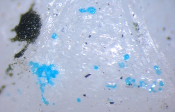
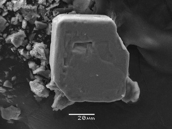
Among more than fifty arsenates known in the Arsenatnaya fumarole, axelite is one of the rarest minerals. It was found in only a few specimens. The specimen which became the holotype (sample #4673) was collected by us in July 2015 from a pocket ~1.5 m below the day surface, within the so-called polymineralic zone of the fumarole (zone IV: Shchipalkina et al., Reference Shchipalkina, Pekov, Koshlyakova, Britvin, Zubkova, Varlamov and Sidorov2020). The temperature measured using a chromel–alumel thermocouple in this pocket during sampling was 420°C. Axelite occurs as tabular, quadratic or, more typically, rectangular (flattened on {001} and sometimes slightly elongated along [100]) or stronger distorted crystals up to 0.02 × 0.1 × 0.1 mm. The crystals, typically with uneven surfaces, are formed by pinacoidal faces {001} and prismatic faces {100} and {110} (Figs 1 and 2). They are separate or combined in interrupted crusts up to 0.4 mm across overgrowing sylvite together with two other Na–Cu arsenates, arsmirandite Na18Cu12Fe3+O8(AsO4)8Cl5 and bradaczekite NaCu4(AsO4)3 (Fig. 1). Associated minerals also include halite, johillerite, tilasite, ericlaxmanite, lammerite, hematite, tenorite, cassiterite, pseudobrookite, aphthitalite-group sulfates, anhydrite, fluoborite, sanidine (As-bearing variety) and fluorophlogopite.
Numerous sky-blue crystals and crystal clusters of axelite, dark olive-green crystal crust of arsmirandite and separate very dark blue prismatic crystals of bradaczekite on the surface of a coarse sylvite crystal. Field of view width is 2.0 mm. Sample # Tolb-4673, photo: I.V. Pekov & A.V. Kasatkin.

Scanning electron microscopy image of a typical crystal of axelite. Sample # Tolb-4673.

Axelite appears to be a ‘classic’ fumarolic mineral. We consider that it was deposited directly from hot gas as volcanic sublimate at the temperatures not lower than 420–450°C.
Physical properties and optical data
Axelite is transparent, sky-blue, with white streak and vitreous lustre. The mineral is brittle; cleavage or parting was not observed. The fracture is uneven. Density calculated using the empirical formula and unit-cell volume found from single-crystal X-ray diffraction data is 3.662 g cm–3.
Axelite is optically uniaxial (–), ɛ = 1.650(4), ω = 1.678(4) (589 nm). In plane-polarised light, the mineral demonstrates weak pleochroism with the following absorption scheme: E (green) > O (light green).
Chemical composition
The chemical composition of axelite was determined using a Jeol JSM–6480LV scanning electron microscope equipped with an INCA-Wave 500 wavelength-dispersive spectrometer (Laboratory of Analytical Techniques of High Spatial Resolution, Department of Petrology, Moscow State University), with an acceleration voltage of 20 kV, a beam current of 20 nA, and a 3 μm beam diameter.
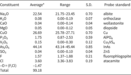
The chemical data are given in Table 1. Contents of other elements with atomic numbers >6 were below detection limits. The empirical formula calculated on the basis of O+F+Cl = 36 atoms per formula unit (apfu) is Na14.37K0.03Ca0.01Mg0.02Cu6.63P0.49V0.03As7.59S0.01O32.36F1.63Cl2.01. The simplified formula of axelite, written taking into account the structure data (see below), is Na14(Cu,Na)7[(As,P)O4]8(F,O)2Cl2. The idealised formula is Na14Cu7(AsO4)8F2Cl2 which requires Na2O 21.84, CuO 28.03, As2O5 46.26, F 1.91, Cl 3.57, –O=(F,Cl) –1.61, total 100 wt.%.
Chemical composition (wt. %) of axelite.

*Averaged for five spot analyses.
S.D. – standard deviation
X-ray crystallography and crystal structure determination details
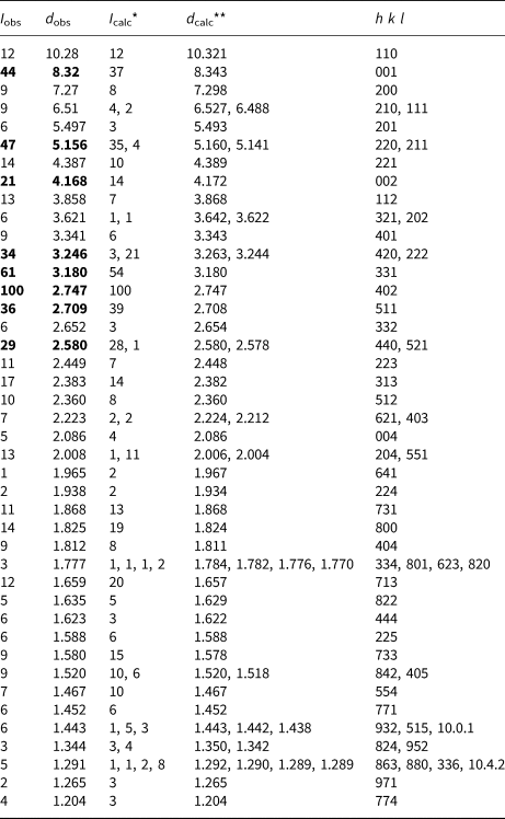
Powder X-ray diffraction (XRD) data for axelite (Table 2) were collected with a Rigaku R-AXIS Rapid II diffractometer equipped with a cylindrical image plate detector (radius 127.4 mm) using Debye-Scherrer geometry, CoKα radiation (rotating anode with VariMAX microfocus optics), 40 kV, 15 mA and an exposure time of 15 min. Angular resolution of the detector is 0.045 2θ (pixel size = 0.1 mm). The data were integrated using the software package Osc2Tab (Britvin et al., Reference Britvin, Dolivo-Dobrovolsky and Krzhizhanovskaya2017). The tetragonal unit cell parameters refined from the powder data are: a = 14.604(2), с = 8.348(2) Å and V = 1780(1) Å3.
Powder X-ray diffraction data (d in Å) of axelite.

* For the calculated pattern, only reflections with intensities ≥1 are given; ** for the unit-cell parameters calculated from single-crystal data; the strongest reflections are marked in bold type.
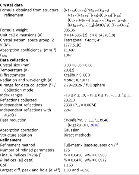
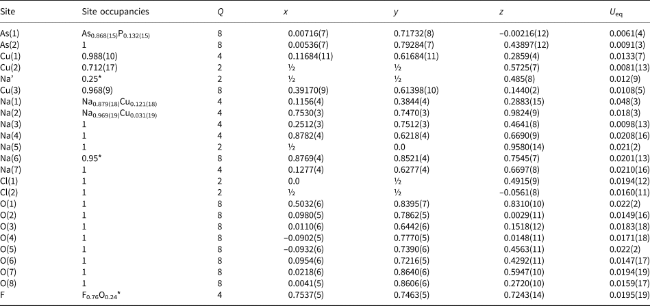
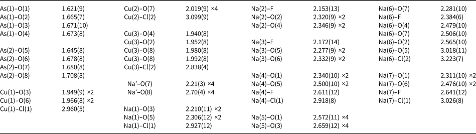
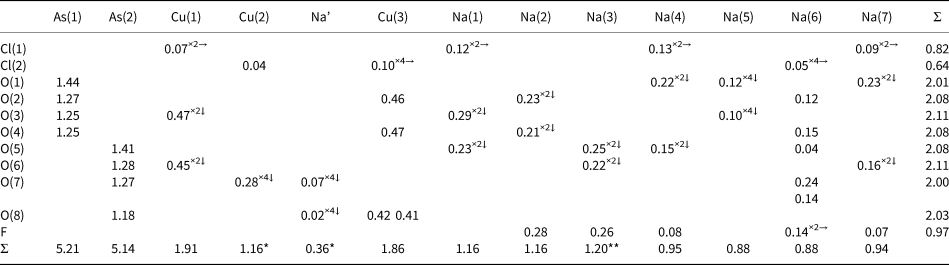
A single-crystal XRD study of axelite was carried out using an Xcalibur S diffractometer equipped with a CCD detector (MoKα radiation). A full sphere of three-dimensional data was collected. Intensity data were corrected for Lorentz and polarisation effects. The crystal structure of the mineral was solved by direct methods and refined using the SHELX software package (Sheldrick, Reference Sheldrick2015) to R = 0.0450 on the basis of 2247 independent reflections with I > 2σ(I). Crystal data, data collection information and structure refinement details are given in Table 3, coordinates and equivalent displacement parameters of atoms in Table 4, selected interatomic distances in Table 5, and bond valence calculations in Table 6. The crystallographic information file has been deposited with the Principal Editor of Mineralogical Magazine and is available as Supplementary material (see below).
Crystal data, data collection information and structure refinement details for axelite.

*Z = 2 for the idealised formula with whole-number coefficients, Na14Cu7(AsO4)8F2Cl2.
Сoordinates and equivalent displacement parameters (U eq, in Å2) of atoms, site multiplicities (Q) and site occupancies for axelite.

*Fixed on the last stage of the refinement for charge balance.
A small amount of Cu2+ could also be present as an admixture in the Na(3) site (possibly accompanied with a vacancy). In this case, the highest peak on the difference-Fourier synthesis (1.83e/Å3) with x = 0.2556, y = 0.2444 and z = 0.2211 could be considered as an additional ligand for Cu2+ in the Na(2) and Na(3) sites slightly occupied by O2–. Interatomic distances between this additional ligand and the Na(2) and Na(3) sites are ~2.00 and 2.03 Å.
Selected interatomic distances (Å) in the structure of axelite.

Bond valence calculations for axelite.

Bond-valence parameters were taken from Gagné and Hawthorne (Reference Gagné and Hawthorne2015); for Cu–Cl, Na–Cl and Na–F the parameters were taken from Brese and O'Keeffe (Reference Brese and O'Keeffe1991). For (F,O) site parameters of F were used. Site occupancies (Table 4) were taken into account.
*The Cu(2) site could possibly accommodate a small amount of Na. The Na’ site is located near Cu(2) [Cu(2)–Na’ = 0.73(4) Å and this makes impossible their simultaneous filling] and could contain a small amount of Ca2+. **The Na(3) site could contain admixed Cu2+ with simultaneous presence of vacancy (see footnote of Table 4).
Discussion
No mineral or synthetic compound closely related to axelite is found in literature and databases: it represents a novel structure type. In addition, being a H-free mineral, axelite is distantly related, in both structural and chemical aspects, to lavendulan-like hydrous arsenates and phosphates [primarily Cl-bearing species mahnertite (Na,Ca,K)Cu3(AsO4)2Cl⋅5H2O (Pushcharovsky et al., Reference Pushcharovsky, Zubkova, Teat, MacLean and Sarp2004), zdenĕkite NaPbCu5(AsO4)4Cl⋅5H2O (Zubkova et al., Reference Zubkova, Pushcharovsky, Sarp, Teat and MacLean2003), lavendulan NaCaCu5(AsO4)4Cl⋅5H2O and sampleite NaCaCu5(PO4)4Cl⋅5H2O (Giester et al., Reference Giester, Kolitsch, Leveret, Turner and Williams2007)] and analogous natural and synthetic compounds (Kiriukhina et al., Reference Kiriukhina, Yakubovich, Shvanskaya, Volkov, Dimitrova, Simonov, Volkova and Vasiliev2022 and references therein). It is noteworthy, however, all these hydrous minerals of supergene origin differ strongly from axelite in unit-cell dimensions, powder XRD and the majority of physical properties.
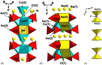
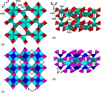
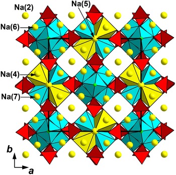
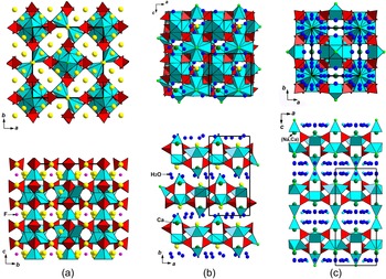
In the axelite structure (Figs 3, 4, 5 and 6a), the important unit is the heteropolyhedral chain running along the с axis and built by clusters formed by CuO4Cl square pyramids connected with AsO4 tetrahedra. Topologically the same clusters were reported in lavendulan-type minerals and structurally related compounds (Giester et al., Reference Giester, Kolitsch, Leveret, Turner and Williams2007 and references therein) and are shown in Fig. 6b,c. The cluster in axelite consists of four Cu(3)O4Cl pyramids linked to each other via common O–Cl edges (four O atoms and one common Cl atom) thus forming a [Cu4O12Cl] unit. Eight AsO4 tetrahedra share vertices with this unit; four of them, As(2)O4 tetrahedra, are further bound via common vertices with the tetragonal base of the fifth Cu(2)O4Cl square pyramid that shares a common Cl atom with the neighbouring [Cu4O12Cl] unit. Thus the heteropolyhedral chains (Fig. 3a) are formed. In contrast to lavendulan-type minerals and the majority of the structurally related compounds in which, topologically, the same clusters consisting of five tetragonal pyramids and eight T 5+O4tetrahedra (T = As, P) are connected to form heteropolyhedral layers, in axelite the linkage of the fifth Cu(2)O4Cl square pyramid to the neighbouring [Cu4O12Cl] unit via the common Cl vertex allows the heteropolyhedral chains to be considered as the main building block. Among lavendulan-like compounds, a similar linkage of the fifth tetragonal pyramid to the neighbouring cluster was found in Ba(VO)Cu4(PO4)4 (tetragonal, P4212, a = 9.5598, c = 7.160 Å: Meyer and Müller-Buschbaum, Reference Meyer and Müller-Buschbaum1997) (Fig. 4c,d), synthetic phosphates isostructural with this compound A(TiO)Cu4(PO4)4 (A = Ba, Sr and Pb) (Kimura et al., Reference Kimura, Sera and Kimura2016, Reference Kimura, Toyoda, Babkevich, Yamauchi, Sera, Nassif, Rønnow and Kimura2018), and structurally close K(NbO)Cu4(PO4)4 (tetragonal, P4/nmm, a = 9.6865, c = 7.1530 Å: Kimura et al., Reference Kimura, Urushihara, Asaka, Toyoda, Miyake, Tokunaga and Kimura2020). Adjacent chains in axelite are connected via common vertices of AsO4 tetrahedra with Cu(1)2O8Cl dimers built by two Cu(1)-centred square pyramids with the common Cl vertex thus forming a heteropolyhedral pseudo-framework (Fig. 4a,b). All Cu2+ cations in axelite have, due to the Jahn-Teller effect, [4+1] coordination and centre tetragonal pyramids. In each of these polyhedra, the square base is built by O atoms with Cu–O distances lying in the range from 1.94 to 2.02 Å, whereas the apical ligand is a Cl atom and Cu–Cl distances vary from 2.84 to 3.10 Å (Table 5). For the Cu(1) and Cu(3) sites, occupancy factors (s.o.f.) are close to 1.0 (0.99 and 0.97, respectively), however the Cu(2) site is filled only to 71%. An additional Na’ site with s.o.f. 0.25 is localised near Cu(2) from the difference-Fourier synthesis [Cu(2)–Na’ = 0.73 Å thus these positions cannot be filled simultaneously]. This site is located inside the chain, directly on its axis, between four As(2)O4 tetrahedra and the [Cu4O12Cl] unit, and centres a slightly distorted cube Na'O8, which shares four common edges with As(2)O4 tetrahedra and four edges with Cu(3)O4Cl pyramids. The Na(6) sites are also located inside the chain but around its axis, at the same height as the Cu(2)-centred pyramid (Fig. 3a). They centre seven-fold polyhedra Na(6)O5FCl. The Na(1) site shows a distinct partial substitution of Na by Cu (Tables 4 and 6) and centres a square pyramid with four O atoms in the square base and Na(1)–O distances in the range from 2.21 to 2.30 Å and an elongated fifth bond Na(1)–Cl(1) = 2.93 Å. Two Na(1)O4Cl pyramids share common O–Cl(1) edges with two Cu(1)O4Cl pyramids thus forming topologically the same cluster [Na2Cu2O12Cl] as was determined for Cu(3)-centred polyhedron. Eight AsO4 tetrahedra share vertices with this unit in the same way as described above for the heteropolyhedral chains. The Na(5) site centres a slightly distorted cube sharing four common edges with As(1)O4 tetrahedra, two edges with two Cu(1)O4Cl pyramids and two edges with two Na(1)O4Cl pyramids. The position of Na(5)-centred cubes is similar with the location of Na’-centred cubes inside the above-described chains (Fig. 3a). Thus, interrupted heteropolyhedral chains topologically close to those described above and shown in Fig. 3a, however, do not contain additional Cu(2)-centred pyramids. The Na(4) and Na(7) sites centre distorted NaO4FCl octahedra and are located inside these interrupted chains around their axes (Fig. 3b), similarly to Na(6) cations (Fig. 3a). The Na(2) and Na(3) sites are located in channels of the heteropolyhedral pseudo-framework and centre square pyramids with four elongated Na–O bonds, forming the base of the pyramids, and one short Na–F bond (Table 5). Adjacent Na(2)- and Na(3)-centred pyramids share common F vertices to form dimers (Fig. 3c). The structures of axelite (tetragonal) and two distantly related hydrous copper chloro-arsenates lavendulan (monoclinic, pseudotetragonal: Giester et al., Reference Giester, Kolitsch, Leveret, Turner and Williams2007) and mahnertite (tetragonal: Pushcharovsky et al., Reference Pushcharovsky, Zubkova, Teat, MacLean and Sarp2004) are compared in Fig. 6. In the layered structures of lavendulan (Fig. 6b), zdenekite, sampleite (Giester et al., Reference Giester, Kolitsch, Leveret, Turner and Williams2007), richelsdorfite Ca2Cu5SbCl(OH)6(AsO4)4⋅6H2O (Süsse and Tillmann, Reference Süsse and Tillmann1987) and synthetic Na2Li0.75(Cs,K)0.5[Cu5(PO4)4Cl]⋅3.5(H2O,OH) (Kiriukhina et al., Reference Kiriukhina, Yakubovich, Shvanskaya, Volkov, Dimitrova, Simonov, Volkova and Vasiliev2022), Na5ACu4(AsO4)4Cl2 (A = Rb and Cs) (Hwu et al., Reference Hwu, Ulutagay-Kartin, Clayhold, Mackay, Wardojo, O'Connor and Krawiec2002), Na3Cu5(PO4)4F⋅4H2O (Yue et al., Reference Yue, Ouyang, Cui, Yin, Xiao, Wang, Liu, Wang, Xia, Huang and He2018) and Sr2Cu5(PO4)4X 2⋅8H2O (X = Cl and, Br) (Qiu et al., Reference Qiu, He, Cui, Chen and Tang2017), the neighbouring heteropolyhedral layers are shifted relative to each other. In mahnertite the neighbouring layers are connected with each other via the fifth vertices of the Cu-centred pyramids which do not belong to the [Cu4O12Cl] clusters and their bases share all vertices with AsO4 tetrahedra linked with the clusters forming a heteropolyhedral pseudo-framework (Fig. 6c). The same linkage of the heteropolyhedral layers was reported for the structure of a microporous potassium vanadyl phosphate analogue of mahnertite K2.5Cu5Cl(PO4)4(OH)0.5(VO2)⋅H2O (tetragonal: Yakubovich et al., Reference Yakubovich, Steele, Kiriukhina and Dimitrova2015). In andyrobertsite KCdCu5(AsO4)4(AsO2(OH)2)⋅2H2O (monoclinic: Cooper and Hawthorne, Reference Cooper and Hawthorne2000) and calcioandyrobertsite–2O KCaCu5(AsO4)4(AsO2(OH)2)⋅2H2O (orthorhombic: Sarp and Černy, Reference Sarp and Černy2004), the layers are linked via additional As-centred tetrahedra. In axelite topologically close layers [in the case when Na(1)-centred tetragonal pyramids are also considered as the cluster-forming polyhedra] are linked via the tetragonal pyramids which do not participate in the clusters. However, in contrast with Ba(VO)Cu4(PO4)4 (Meyer and Müller-Buschbaum, Reference Meyer and Müller-Buschbaum1997), phosphates A(TiO)Cu4(PO4)4 (A = Ba, Sr and Pb) (Kimura et al., Reference Kimura, Sera and Kimura2016, Reference Kimura, Toyoda, Babkevich, Yamauchi, Sera, Nassif, Rønnow and Kimura2018) and K(NbO)Cu4(PO4)4 (Kimura et al., Reference Kimura, Urushihara, Asaka, Toyoda, Miyake, Tokunaga and Kimura2020), only one half of these pyramids are present in axelite (Fig. 4): the clusters with Na(1)-centred pyramids are not connected with the additional tetragonal pyramids.
Fragments of the crystal structure of axelite: (a) heteropolyhedral chain built by [Cu(3)4O12Cl] units, AsO4 tetrahedra and Cu(2)O4Cl square pyramids; an additional Na’ site is shown, its coordination polyhedron is semi-transparent; (b) a fragment consisting of [Na2Cu2O12Cl] clusters, AsO4 tetrahedra and Na(5)O8 cubes; (c) dimers built by Na(2) and Na(3) square pyramids.

Heteropolyhedral Cu–As–O–Cl pseudo-framework in the structure of axelite in two projections (a,b) and Cu–P–V–O pseudo-framework in Ba(VO)Cu4(PO4)4 (c,d, drawn after Meyer and Müller-Buschbaum, Reference Meyer and Müller-Buschbaum1997). The unit cells are outlined.

The crystal structure of axelite in ab projection with Na(1) sites shown as polyhedra (yellow). The unit cell is outlined.

Crystal structures of (a) axelite Na14Cu7(AsO4)8F2Cl2, (b) lavendulan NaCaCu5(AsO4)4Cl⋅5H2O (drawn after Giester et al., Reference Giester, Kolitsch, Leveret, Turner and Williams2007), and (c) mahnertite (Na,Ca)Cu3(AsO4)2Cl⋅5H2O (drawn after Pushcharovsky et al., Reference Pushcharovsky, Zubkova, Teat, MacLean and Sarp2004), each in two projections. Cu-centred polyhedra are light blue (additional Cu sites in mahnertite are light-blue circles), AsO4 tetrahedra are red, Cl atoms are light-green circles. The unit cells are outlined.

The crystal chemical formula of a crystal of axelite used for the structure determination (see Table 4) is Na(1)(Na0.88Cu0.12)2 Na(2)(Na0.97Cu0.03)2 Na(3)Na2 Na(4)Na2 Na(5)Na Na(6)(Na0.95□0.05)4 Na(7)Na2 Cu(1)(Cu0.99□0.01)2 {Na’(□0.75Na0.25)Cu(2)(Cu0.71□0.29)} Cu(3)(Cu0.97□0.03)4 [As(1)(As0.87P0.13)O4]4 [As(2)AsO4]4 Cl(1)Cl Cl(2)Cl F(F0.76O0.24)2 (Z = 2). This formally corresponds to the composition Na14.75Cu6.87P0.52As7.48O32.48F1.52Cl2.00 that slightly differs, mainly in part of the Na:Cu ratio, from the empirical formula Na14.37K0.03Ca0.01Mg0.02Cu6.63P0.49V0.03As7.59S0.01O32.36F1.63Cl2.01 calculated from averaged electron microprobe data (Table 1). It is probably caused by slight ambiguity in the determination of content of several cationic sites: Na(1), Na(2) and, especially, adjacent and, therefore, partially occupied Na’ and Cu(2). Thus we prefer (taking also into account that the Na’ site is definitely vacancy-dominant) to use data from the electron microprobe which gives a whole-number coefficient of 14 for Na in the simplified formula of axelite: Na14Cu7(AsO4)8F2Cl2 (Z = 2).
Acknowledgements
We thank Giuseppina Balassone, Oleg Siidra, two anonymous referees and Associate Editor Elena Zhitova for their valuable comments. The mineralogical and crystal chemical studies of axelite by IVP, NVZ and DYP were supported by the Russian Science Foundation, grant no. 19-17-00050. The technical support by the SPbSU X-Ray Diffraction Resource Center in the powder XRD study is acknowledged.
Supplementary material
To view supplementary material for this article, please visit https://doi.org/10.1180/mgm.2022.120
Competing interests
The authors declare none.