


Introduction
In 2019, in the pyrometamorphic Hatrurim Complex rocks in the Negev Desert, Israel, we discovered a unique phosphide-bearing breccia, in which osbornite, TiN, was found in situ (Galuskin et al., Reference Galuskin, Galuskina, Kamenetsky, Ye, Kusz and Zieliński2022; Fig. 1a,b; Fig. 2a,b). Osbornite is an indicator of the super-reduced conditions of rock genesis (Griffin et al. Reference Griffin, Gain, Saunders, Alard, Shaw, Toledo and O'Reilly2021a). It is a mineral typical of enstatite meteorites (Bannister, Reference Bannister1941; Weisberg et al., Reference Weisberg, Prinz and Nehru1988; Grokhovsky, Reference Grokhovsky2006; Leitner et al., Reference Leitner, Vollmer and Hoppe2018) and encountered extremely rarely on Earth. All ‘terrestrial’ findings of osbornite are associated exclusively with mineral separates of bulk geological samples (mineral fractions) (Tatarincev et al., Reference Tatarintsev, Sandomirskaya and Tsumbal1987; Dobrzhinetskaya et al., Reference Dobrzhinetskaya, Wirth, Yang, Hutcheon, Weber and Green2009; Xu et al., Reference Xu, Yang, Guo and Xiong2013; Xiong et al., Reference Xiong, Griffin, Huang, Gain, Toledo, Pearson and O'Reilly2017, Reference Xiong, Xu, Mugnaioli, Gemmi, Wirth, Grew and Robinson2020, Reference Xiong, Xu, Mugnaioli, Gemmi, Wirth, Grew and Robinson2022; Griffin et al., Reference Griffin, Gain, Adams, Huang, Saunders, Toledo, Pearson and O'Reilly2016a, Reference Griffin, Gain, Adams, Toledo, Pearson and O'Reilly2016b, Reference Griffin, Gain, Huang, Saunders, Shaw, Toledo and O'Reilly2019a, Reference Griffin, Gain, Saunders, Cámara, Bindi, Spartà, Toledo and O'Reilly2021b; Silaev et al., Reference Silaev, Karpov, Anikin, Vergasova, Fillipov and Tarasov2019; Yatsenko et al., Reference Yatsenko, Skublov, Levashova, Galankina and Bekesha2020, Reference Yatsenko, Galankina, Marin and Skublovb2021). A skeletal osbornite occurring as inclusions in corundum from the alluvial sediments of Mt Carmel, North Israel, is the best known terrestrial osbornite (Griffin et al., Reference Griffin, Gain, Saunders, Alard, Shaw, Toledo and O'Reilly2021a). Mineral separates (concentrates) mined by the Shefa Yamim Company (https://www.shefagems.com) as a result of exploration for gemstones in alluvial deposits are a source of this corundum. Osbornite in the corundum aggregates is associated with unusual super-reduced minerals represented by nitrides, carbides, silicides, FeSi(±Ti,Cr,V)-alloys, and Ti3+-bearing oxides (Griffin et al., Reference Griffin, Gain, Adams, Toledo, Pearson and O'Reilly2016b, Reference Griffin, Gain, Bindi, Toledo, Cámara, Saunders and O'Reilly2018, Reference Griffin, Gain, Huang, Saunders, Shaw, Toledo and O'Reilly2019a,Reference Griffin, Toledo and O'Reillyb, Reference Griffin, Gaina, Cámara, Bindi, Shaw, Alard, Saunders, Huang, Toledo and O'Reilly2020a,Reference Griffin, Gain, Saunders, Bindi, Alard, Toledo and O'Reillyb, Reference Griffin, Gain, Saunders, Alard, Shaw, Toledo and O'Reilly2021a,Reference Griffin, Gain, Saunders, Cámara, Bindi, Spartà, Toledo and O'Reillyb; Bindi et al., Reference Bindi, Cámara, Gain, Griffin, Huang, Saunders and Toledo2020).
Localities of osbornite findings in Israel. (a) Negev Desert, Hatrurim Basin – the biggest area of the Hatrurim Complex pyrometamorphic rock outcrops in Israel. The location of (b) is shown in the frame. (b) Outcrop of low-temperature rocks of the Hatrurim Complex along the road Arad–Dead Sea, where phosphide-bearing breccia with osbornite was found (grey fragments of rock shown by arrow). A breccia fragment consisting of grey homogeneous clasts of the host rock cemented by gehlenite paralava is shown in the inset. (c) Typical view of re-cultivated fields after exploration of alluvial deposit by the Shefa Yamim Company. The numbers of the exploration drilling section based on the Shefa Yamim annual 2019 report are shown (Supplementary Fig. S1). (d) Сurrent state of the Kishon River in the area of alluvial deposit exploration by the Shefa Yamim Company. All images were taken in 2022.

Morphology of osbornite found in Israel. (a, b) Osbornite in phosphide-bearing breccia of the Hatrurim Complex: (a) xenomorphic grain with rutile rim; and (b) crystal with morphological elements of skeletal growth. (c, d) Skeletal crystals in sapphire from Carmel confined to glass inclusions and associated with carmeltazite and Ti3+-bearing oxides: (c) typical rounded surfaces of skeletal crystals formed from the melt; and (d) emulsion of osbornite near its skeletal crystals. Brg = barringerite, Cal = calcite, Crn = corundum, Ctz = carmeltazite, Fe = native iron, Gls = glass, Hgr = hydrogrossular, Obn = osbornite, Ssi = sassite-like mineral and Tta = tistarite.

This corundum with inclusions of super-reduced minerals obtained the trademark ‘Carmel sapphire’ and has been the host of 10 new mineral species discovered in the last five years and approved by the Commission on New Minerals, Nomenclature and Classification (CNMNC) of the International Mineralogical Association (IMA): carmeltazite (IMA2018-103) ZrAl2Ti4O11 (Griffin et al., Reference Griffin, Gain, Bindi, Toledo, Cámara, Saunders and O'Reilly2018); kishonite (IMA2020-023) VH2 (Bindi et al., Reference Bindi, Cámara, Gain, Griffin, Huang, Saunders and Toledo2020); oreillyite (IMA 2020-030a) Cr2N (Bindi et al., Reference Bindi, Cámara, Gain, Griffin, Huang, Saunders and Toledo2020); griffinite (IMA 2021-110) Al2TiO5 (Ma et al., Reference Ma, Bindi, Cámara and Toledo2022a); magnéliite (IMA2021-111) Ti3+2Ti4+2O7 (Ma et al., Reference Ma, Griffin, Bindi, Cámara and Toledo2022b); ziroite (IMA2022-013) ZrO2 (Ma et al., Reference Ma, Griffin, Bindi, Cámara and Toledo2022c); sassite (IMA2022-014) Ti3+2Ti4+O5 (Ma et al., Reference Ma, Griffin, Bindi, Cámara and Toledo2022d); mizraite-(Ce) (IMA2022-027) Ce(Al11Mg)O19 (Ma et al., Reference Ma, Griffin, Bindi, Cámara and Toledo2022e); toledoite (IMA2022-036) TiFeSi (Ma et al., Reference Ma, Griffin, Bindi and Cámara2022f); and yeite (IMA2022-079) TiSi (Ma et al., Reference Ma, Griffin, Bindi, Cámara and Toledo2023). Grain aggregates of Carmel sapphire (and also hibonite and moissanite) from Carmel have been the objects of numerous scientific publications (Griffin et al., Reference Griffin, Gain, Adams, Huang, Saunders, Toledo, Pearson and O'Reilly2016a,Reference Griffin, Gain, Adams, Toledo, Pearson and O'Reillyb, Reference Griffin, Gain, Bindi, Toledo, Cámara, Saunders and O'Reilly2018, Reference Griffin, Gain, Huang, Saunders, Shaw, Toledo and O'Reilly2019a,Reference Griffin, Toledo and O'Reillyb, Reference Griffin, Gaina, Cámara, Bindi, Shaw, Alard, Saunders, Huang, Toledo and O'Reilly2020a,Reference Griffin, Gain, Saunders, Bindi, Alard, Toledo and O'Reillyb, Reference Griffin, Gain, Saunders, Alard, Shaw, Toledo and O'Reilly2021a,Reference Griffin, Gain, Saunders, Cámara, Bindi, Spartà, Toledo and O'Reillyb, Reference Griffin, Gain, Saunders, Huang, Alard, Toledo and O'Reilly2022; Xiong et al., Reference Xiong, Griffin, Huang, Gain, Toledo, Pearson and O'Reilly2017; Dobrzhinetskaya et al., Reference Dobrzhinetskaya, Mukhin, Wang, Wirth, O'Bannon, Zhao, Eppelbaum and Sokhonchuk2018; Cámara et al., Reference Cámara, Bindi, Pagano, Pagano, Gain and Griffin2019; Bindi et al., Reference Bindi, Cámara, Griffin, Huang, Gain, Toledo and O'Reilly2019, Reference Bindi, Cámara, Gain, Griffin, Huang, Saunders and Toledo2020; Huang et al., Reference Huang, Xiong, Gain, Griffin, Murphy, Shiryaev, Li, Toledo, Tomshin and O'Reilly2020; Stan et al., Reference Stan, O'Bannon III, Mukhin, Tamura and Dobrzhinetskaya2020; Oliveira et al., Reference Oliveira, Griffin, Gain, Saunders, Shaw, Toledo, Afonso and O'Reilly2021; Lu et al., Reference Lu, Griffin, Huang, Hong-Kun Dai, Castillo-Oliver and O'Reilly2022), in which the authors present the highly controversial hypothesis that the corundum rock with super-reduced minerals formed at the crust–mantle boundary as a result of CH4+H2 fluid flowing through the magmatic melt.
In the article “Nitrogen under super-reducing conditions: Ti oxynitride melts in xenolithic corundum aggregates from Mt Carmel (N. Israel)”, Griffin at al. (Reference Griffin, Gain, Saunders, Cámara, Bindi, Spartà, Toledo and O'Reilly2021b) described skeletal osbornite crystals, which are characterised by the variable composition forming solid solutions with TiC and TiO. The skeletal osbornite crystals are usually accompanied by an emulsion of osbornite, which the authors interpret as “reflect[ing] the expulsion of N from the silicate melts during the eruption of the host magma” (Griffin et al., Reference Griffin, Gain, Saunders, Cámara, Bindi, Spartà, Toledo and O'Reilly2021b).
Recently, the natural origin of Carmel sapphire has been contested (Litasov et al., Reference Litasov, Kagi and Bekker2019a; Ballhaus et al., Reference Ballhaus, Helmy, Fonseca, Wirth, Schreiber and Jöns2021), therefore comparing in situ osbornite from the Negev Desert with the skeletal osbornite crystals from the Carmel sapphire aggregates, and also with osbornite from ‘electrocorundum’ (fused alumina) became a prority. In 2022 we visited the Carmel locality at the Kishon River, Northern Israel (Fig. 1c,d). We succeeded in obtaining original material containing Carmel sapphire, which includes separated grains up to 100 μm in size, and also a small fragment ca. 7 mm, the so-called ‘white breccia’ with Carmel sapphire. Griffin et al. (Reference Litasov, Bekker and Kagi2019b) describe ‘white breccia’ with Carmel sapphire as follows: “The CS [Carmel sapphire] grains from the pyroclastic deposits, and even from the alluvial beds, commonly are coated in glass, but even more commonly by a fine-grained white breccia, consisting of carbonate-cemented volcanic ash. This cement is a result of eruption of the ashes on a shallow marine carbonate platform, and the subsequent covering of the ash beds by thick carbonate sequences.” Griffin and co-authors (Griffin et al., Reference Griffin, Gain, Adams, Toledo, Pearson and O'Reilly2016b, Reference Griffin, Gain, Bindi, Toledo, Cámara, Saunders and O'Reilly2018, Reference Griffin, Gain, Huang, Saunders, Shaw, Toledo and O'Reilly2019a,Reference Griffin, Toledo and O'Reillyb, Reference Griffin, Gaina, Cámara, Bindi, Shaw, Alard, Saunders, Huang, Toledo and O'Reilly2020a,Reference Griffin, Gain, Saunders, Bindi, Alard, Toledo and O'Reillyb, Reference Griffin, Gain, Saunders, Alard, Shaw, Toledo and O'Reilly2021a,Reference Griffin, Gain, Saunders, Cámara, Bindi, Spartà, Toledo and O'Reillyb) were not specific about the cement composition or corundum-free fragments of rock in the ‘white breccia’. On only one occasion the amorphous carbon is recorded in the cement of the ‘white breccia’ (Xiong et al., Reference Xiong, Griffin, Huang, Gain, Toledo, Pearson and O'Reilly2017).
In this paper we present a comparative study of osbornite from the Negev, Carmel and ‘electrocorundum’. We present the results of a mineralogical study of the ‘white breccia’ with Carmel sapphire, which irrefutably indicate its anthropogenic origin. Our conclusions regarding the anthropogenic origin of Carmel sapphire are confirmed by the analysis of the growth inhomogeneity of Carmel sapphire and synthetic corundum using cathodoluminescence (CL).
An optical microscope, scanning electron microscopes Quanta 250/EDS and Philips XL30/EDS/CL, and Raman spectrometer WITec alpha 300R were the main equipment used to investigate the ‘white breccia’. In this paper we use mineral names describing their synthetic analogues.
Israeli osbornite
Osbornite from the phosphide-bearing breccia of the pyrometamorphic Hatrurim Complex, found in the Negev Desert was studied by us and the results of that investigation have been published recently (Galuskin et al., Reference Galuskin, Galuskina, Kamenetsky, Ye, Kusz and Zieliński2022). The mineral is represented by xenomorphic grains that are homogenous in composition (Fig. 2a), and more rarely by crystals with elements of skeletal growth (Fig. 2b). Osbornite is usually associated with schreibersite, barringerite, schreibersite–iron eutectic and native iron (Galuskin et al., Reference Galuskin, Kusz, Galuskina, Książek, Ye and Zieliński2023). Its composition and structure correspond to stoichiometric TiN (Galuskin et al., Reference Galuskin, Galuskina, Kamenetsky, Ye, Kusz and Zieliński2022).
Osbornite from Carmel sapphire grains forms skeletal crystals, which as a rule are confined to glass inclusions and associated with the super-reduced minerals (Fig. 2c,d; Griffin et al., Reference Griffin, Gain, Bindi, Toledo, Cámara, Saunders and O'Reilly2018, Reference Griffin, Gain, Saunders, Cámara, Bindi, Spartà, Toledo and O'Reilly2021b). Griffin et al. (Reference Griffin, Gain, Saunders, Cámara, Bindi, Spartà, Toledo and O'Reilly2021b) underlines the nonstable composition of osbornite from Carmel sapphire presented by the phases of the TiN–TiC–TiO system, however they do not provide data on the composition trends and zonation of these skeletal crystals. Our investigation of numerous skeletal crystals from Carmel sapphire showed that they exhibit regular zonation: the centrum of osbornite skeletal crystals is enriched in C, whereas in the rim the N(+O) content increases, as is reflected in the optical properties and well manifested under reflected light (Fig. 3). In the Raman spectra of osbornite from Carmel sapphire, the presence of C impurity (>1–2 wt.%) is displayed in the appearance of a band at 685 cm–1 (Fig. 3e; Escobar-Alarcona et al., Reference Escobar-Alarcon, Medina, Romero, Fernandez and Solis-Casados2011).
(a,b) Carmel sapphire grain with inclusions of skeletal osbornite crystals: (a) back-scattered electron (BSE) image, fragment magnified in (c) and (d) is shown in the frame; and (b) cathodoluminescence, growing inhomogeneity of corundum is visible. (c,d) Cross-section perpendicular to the skeletal crystal elongation of osbornite: (c) BSE image; and (d) reflected light image showing points 1 and 2 for Raman analysis. The central part of the skeletal crystal enriched in С has a darker, brownish colour in (d). (e) Raman spectra for osbornite from location 1; and (f) Raman spectra for osbornite from location 2 marked in (d). Crn = corundum, Ji = jingsuiite, Obn = osbornite and Sp = spinel.

The osbornite inclusions investigated from the synthetic corundum from slags of the fused alumina production (Supplementary Fig. S2) have zonation analogous to the zonation of osbornite from Carmel sapphire: the centrum is enriched in С and the rim in N(+O) (Supplementary Fig. S3).
‘White breccia’
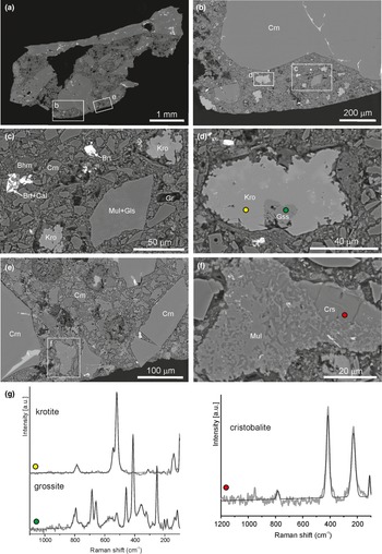
A fragment of the ‘white breccia’ (3×4×7 mm in size, from which a polished mount was prepared) was investigated (Supplementary Fig. S4). In addition to the blue and light-brown fragments of Carmel sapphire grains and aggregates containing a typical super-reduced mineral association, which has been described several times by Griffin and co-authors (Supplementary Figs S5–S7; Griffin et al., Reference Griffin, Gain, Adams, Toledo, Pearson and O'Reilly2016b, Reference Griffin, Gain, Bindi, Toledo, Cámara, Saunders and O'Reilly2018, Reference Griffin, Gain, Huang, Saunders, Shaw, Toledo and O'Reilly2019a,Reference Griffin, Toledo and O'Reillyb, Reference Griffin, Gaina, Cámara, Bindi, Shaw, Alard, Saunders, Huang, Toledo and O'Reilly2020a,Reference Griffin, Gain, Saunders, Bindi, Alard, Toledo and O'Reillyb, Reference Griffin, Gain, Saunders, Alard, Shaw, Toledo and O'Reilly2021a,Reference Griffin, Gain, Saunders, Cámara, Bindi, Spartà, Toledo and O'Reillyb), in the white cement we recognise corundum-free fragments 50–200 μm in size. Primarily aggregates of krotite, CaAl2O4, with grossite, CaAl4O7, and diaoyudaoite, NaAl11O17, inclusions, and also angular grains presented as mullite in glass or cristobalite (Fig. 4; Supplementary Fig. S8). It should be noted that some Carmel sapphire inclusions in the ‘white breccia’ intergrowing with dmisteinbergite, CaAl2Si2O8, crystals look ‘very fresh’ and unaltered (Supplementary Fig. S8a).
(a–f) BSE images. (a) Typical view of ‘white breccia’ fragments, with the location of magnified images (b) and (e) marked. (b) Fragment of breccia with krotite and mullite debris in glass, frames are shown for areas magnified in (c) and (d). (c) Glass angular grain with mullite symplectites. (d) Krotite grain with grossite inclusion, points of Raman spectra measurements are shown by yellow (krotite) and green (grossite) circles. (e) Fragment of ‘white breccia’, with area magnified in (f) indicated. (f) Cristobalite grain with mullite inclusions and red spot indicating the point of Raman spectrum measurement. (g) Raman spectra of minerals marked in (d) and (f). Bhm = böhmite-like mineral, Brt = baryte, Cal = calcite, Crs = cristobalite, Gr = graphite, Gls = glass, Crn = corundum, Gss = grossite, Kro = krotite and Mul = mullite.

In the ‘white breccia’, a rounded porous böhmite-like mineral with ‘cauliflower’ morphology and graphite-like mineral segregations were identified (Fig. 5).
Böhmite-like and graphite-like phases in ‘white breccia’. (a) General view of mounted specimen, fragment magnified in (b) is shown in the frame. (b) Breccia fragment enriched in grains of graphite-like substance, areas magnified in (c) and (d) are outlined by frames. (c) Graphite-like grain with calcite rim; yellow spot location of Ramana spectrum measurement. (d) Böhmite-like mineral; green spot shows a place of EDS spectrum measurement. (e) Raman spectrum of graphite-like mineral and its image in the reflected light. (f) EDS spectrum of böhmite-like phase. Bhm = böhmite-like mineral, Cal = calcite, Fe = native iron, Gr = graphite, Gls = glass, Crn = corundum, Mul = mullite.

A graphite-like substance in the form of a thin emulsion is also present as inclusions in glass from Carmel sapphire. It is practically invisible in optical or electron microscopes, however in the Raman spectra of the glass, characteristic bands of disordered graphite-like substance appear (Fig. 6).
Ti-mineral inclusions in glass of ‘white breccia’. (a) Reflected light and (b) BSE images; the fragment magnified in (c) is shown in the frame. (c) Sassite-like mineral, carmeltazite and spinel intergrowth, fragment magnified in (d) is shown in the frame. (d) Glass with striated surface, which appears as a result of re-polishing of the mount with ‘white breccia’ to a depth of ~0.5 mm. At this depth, the mount sample had only been lightly impregnated with epoxy and so numerous microscopic corundum grains were plucked out and striated the surface of the minerals. Coloured spots show locations of EDS spectra measurement given in (f). (e) Raman spectra of glass with graphite-like phase, inset optical image with white spot shows the place of Raman analysis. (f) EDS spectra of glass (yellow) and spinel (pink). Crn = corundum, Ctz = carmeltazite, Dsb = dmisteinbergite, Gls = glass, Obn = osbornite, Sp = spinel and Tta = tistarite.

An occurrence of a Zr-bearing glass lamella, 2–3 μm in thickness, after re-polishing was unexpected (Fig. 7). It is not probable that this thin, brittle glass lamella was added to the mount during repeated grinding on diamond powder; rather, the lamella was in the ‘white breccia’ cement originally.
(a) Lamella of Zr-bearing glass in the ‘white breccia’, fragment magnified in (b) is shown in the frame. (b) The lamella is ~3 μm in thickness, spots show site of Raman and EDS spectra measurements. (c) Raman spectrum of Zr-bearing glass. (d) EDS spectrum of Zr-bearing glass. Bhm = böhmite-like mineral, Cal = calcite, Gr = graphite, Crn = corundum, Gls = glass, Kro = krotite and Fe = iron.

As noted above, the predominant type of debris in the investigated ‘white breccia’ is corundum. Larger corundum aggregates contain silica glass that is typical for the Carmel sapphire inclusion of super-reduced minerals (Supplementary Figs S5–S7). Silica melt is usually squeezed out between corundum grains (Xiong et al., Reference Xiong, Griffin, Huang, Gain, Toledo, Pearson and O'Reilly2017; Fig. 3a), whereas in the corundum ball-like voids (gaseous) are observed (Fig. 5c, 7b). The size of the angular fragments of corundum in the investigated sample of the ‘white breccia’ varies from 7 mm to 1 μm or less; there is no sorting of grains by size (Fig. 4c,e; 5c; 7a,b; Supplementary Figs S4, S6D, S7B). There are fragments of ‘cracked’ corundum, which probably underwent a shock thermal effect (Fig. 8).
(a) General view of the ‘white breccia’ fragment, the area magnified in (b) is shown in frame. (b) ‘Cracked’ corundum grain. Coloured spots show points of EDS analyses. (c–f) EDS spectra: (c) calcite; (d) böhmite-like mineral; (e) cement; and (f) corundum. Bhm = böhmite-like mineral, Cal = calcite and Crn = corundum.

The cement of the ‘white breccia’ is mainly represented by Al-hydroxides (bauxite). It is clearly visible in the X-ray maps of this element (Fig. 9) and confirmed by the energy-dispersive spectroscopy (EDS) analytical data (Fig. 8). Calcite distributed in the breccia is very inhomogeneous (Fig. 7b; Supplementary Fig. S8B), as reflected in the X-ray maps of Ca, C and O (Fig. 9), and typically the mineral forms separate grains (Fig. 8a,b; 10a). In calcite grains, relics of krotite are observed, which indicates that most of the calcite formed as a result of the carbonatisation of Ca-aluminates (Fig. 10b). Thus, the ‘white breccia’ can be ideally presented as angular grains of Carmel sapphire from 1 μm to 7 mm that are not sorted by size and are cemented by bauxite material.
BSE image of ‘white breccia’. The colour scale in top right indicates the relative element concentration for the X-ray maps of the chosen elements.

(a) Calcite grains in ‘white breccia’. (b) Calcite grain with krotite relics, green spot shows the location of EDS and Raman spectra measurements. (c) EDS and (d) Raman spectrum of krotite. Bhm = böhmite-like mineral, Cal = calcite, Crn = corundum, Kro = krotite.

A comparison of the internal structure of Carmel sapphire and synthetic corundum
For investigation two random grains were selected: (1) pink corundum from a slag sample (Supplementary Fig. S2, Fig. 11a); and (2) light-brown corundum from the ‘white breccia’ (Fig. 11d). In both grains of corundum there are glass and super-reduced mineral inclusions, which in the back-scattered electron images are distinguished by their lighter colour (Fig. 11b,e). Our investigation shows that aggregates of Carmel sapphire and pink corundum have similar growth inhomogeneity, as is clearly visible in the CL images (Fig. 11c,f).
Comparison of growth inhomogeneity of grain aggregates of synthetic corundum (a–c) and Carmel sapphire (d–f). (a, d) – optical images; (b, e) – BSE images; (c, f) – CL images.

Discussion
The composition and morphology of inclusions in the corundum from the Mt Carmel ‘white breccia’ (Supplementary Figs S5–S7) correspond well to the super-reduced minerals in the Carmel sapphire described by Griffin et al. (2016b, 2018, 2019a,b, 2020a,b, 2021a,b). The photos of ‘white breccia’, mentioned by Griffin et al. (Reference Griffin, Toledo and O'Reilly2019b) as a source of Carmel sapphire, are publicly available (https://www.mining.com/new-mineral-found-inside-gemstones, May 2023 – see Supplementary Fig. S9). We consider that the samples studied of the ‘white breccia’ from Carmel are identical to those containing Carmel sapphire.
The anthropogenic genesis of Carmel sapphire was proposed by Litasov et al. (Reference Litasov, Kagi and Bekker2019a) on the basis of comparison of super-reduced inclusions in Carmel sapphire with inclusions in fused alumina. Ballhaus et al. (Reference Ballhaus, Helmy, Fonseca, Wirth, Schreiber and Jöns2021), with reference to the thermodynamic analysis, have shown that super-reduced phases from Carmel sapphire cannot be stable in the conditions of the mantle–crustal boundary, where, in the opinion of Griffin and co-authors, Carmel sapphire was formed (Reference Griffin, Gain, Huang, Saunders, Shaw, Toledo and O'ReillyGriffin et al., 2019а, Reference Griffin, Gaina, Cámara, Bindi, Shaw, Alard, Saunders, Huang, Toledo and O'Reilly2020а). Ballhaus et al. (Reference Ballhaus, Helmy, Fonseca, Wirth, Schreiber and Jöns2021), following Litasov et al. (Reference Litasov, Kagi and Bekker2019a), conclude that Carmel sapphire has an anthropogenic origin. We agree with these authors and provide additional evidence below, which confirms that Carmel sapphire has an anthropogenic origin.
(1) The ‘white breccia’ is composed of unsorted angular corundum grains and aggregates from 1 μm to 7 mm in size (Fig. 4c,e; 5c; 7a,b; Supplementary Figs S4, S6D, S7B). The debris are cemented by bauxite-like material (Al-hydroxides), which also forms aggregates with ‘cauliflower’ morphology up to 200 μm in size – characteristic of calcined bauxite (Fig. 5d). Calcite does not represent the breccia cement, and commonly occurs as separated grains, which contain relics of the high-temperature aluminate: krotite (Fig. 10). Thus the ‘white breccia’ is not a ‘carbonate-cemented volcanic ash’ and it could not form in marine conditions as is suggested by Griffin et al. (Reference Griffin, Toledo and O'Reilly2019b). We think that the Carmel sapphire grains were cemented by calcined bauxite used as a component of the furnace mixture for fused alumina production. The ‘white breccia’ is therefore comprised of the mixed waste of fused alumina production with the material of the furnace mixture. The discovery of Zr-bearing glass used in the production of special laboratory glassware confirms that the ‘white breccia’ is a mixed waste.
(2) Phases typical for slags of fused alumina production and metallurgical slags were identified in the ‘white breccia’ (Erokhin et al., Reference Yu, Khiller and Zakharova2017; Litasov et al., Reference Litasov, Kagi and Bekker2019a). Krotite fragments, commonly with grossite and diaoyudaoite inclusions, are distributed in breccia irregularly (Fig. 4b,c; 7a). Krotite and grossite were discovered and described from meteorites (Weber and Bischoff, Reference Weber and Bischoff1994; Ma et al., Reference Ma, Kampf, Connolly, Beckett, Rossman, Sweeney Smith and Schrader2011), and terrestrial examples are questionable and require verification. For example, the mineral described by Shulamite Gross with the formula CaAl4O7 from a pyrometamorphic rock of the Hatrurim Complex is not grossite, as follows from Gross's original description (Gross, Reference Gross1977). This mineral was identified only by three lines of the diffraction pattern and is characterised by low birefringence, whereas krotite has high birefringence, δ = 0.027. Most likely, it was a product of the hydration of ye'elimite (Ca4Al6(SO4)O12) or brownmillerite (Ca2AlFe3+O5).
Glass fragments with inclusions of mullite crystals are typical for slags from fused alumina production (Hemberger et al., Reference Hemberger, Presser, Zimmermann, Krause and Nickel2009). We found such fragments with mullite in the ‘white breccia’ (Fig. 4c,f; Supplementary Fig. S8). It was not surprising to find grains in the ‘white breccia’ from Carmel sapphire inclusions of a graphite-like material which forms both separated aggregates and an emulsion in silicate glass. We consider that the graphite-like material is the remains of thermally modified coal coke added to the furnace mixture to reduce Fe and Si.
(3) In meteorites and in breccia of the Hatrurim Complex, osbornite associates with iron phosphides and silicates (Bannister, Reference Bannister1941; Weisberg et al., Reference Weisberg, Prinz and Nehru1988; Galuskin et al., Reference Galuskin, Galuskina, Kamenetsky, Ye, Kusz and Zieliński2022, Reference Galuskin, Kusz, Galuskina, Książek, Ye and Zieliński2023). There is no mechanism for the simultaneous removal of Fe and Si from natural melt systems. In the technological process of fused alumina production at temperatures >2000°С there is an almost complete removal of Si and Fe in the form of a melt, which has a lower melting point than corundum, and is gravitationally accumulated in the lower part of the electric furnace in the form of a slag. Very small droplets of the Si–Fe melt remain suspended in the corundum melt and are captured during its crystallisation. Carmel sapphire usually has numerous tiny spherical inclusions of the Si–Fe (±Ti, Cr, Mn…) alloy (Supplementary Fig. S5A), indicating that the removal of Si and Fe occurred at temperatures >2000°С, which is unrealistic for natural magma.
(4) In natural objects (meteorites and pyrometamorphic breccia), osbornite does not form skeletal forms (Fig. 2a,b; Bannister, Reference Bannister1941; Weisberg et al., Reference Weisberg, Prinz and Nehru1988; Galuskin et al., Reference Galuskin, Galuskina, Kamenetsky, Ye, Kusz and Zieliński2022), whose formation from the melt requires extremely rapid quenching of the melt/fluid. Natural osbornite is represented by grains and crystals of homogeneous composition, whereas osbornite from Carmel sapphire, which forms skeletal forms, has a chemical zonation characteristic of fused alumina osbornite with enrichment of central zones in carbon (Fig. 3; Supplementary Fig. S3B, D). Also, in Carmel sapphire an overgrowth of the early-forming khamrabaevite, TiC, by osbornite is observed (Supplementary Fig. S6D; Xiong et al., 2017). TiC has a higher melting point than TiN, which determines the sequence of crystallisation: titanium carbide → titanium nitride with decreasing temperature of the corundum melt.
(5) A very similar type of growth heterogeneity (zonal-sectoral structure) of Carmel sapphire and synthetic ‘electrocorundum’ (Fig. 11) indicates that the crystallisation of the corundum melt proceeded according to a similar scenario in both cases.
In conclusion, our investigation of the ‘white breccia’, osbornite and associated minerals allow us to conclude unambiguously an anthropogenic origin of Carmel sapphire. Therefore, all new minerals previously described in Carmel sapphire should be discredited by the IMA–CNMNC as they do not meet the basic criterion of a mineral which has a natural origin.
Acknowledgements
Investigations were supported by the National Science Center of Poland Grant [grant number 2021/41/B/ST10/00130]. We are grateful to Prof. V. Kahlenberg for a donated sample of slag with corundum. The authors thank Sergey Britvin and Chris Ballhaus for their careful review that improved the early version of the manuscript.
Supplementary material
The supplementary material for this article can be found at https://doi.org/10.1180/mgm.2023.25
Competing interests
The authors declare none.






Target article
Evidence of the anthropogenic origin of the ‘Carmel sapphire’ with enigmatic super-reduced minerals
Author response
Reply to the discussion of Galuskin and Galuskina (2023) “Evidence of the anthropogenic origin of the ‘Carmel sapphire’ with enigmatic super-reduced minerals” by Griffin et al. (2023)