Introduction
Alkaline button batteries represent less than 2 per cent of foreign bodies ingested by children.Reference Yardeni, Yardeni, Coran and Golladay1 However, the increasing use of electrical toys by smaller children may have raised the incidence of accidental battery ingestion in this age group, particularly in children under three years of age.Reference Chan, Chang, Kao, Liao, Liaw and Chiu2 Most of these accidental ingestions have an uneventful course. However, impaction of a button battery in the oesophagus is a strong predictor of severe morbidity, including tracheoesophageal fistula (TOF).Reference Litovitz3 One study reported that 3 per cent of ingested button batteries larger than 20 mm were associated with significant oesophageal injury.Reference Yardeni, Yardeni, Coran and Golladay1
Interval surgical closure is the conventional management for acquired TOF, after initial endoscopic removal of the foreign body. Most acquired TOFs do not close spontaneously and surgical closure is required, which may be performed endoscopically or by open surgical repair.Reference Birman and Beckenham4, Reference Slamon, Hertzog, Penfil, Raphaely, Pizarro and Derby5 The complexity of the procedure, the age of the patient, the rarity of the condition and the paucity of literature (even from tertiary referral centres) makes these procedures challenging. The reported incidence of complications is high, with a risk of recurrent laryngeal nerve injury, tracheal stenosis, recurrent fistula and mortality.
Here, we describe the first reported case in which planned conservative management was successful in achieving the spontaneous closure of a persistent acquired TOF secondary to chemical trauma.
Case report
A 15-month-old boy was referred to the Sheffield Children's Hospital with a six-day history of non-specific cough associated with poor feeding. On examination, he was stridulous and did not respond to medical management with steroid and adrenaline nebulisers.
A plain X-ray of the neck revealed a coin-like, radio-paque foreign body in the upper oesophagus.
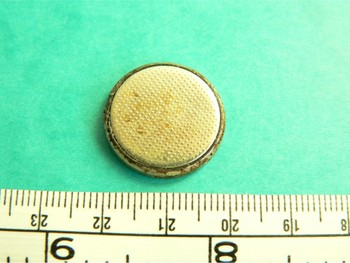
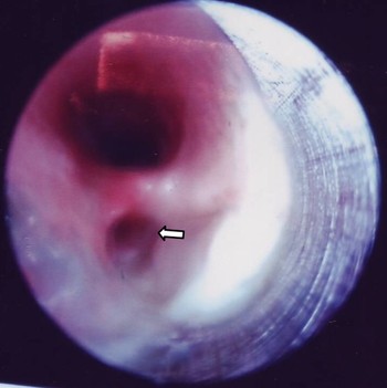
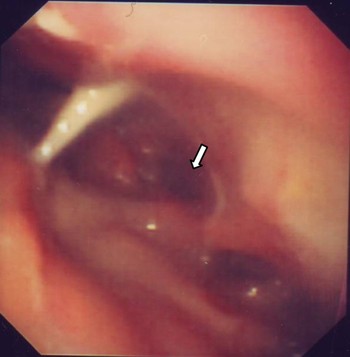
Emergency endoscopy of the upper aerodigestive tract was performed, and a large (21 mm diameter) disc battery was removed from just below the crico-pharyngeal junction (Figure 1). The trachea was found to be partially obstructed by necrotic tissue. On removal of this necrotic tissue, a large, oval-shaped TOF was discovered (Figure 2). Surgical tracheostomy was performed and a Shiley size 4 tube was inserted. The surgical gastroenterology team performed oesophagoscopy and confirmed the extensive nature of the fistula and mucosal erosion (Figure 3). Gastrostomy was performed under combined endoscopic and laparoscopic vision, and a 16 F Carflo Percutaneous endoscopic gastrostomy (PEG) tube was placed.
The 21 mm diameter disc battery removed from the oesophagus, just below the level of the crico-pharyngeal junction.

Rod endoscopic view using a rigid bronchoscope at the level of the upper trachea, showing blockage of the main tracheal lumen with necrotic tissue, and a large tracheoesophageal fistula (arrow) just below the level of the crico-pharyngeal junction.

Flexible oesophagoscopic view at the level of the crico-pharyngeal junction, confirming tracheoesophageal fistula development (arrow) and mucosal injury.

The patient was monitored in the intensive treatment unit for the first two days post-operatively. The post-operative period was uneventful. Intravenous antibiotics (co-amoxiclav) were continued for two weeks, and nutrition was maintained by gastrostomy feeding. The patient was finally discharged home 17 days after admission.
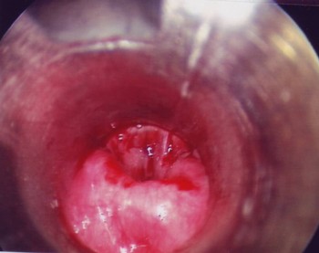
Follow-up bronchoesophagoscopy performed four weeks after discharge showed spontaneous closure of the fistula (Figure 4). A contrast swallow study in the same week confirmed free flow of contrast from the pharynx to the stomach, without any leakage.
Follow up bronchoesophagoscopy six weeks after battery removal, showing spontaneous closure of tracheoesophageal fistula.

On the second follow-up endoscopy, six weeks after the first follow-up endoscopy, spontaneous closure of the fistula was again confirmed.
A chest infection delayed the decannulation process. Decannulation was finally achieved four weeks post-discharge.
Further aerodigestive tract endoscopy, conducted jointly by the head and neck and the surgical gastroenterology teams three months after the second follow-up endoscopy, again confirmed complete resolution of the fistula. The gastrostomy tube was removed at this point.
The child was closely supported by the speech and language therapists to maintain his voice and swallowing.
The child was finally discharged from routine ENT clinic review a year after the incident, in good health with a healthy trachea, good quality of voice, and satisfactory swallowing and oesophageal motility.
Discussion
We reviewed the English language literature using the Medline (through PubMed; 1950–2009), EMBASE (1980–2009) and Ovid (1958–2009) databases, using the search terms ‘tracheooesophageal fistula’ (and spelling variants), ‘spontaneous closure’ and ‘conservative management’. This yielded a total of three documented cases in which conservative management had been attempted.Reference Grisel, Richter, Casper and Thompson6–Reference Senthilkumaran, Crankson and Yousef8 Grisel et al. raised the possibility of late recurrence of fistula after a period of conservative management, dealt with by surgical closure.Reference Grisel, Richter, Casper and Thompson6
The paediatric clinical presentation of an impacted oesophageal foreign body is variable. The event often goes unnoticed by the child's parents (as in our case), resulting in delayed diagnosis and intervention.Reference Slamon, Hertzog, Penfil, Raphaely, Pizarro and Derby5 The interval between ingestion of the foreign body and its passage in the stool is usually not more than five days; therefore, any unresolved suspicion of battery ingestion and possible impaction for longer than five days raises the risk of fistula formation.Reference Chan, Chang, Kao, Liao, Liaw and Chiu2 The clinical history may mimic an episode of respiratory tract infection, and the child may present with airway compromise, as in our patient.Reference Slamon, Hertzog, Penfil, Raphaely, Pizarro and Derby5, Reference Wall, Nadel and Handler9 On plain radiological images, a disc battery is indistinguishable from a simple coin.Reference Bernstein, Burrows and Saunders10 A high index of clinical suspicion is essential. An urgent X-ray and endoscopic removal of the foreign body will allow assessment of any alkaline burn, so that treatment can be tailored accordingly.
• Tracheoesophageal rest (bypassing the site of a tracheoesophageal fistula (TOF) via gastrostomy and tracheostomy) should be the first line of management for acquired TOF in the paediatric age group, to permit closure by secondary intention
• The risks and benefits of early surgical closure must be discussed with experienced colleagues, and the child's parents should be involved in the decision
• Close follow up over a year, with serial endoscopic and radiographic monitoring, is paramount before assuming fistula resolution
Conclusion
Tracheoesophageal rest (bypassing the TOF site by gastrostomy and tracheostomy) should be the first line of management for acquired TOF in the paediatric age group, to permit closure by secondary intention. The risks and benefits of early surgical closure must be discussed with experienced colleagues, and the child's parents should be involved in the decision. Late recurrence of TOF following conservative management is a potential problem. Close follow up over a year, with serial endoscopic and radiographic monitoring, is paramount before assuming fistula resolution. The rarity of this condition precludes a randomised trial.