Introduction
Schizophrenia recognizes a strong genetic liability (McGuffin et al. Reference McGuffin, Riley and Plomin2001). Genetic risk for schizophrenia is also associated with behavioural and physiological correlates of brain processing of different environmental stimuli (Egan et al. Reference Egan, Goldberg, Gscheidle, Weirich, Rawlings, Hyde, Bigelow and Weinberger2001a;Callicott et al. Reference Callicott, Mattay, Verchinski, Marenco, Egan and Weinberger2003b). Previous studies have consistently indicated that behavioural performance at cognitive tasks in non-affected relatives of patients with schizophrenia is impaired relative to controls from the general population (Egan et al. Reference Egan, Goldberg, Gscheidle, Weirich, Rawlings, Hyde, Bigelow and Weinberger2001a, Reference Egan, Goldberg, Kolachana, Callicott, Mazzanti, Straub, Goldman and Weinbergerb; Chen et al. Reference Chen, Rice, Thompson, Barch and Csernansky2009). Consistently, abnormal brain activity during cognition in healthy siblings of patients with schizophrenia has been demonstrated in several studies (Callicott et al. Reference Callicott, Egan, Mattay, Bertolino, Bone, Verchinksi and Weinberger2003a; Woodward et al. Reference Woodward, Tibbo and Purdon2007; Delawalla et al. Reference Delawalla, Csernansky and Barch2008).
Emotion abnormalities play a prominent role in this brain disorder with an important impact on patients' behaviour in everyday life, but the contribution of genetic risk to this class of symptoms is less clear. More specifically, these abnormalities include deficits in emotion perception (Chan et al. Reference Chan, Li, Cheung and Gong2010) and emotion control (Henry et al. Reference Henry, Green, de Lucia, Restuccia, McDonald and O'Donnell2007). Previous imaging studies have revealed aberrant functional responses of brain areas strongly associated with emotion processing, in particular: the amygdala, implicated in processing of emotionally aversive stimuli (Breiter et al. Reference Breiter, Etcoff, Whalen, Kennedy, Rauch, Buckner, Strauss, Hyman and Rosen1996; Morris et al. Reference Morris, Frith, Perrett, Rowland, Young, Calder and Dolan1996; LaBar et al. Reference LaBar, Gatenby, Gore, LeDoux and Phelps1998; Yang et al. Reference Yang, Menon, Eliez, Blasey, White, Reid, Gotlib and Reiss2002; Suslow et al. Reference Suslow, Ohrmann, Bauer, Rauch, Schwindt, Arolt, Heindel and Kugel2006; Adolphs, Reference Adolphs2008, Reference Adolphs2010; Blasi et al. Reference Blasi, Hariri, Alce, Taurisano, Sambataro, Das, Bertolino, Weinberger and Mattay2009a); the prefrontal cortex (PFC), involved in cognitive evaluation of stimuli and modulation of emotional responses (Hariri et al. Reference Hariri, Mattay, Tessitore, Fera and Weinberger2003; Ochsner & Gross, Reference Ochsner and Gross2005; Tessitore et al. Reference Tessitore, Hariri, Fera, Smith, Das, Weinberger and Mattay2005; Williams et al. Reference Williams, Das, Liddell, Kemp, Rennie and Gordon2006); and the striatum (Gur et al. Reference Gur, McGrath, Chan, Schroeder, Turner, Turetsky, Kohler, Alsop, Maldjian, Ragland and Gur2002; Williams et al. Reference Williams, Das, Harris, Liddell, Brammer, Olivieri, Skerrett, Phillips, David, Peduto and Gordon2004; Das et al. Reference Das, Kemp, Flynn, Harris, Liddell, Whitford, Peduto, Gordon and Williams2007; Li et al. Reference Li, Chan, McAlonan and Gong2010), important for avoidance learning (Jensen et al. Reference Jensen, McIntosh, Crawley, Mikulis, Remington and Kapur2003; Menon et al. Reference Menon, Jensen, Vitcu, Graff-Guerrero, Crawley, Smith and Kapur2007), avoidance responses to threatening stimuli (Schlund et al. Reference Schlund, Siegle, Ladouceur, Silk, Cataldo, Forbes, Dahl and Ryan2010) and learning behavioural actions allowing control over fearful responses (Delgado et al. Reference Delgado, Jou, LeDoux and Phelps2009). This literature consistently suggests abnormal physiology associated with emotion processing in patients with schizophrenia. Furthermore, altered activity of the striatum, amygdala and PFC during emotion processing and in other cognitive domains has also been reported in subjects at greater risk for schizophrenia, suggesting genetic implications (Brunelin et al. Reference Brunelin, d'Amato, Van Os, Costes, Suaud Chagny and Saoud2010; van Buuren et al. Reference van Buuren, Vink, Rapcencu and Kahn2011; Zanbelt et al. Reference Zandbelt, van Buuren, Kahn and Vink2011). However, the few behavioural (Toomey et al. Reference Toomey, Seidman, Lyons, Faraone and Tsuang1999; Bolte & Poustka, Reference Bolte and Poustka2003; Gur et al. Reference Gur, Nimgaonkar, Almasy, Calkins, Ragland, Pogue-Geile, Kanes, Blangero and Gur2007) and imaging studies (Habel et al. Reference Habel, Klein, Shah, Toni, Zilles, Falkai and Schneider2004; Rasetti et al. Reference Rasetti, Mattay, Wiedholz, Kolachana, Hariri, Callicott, Meyer-Lindenberg and Weinberger2009; van Buuren et al. Reference van Buuren, Vink, Rapcencu and Kahn2011) investigating the potential effects of genetic risk on emotion abnormalities in schizophrenia have provided somewhat conflicting results.
The potential link between schizophrenia and anomalies in emotion processing may be represented by dopamine (DA) neurotransmission. In this regard, credited models suggest that DA neurotransmission is dysregulated in schizophrenia (Weinberger, Reference Weinberger1987; Grace, Reference Grace1991), and consistent literature indicates that DA is involved in emotion processing (Badgaiyan et al. Reference Badgaiyan, Fischman and Alpert2009). In particular, several studies have indicated that DA biases affective responses via modulation of functional dynamics of key brain nodes, such as the amygdala, striatum and PFC (Karreman & Moghaddam, Reference Karreman and Moghaddam1996; Grace, Reference Grace2000; Grace & Rosenkranz, Reference Grace and Rosenkranz2002; LeDoux, Reference LeDoux2003;Phillips et al. Reference Phillips, Drevets, Rauch and Lane2003a; Badgaiyan et al. Reference Badgaiyan, Fischman and Alpert2009). Of note, other data have also revealed that genetic factors can modulate DA neurotransmission. Individual variability in genes involved in DA metabolism is associated with differential brain physiology and behaviour during emotion processing (Blasi et al. Reference Blasi, Lo Bianco, Taurisano, Gelao, Romano, Fazio, Papazacharias, Di Giorgio, Caforio, Rampino, Masellis, Papp, Ursini, Sinibaldi, Popolizio, Sadee and Bertolino2009b). In particular, a genetically determined function of the DA inactivator catechol-O-methyltransferase (COMT) has been investigated in depth (Lachman et al. Reference Lachman, Papolos, Saito, Yu, Szumlanski and Weinshilboum1996; Mannisto & Kaakkola, Reference Mannisto and Kaakkola1999) with particular regard to the COMT Val158Met polymorphism, which is associated with differential COMT activity (Chen et al. Reference Chen, Lipska, Halim, Ma, Matsumoto, Melhem, Kolachana, Hyde, Herman, Apud, Egan, Kleinman and Weinberger2004; for a review, see Dickinson et al. Reference Dickinson and Elvevag2009). This single nucleotide polymorphism in the COMT gene (rs4680, G > A) results in a valine (Val) to methionine (Met) substitution at codon 158 (Val158Met) (Lachman et al. Reference Lachman, Papolos, Saito, Yu, Szumlanski and Weinshilboum1996; Mannisto & Kaakkola, Reference Mannisto and Kaakkola1999). This substitution is functional in that the Val allele is associated with greater enzymatic activity relative to the Met allele (Lachman et al. Reference Lachman, Papolos, Saito, Yu, Szumlanski and Weinshilboum1996; Mannisto & Kaakkola, Reference Mannisto and Kaakkola1999; Chen et al. Reference Chen, Lipska, Halim, Ma, Matsumoto, Melhem, Kolachana, Hyde, Herman, Apud, Egan, Kleinman and Weinberger2004).
Previous studies have suggested that COMT Val158Met is associated with cognitive and emotional behaviour and related brain activity in animal models and in humans (Gogos et al. Reference Gogos, Morgan, Luine, Santha, Ogawa, Pfaff and Karayiorgou1998; Woo et al. Reference Woo, Yoon and Yu2002; Enoch et al. Reference Enoch, Xu, Ferro, Harris and Goldman2003; Rujescu et al. Reference Rujescu, Giegling, Gietl, Hartmann and Moller2003; Tsai et al. Reference Tsai, Hong, Yu and Chen2004; Volavka et al. Reference Volavka, Kennedy, Ni, Czobor, Nolan, Sheitman, Lindenmayer, Citrome, McEvoy and Lieberman2004; Reuter & Hennig, Reference Reuter and Hennig2005; Smolka et al. Reference Smolka, Schumann, Wrase, Grusser, Flor, Mann, Braus, Goldman, Buchel and Heinz2005; Stein et al. Reference Stein, Fallin, Schork and Gelernter2005; Drabant et al. Reference Drabant, Hariri, Meyer-Lindenberg, Munoz, Mattay, Kolachana, Egan and Weinberger2006; Heinz & Smolka, Reference Heinz and Smolka2006). The direction of the COMT Val158Met effect within the cognitive domain has been fairly consistent (Mier et al. Reference Mier, Kirsch and Meyer-Lindenberg2010) in healthy subjects. Within the emotion-processing context, the available studies in healthy subjects have reported COMT Val158Met genotype modulation of activity in the PFC, amygdala and striatum, even though the direction of the effect has not always been the same across different experiments (Smolka et al. Reference Smolka, Schumann, Wrase, Grusser, Flor, Mann, Braus, Goldman, Buchel and Heinz2005; Drabant et al. Reference Drabant, Hariri, Meyer-Lindenberg, Munoz, Mattay, Kolachana, Egan and Weinberger2006; Tan et al. Reference Tan, Chen, Goldberg, Mattay, Meyer-Lindenberg, Weinberger and Callicott2007; Schmack et al. Reference Schmack, Schlagenhauf, Sterzer, Wrase, Beck, Dembler, Kalus, Puls, Sander, Heinz and Gallinat2008; Dreher et al. Reference Dreher, Kohn, Kolachana, Weinberger and Berman2009; Domschke et al. Reference Domschke, Baune, Havlik, Stuhrmann, Suslow, Kugel, Zwanzger, Grotegerd, Sehlmeyer, Arolt and Dannlowski2012). In this regard, one of the factors explaining at least part of these discrepancies may have been the task used to probe emotion processing. On the other hand, an effect of different genetic architecture in different ethnicities on these discrepancies cannot be excluded either.
Other studies have focused on potential differential effects of COMT Val158Met genotype in healthy subjects and in patients with schizophrenia. These studies have demonstrated that the neurophysiology of brain structures involved by verbal fluency and memory encoding tasks is differentially predicted by COMT Val158Met genotype in patients and controls (Prata et al. Reference Prata, Mechelli, Fu, Picchioni, Kane, Kalidindi, McDonald, Howes, Kravariti, Demjaha, Toulopoulou, Diforti, Murray, Collier and McGuire2009; Di Giorgio et al. Reference Di Giorgio, Caforio, Blasi, Taurisano, Fazio, Romano, Ursini, Gelao, Bianco, Papazacharias, Sinibaldi, Popolizio, Bellomo and Bertolino2011). In particular, these imaging studies have indicated that the direction of COMT Val158Met genotype effect on brain activity is opposite in patients with schizophrenia compared with that in healthy subjects suggesting a genotype by genetic architecture (diagnosis) interaction. A possible interpretation of these data is that the pathophysiology of schizophrenia and COMT Val158Met genotype both alter DA signalling, and that their interaction on brain DA tone may lead to the reported interaction on brain physiology. This interpretation, however, may suffer from the potential of state variables like treatment with antipsychotics confounding the interaction. If these results were not because of state variables associated with being ill, a similar interaction in DA-modulated brain regions such as the amygdala, PFC and striatum during emotion processing can be hypothesized in subjects at greater risk for schizophrenia. Furthermore, in order to control for potential confounds of state variables associated with schizophrenia and knowing that siblings share on average 50% of genetic variation with their ill probands, it is relevant to investigate brain responses during emotion processing also in healthy siblings of patients with this brain disorder.
The aim of the present study was to demonstrate that genetic risk for schizophrenia and its interaction with COMT Val158Met is associated with emotion-processing physiology. With this purpose, we used functional magnetic resonance imaging (fMRI) to investigate brain activity in schizophrenia patients, healthy siblings of patients with schizophrenia, and healthy individuals during implicit and explicit evaluation of emotional stimuli with negative valence. These stimuli strongly activate the emotional network, including the amygdala, PFC and striatum, which have also been previously associated with modulation by COMT Val158Met genotype and genetic risk for schizophrenia (Smolka et al. Reference Smolka, Schumann, Wrase, Grusser, Flor, Mann, Braus, Goldman, Buchel and Heinz2005; Tan et al. Reference Tan, Chen, Goldberg, Mattay, Meyer-Lindenberg, Weinberger and Callicott2007; Brunelin et al. Reference Brunelin, d'Amato, Van Os, Costes, Suaud Chagny and Saoud2010; van Buuren et al. Reference van Buuren, Vink, Rapcencu and Kahn2011). Based on the previous considerations and on the aforementioned findings, we hypothesized an interaction between COMT Val158Met genotype and risk for schizophrenia during emotion processing. In particular, we specifically hypothesized that COMT Val158Met might differentially affect patterns of brain responses in the amygdala, PFC and striatum during emotion processing in patients, unaffected siblings and healthy subjects.
Method
Subjects
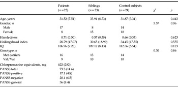
A total of 25 patients with schizophrenia (17 males, mean age 31.5 years, s.d. = 7.5 years), 23 unaffected siblings (eight males, mean age 33.9 years, s.d. = 8.7 years) and 24 normal control subjects (14 males, mean age 31.8 years, s.d. = 3.3 years) were enrolled in the study (Table 1). All participants were white Caucasians from the province of Bari. The Structured Clinical Interview for DSM-IV (First et al. 1996) was used to confirm diagnosis of schizophrenia for patients and to exclude any Axis I psychiatric disorder for siblings and healthy subjects. Exclusion criteria were presence of any neurological or medical condition revealed by clinical and magnetic resonance imaging evaluation, presence of head trauma with loss of consciousness and drug abuse within the past 6 months. All patients were on stable pharmacological treatment with antipsychotics. Symptoms were evaluated using the Positive and Negative Syndrome Scale (PANSS; Kay et al. Reference Kay, Fiszbein and Opler1987). Furthermore, socio-economic status was measured with the Hollingshead index (Hollingshead & Redlich, Reference Hollingshead and Redlich1958), handedness with the Edinburgh Inventory (Oldfield, Reference Oldfield1971) and pre-morbid intelligence quotient (IQ) with the Italian version of the Wide Reading Achievement Test, revised (Sartori & Colombo, Reference Sartori, Colombo, Vallar, Rusconi and Pinarello1997).
Genetics, demographics and clinical data

IQ, Intelligence quotient; Met, methionine; Val, valine; PANSS, Positive and Negative Syndrome Scale.
Data are given as mean (standard error) or as number of participants.
All subjects provided written informed consent to the study after the procedure was fully explained to them. The protocol was approved by the local Institutional Review Board.
Genotyping
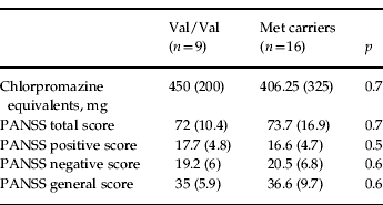
All participants were genotyped for the COMT Val158Met polymorphism, which was determined as a restriction fragment length polymorphism after polymerase chain reaction amplification and digestion with NlaIII, as previously described (Bertolino et al. Reference Bertolino, Caforio, Blasi, De Candia, Latorre, Petruzzella, Altamura, Nappi, Papa, Callicott, Mattay, Bellomo, Scarabino, Weinberger and Nardini2004). COMT genotypes of the three groups of subjects included were as follows: patients Met/Met (n = 6), Val/Met (n = 10), Val/Val (n = 9); siblings Met/Met (n = 3), Val/Met (n = 10), Val/Val (n = 10); and healthy subjects Met/Met (n = 6), Val/Met (n = 8), Val/Val (n = 10). Allele frequencies did not deviate from Hardy–Weinberg equilibrium (p > 0.1). Because of the small number of Met/Met individuals, we grouped subjects carrying at least one Met allele (Met carriers) for further analyses as follows: Met carrier patients (n = 16), Val/Val (n = 9); Met carrier siblings (n = 13), Val/Val (n = 10); healthy subjects: Met carriers (n = 14), Val/Val (n = 10), as in previous studies (Slifstein et al. Reference Slifstein, Kolachana, Simpson, Tabares, Cheng, Duvall, Frankle, Weinberger, Laruelle and Abi-Dargham2008; Colzato et al. Reference Colzato, Waszak, Nieuwenhuis, Posthuma and Hommel2010; Mueller et al. Reference Mueller, Makeig, Stemmler, Hennig and Wacker2011; Nixon et al. Reference Nixon, Prust, Sambataro, Tan, Mattay, Weinberger and Callicott2011) (Table 2).
Clinical characteristics of patients with schizophrenia as a function of COMT genotype
COMT, Catechol-O-methyltransferase; Val, valine; Met, methionine; PANSS, Positive and Negative Syndrome Scale.
Data are given as mean (standard error).
fMRI experimental paradigm
The event-related fMRI paradigm (Blasi et al. Reference Blasi, Lo Bianco, Taurisano, Gelao, Romano, Fazio, Papazacharias, Di Giorgio, Caforio, Rampino, Masellis, Papp, Ursini, Sinibaldi, Popolizio, Sadee and Bertolino2009b) consisted of two runs: each run presented angry, fearful, happy and neutral facial expressions from a validated set of facial pictures (NimStim, http://www.macbrain.org/resources.htm, Tottenham et al. Reference Tottenham, Tanaka, Leon, McCarry, Nurse, Hare, Marcus, Westerlund, Casey and Nelson2009). The order of stimuli was randomly distributed. However, the same stimuli were presented in both runs with the same order. During one run (emotional perceptual processing – implicit processing), subjects identified the gender of each face. In the other run (explicit emotional evaluation – explicit processing), they had to decide whether they would like to ‘approach’ or ‘avoid’ the face. From stimulus appearance, 2 s were allowed for behavioural responses. The presentation of the two runs was counterbalanced across subjects. Each stimulus was presented for 500 ms, with the interstimulus interval randomly varied between 2 and 7 s. The total number of stimuli was 144: 30 angry, 39 fearful, 37 happy and 38 neutral faces. Duration of each run was 6 min 8 s. A fixation crosshair was presented during the interstimulus interval. For the purpose of this study, we focused our analysis on processing of facial stimuli with negative valence (angry and fearful faces) because of their strong association with activity in the emotion network. Furthermore, aversive emotional stimuli have been previously associated with COMT Val158Met effects (Hariri et al. Reference Hariri, Tessitore, Mattay, Fera and Weinberger2002; Smolka et al. Reference Smolka, Schumann, Wrase, Grusser, Flor, Mann, Braus, Goldman, Buchel and Heinz2005; Drabant et al. Reference Drabant, Hariri, Meyer-Lindenberg, Munoz, Mattay, Kolachana, Egan and Weinberger2006).
fMRI data acquisition
fMRI was performed on a GE Signa 3T scanner (General Electric Medical Systems, USA) with a gradient echo-planar imaging sequence (repetition time: 2000 ms; echo time: 28 ms; 26 interleaved slices; thickness: 4 mm; gap: 1 mm; voxel size: 4 × 4×5 mm; scan repetitions: 180; flip angle: 90°; field of view: 24 cm; matrix: 64 × 64). The first four scans were discarded to allow for signal saturation. All stimuli were presented via a back-projection system. A fibre optic response box was used to measure subject preference (and reaction time) for each stimulus: left button for approach/male responses and right button for avoid/female responses.
fMRI data analysis
Statistical Parametric Mapping 5 (SPM5, http://www.fil.ion.ucl.ac.uk/spm) was used for imaging analysis. Images for each subject were realigned, spatially normalized into the Montreal Neurological Institute template (12 parameter affine model) and spatially smoothed (10 mm Gaussian filter). After realignment, datasets were also screened for high quality (scan stability) as demonstrated by small motion correction (<3 mm translation and <3° rotation).
For both runs, we collapsed in a single vector the timing of presentation of facial expressions with negative valence (both angry and fearful faces; for implicit processing correct responses only were used). Vectors were also created using the timing of presentation of all other facial stimuli, but they were not used for further analyses. Residual movement was also modelled as a regressor of no interest. Linear contrasts were used to estimate condition-specific blood oxygen level-dependent (BOLD) activation for each individual and each run relative to aversive facial expressions (aversive facial expressions v. crosshairs).
These individual contrast images were then entered in a random-effects analysis of variance (ANOVA) for group analysis, with COMT Val158Met and diagnosis as predictors and task (implicit v. explicit) as the repeated-measures factor. Given the strong trend for unbalanced gender distribution between groups (see below) and the reported sexually dimorphic effect of COMT on DA levels and behaviour (Gogos et al. Reference Gogos, Morgan, Luine, Santha, Ogawa, Pfaff and Karayiorgou1998; Karayiorgou et al. Reference Karayiorgou, Sobin, Blundell, Galke, Malinova, Goldberg, Ott and Gogos1999; Weinshilboum et al. Reference Weinshilboum, Otterness and Szumlanski1999; Alsobrook et al. Reference Alsobrook, Zohar, Leboyer, Chabane, Ebstein and Pauls2002; Goudreau et al. Reference Goudreau, Maraganore, Farrer, Lesnick, Singleton, Bower, Hardy and Rocca2002; Rybakowski et al. Reference Rybakowski, Borkowska, Czerski and Hauser2002; Kremer et al. Reference Kremer, Pinto, Murad, Muhaheed, Bannoura, Muller, Schulze, Reshef, Blanaru, Gathas, Goichman, Rietschel, Dobrusin, Bachner-Melman, Nemanov, Belmaker, Maier and Ebstein2003; Chen et al. Reference Chen, Lipska, Halim, Ma, Matsumoto, Melhem, Kolachana, Hyde, Herman, Apud, Egan, Kleinman and Weinberger2004; Domschke et al. Reference Domschke, Freitag, Kuhlenbäumer, Schirmacher, Sand, Nyhuis, Jacob, Fritze, Franke, Rietschel, Garritsen, Fimmers, Nöthen, Lesch, Stögbauer and Deckert2004; Ono et al. Reference Ono, Shirakawa, Nushida, Ueno and Maeda2004; Sazci et al. Reference Sazci, Ergul, Kucukali, Kilic, Kaya and Kara2004; Shifman et al. Reference Shifman, Bronstein, Sternfeld, Pisante, Weizman, Reznik, Spivak, Grisaru, Karp, Schiffer, Kotler, Strous, Swartz-Vanetik, Knobler, Shinar, Yakir, Zak and Darvasi2004), gender was used as a covariate of no interest. Analyses were constrained by an inclusive mask generated by combining group activation maps during each task (p < 0.05).
For these fMRI analyses we used a statistical threshold of p < 0.05 false discovery rate (FDR) corrected within three regions of interest in the amygdala, PFC and striatum as identified with the Wake Forest University PickAtlas 1.04 (http://www.rad.wfubmc.edu/fmri), which are brain regions strongly involved in emotion processing and previously found to be associated with genetic load for schizophrenia and/or COMT Val158Met effect (Phillips, Reference Phillips2003; Habel et al. Reference Habel, Klein, Shah, Toni, Zilles, Falkai and Schneider2004; Tunbridge et al. Reference Tunbridge, Harrison and Weinberger2006). The three regions of interest in the amygdala, PFC and striatum were considered as three different regions of interest. BOLD responses were extracted from significant clusters using MarsBar (http://marsbar.sourceforge.net/) to further explore and characterize significant effects. Because we did not have strong a priori hypotheses on other brain regions, we used a FDR whole-brain correction at p < 0.05 for areas outside our regions of interest.
Statistical analyses
We used χ2 to compare demographics and genotype distribution. ANOVA with genotype and diagnosis as categorical predictors was performed to further explore BOLD signal in significant clusters of activity and behavioural differences with Statistica software (StatSoft, Inc., USA). Fisher's least significant difference test was used for post hoc comparisons. To test the relationship between behavioural performance, diagnosis and BOLD fMRI activity, we performed Spearman correlation analyses. Spearman correlation analyses were also performed to evaluate the relationship between BOLD signal, antipsychotic treatment and symptom scores in patients.
Results
Demographics
Groups were matched in terms of age, handedness, socio-economic status, pre-morbid IQ and genotype (all p > 0.1). Since there was a strong statistical trend for unequal distribution of gender across diagnostic groups (χ2 = 5.57, p = 0.06), we covaried all further analyses for gender. Within the patients, genotype groups were matched in terms of clinical variables, including severity of clinical symptoms (PANSS total, general, negative and positive symptoms scores: all p > 0.5) and antipsychotic medication dose (chlorpromazine equivalents: p = 0.7) (Tables 1 and 2).
Behavioural data
Accuracy data during the implicit condition of the task indicated a main effect of diagnosis [F(2, 66) = 10.518, p = 0.0001], with reduced performance in patients relative to siblings and healthy subjects (all p < 0.0002). Reaction time data revealed a main effect of task (p < 0.05, explicit slower than implicit) and an interaction between diagnosis, genotype and task (p < 0.01), with longer reaction times in Met carrier healthy subjects relative to Val/Val healthy subjects (p < 0.01) and Met carrier siblings (p < 0.006). No other significant results were found (all p > 0.1).
Imaging data
Analysis of covariance indicated a main effect of diagnosis (Fig. 1a) in the right amygdala (x = 26, y = −7, z = −22, k = 4, Z = 3.02, FDR-corrected p = 0.03) and an interaction between diagnosis and genotype in the left middle frontal gyrus (Brodmann area 9: x = −41, y = 30, z = 38, k = 11, Z = 3.08, FDR-corrected p = 0.05) (Fig. 2a). Furthermore, there was an interaction between diagnosis, genotype, and task condition in the left putamen (x = −22, y = 8, z = 15, k = 22, Z = 3.46, FDR-corrected p = 0.05). No other significant effects were found. Analysis of BOLD responses extracted from significant clusters was thus performed to better characterize main effects and interactions found with SPM analysis. Parameter estimates in the right amygdala cluster demonstrating a main effect of diagnosis indicated greater activity in healthy subjects relative to both siblings and patient groups (Fisher's test: p < 0.02), who presented similar levels of activity (Fisher's test: p < 0.2) (Fig. 1b). On the other hand, BOLD responses extracted from the prefrontal cluster of the interaction between genotype and diagnosis revealed more complex effects (Fig. 2b). In particular, healthy subjects had again greater activity compared with patients and siblings, but only in the context of Val/Val genotype (Fisher's test: p < 0.01). Furthermore, while in healthy subjects BOLD responses were greater in Val/Val relative to Met carrier individuals (Fisher's test: p < 0.01), an opposite pattern was present in siblings and patients with schizophrenia (Fisher's test: p = 0.1 for patients; Fisher's test: p < 0.04 for siblings), i.e. Met carrier subjects had greater mean activity relative to Val/Val individuals.
(a) Sagittal, coronal and axial sections of the brain showing the cluster associated with the main effect of diagnosis in the right amygdala (x = 26, y = −7, z = −22) during emotion processing. The colour bar represents T score values. (b) Parameter estimates extracted from this cluster. Values are means, with standard errors represented by vertical bars. See text for statistics. AU, Arbitrary units; Scz, schizophrenic; Sib, sibling; NC, normal control.

(a) Sagittal, coronal and axial sections of the brain showing the cluster in the middle frontal gyrus (x = −41, y = 30, z = 38) associated with a genotype × diagnosis interaction. The colour bar represents T score values. (b) Parameter estimates extracted from this cluster. Values are means, with standard errors represented by vertical bars. AU, Arbitrary units; Met, methionine; Val, valine; COMT, catechol-O-methyltransferase; Scz, schizophrenic; Sib, sibling; NC, normal control.
To further characterize the interaction between diagnosis, genotype and task (Fig. 3a), parameter estimates were extracted from the left putamen cluster and investigated outside SPM. This analysis revealed that the effect of diagnosis was particularly evident in the context of explicit processing along with a complete inversion of the difference between patients and healthy subjects based on COMT Val158Met genotype (Val/Val controls >Val/Val patients: Fisher's test p = 0.04; Met carrier patients >Met carrier controls: Fisher's test p = 0.02)), while there was no COMT modulation in siblings (Fig. 3b). No other areas outside the regions of interest reached statistical significance.
(a) Sagittal, coronal and axial sections of the brain showing the cluster associated with a genotype × diagnosis interaction in the left striatum (x = −22, y = 8, z = 15) during implicit and explicit emotion processing. The colour bar represents T score values. (b) Parameter estimates extracted from this cluster. Values are means, with standard errors represented by vertical bars. AU, Arbitrary units; Met, methionine; Val, valine; Scz, schizophrenic; Sib, sibling; NC, normal control.
Relationship of brain activity with symptoms scores and behavioural performance
Spearman analysis did not demonstrate significant correlations between BOLD signal and both chlorpromazine equivalents and symptoms scores (PANSS total, positive, negative and general) in the amygdala, PFC and striatum (all ρ < 0.3; all p > 0.2). Furthermore, there were no significant correlations of BOLD responses in amygdala, PFC and striatum with behavioural performance during the fMRI task (all ρ < 0.1; all p > 0.3).
Discussion
The present results suggest that genetic risk for schizophrenia affects amygdala responses and interacts with COMT Val158Met genotype in modulating brain activity of the emotion network during processing of aversive emotional stimuli. In particular, we found that schizophrenia patients and unaffected siblings have reduced amygdala activity relative to healthy comparison subjects. We also found a diagnosis by COMT genotype interaction in the PFC. Here, the direction of the Val/Met effect in healthy subjects was consistent with earlier studies involving cognitive decisions (Egan et al. Reference Egan, Goldberg, Kolachana, Callicott, Mazzanti, Straub, Goldman and Weinberger2001b; Bilder et al. Reference Bilder, Volavka, Czobor, Malhotra, Kennedy, Ni, Goldman, Hoptman, Sheitman, Lindenmayer, Citrome, McEvoy, Kunz, Chakos, Cooper and Lieberman2002; Ho et al. Reference Ho, Wassink, O'Leary, Sheffield and Andreasen2005; McIntosh et al. Reference McIntosh, Baig, Hall, Job, Whalley, Lymer, Moorhead, Owens, Miller, Porteous, Lawrie and Johnstone2007), whereas in siblings and in patients it was in the opposite direction. Moreover, patients and siblings had reduced activity compared with healthy subjects only in the context of Val/Val genotype. In the putamen, a similar interaction between diagnosis and COMT Val158Met genotype was found, but it was more specifically elicited by explicit processing of aversive emotional stimuli. Different patterns of activity were present in Val/Val and Met carrier individuals as a function of diagnosis.
Our findings of altered amygdala activation in siblings and patients are consistent with studies showing an effect of genetic risk for schizophrenia both in terms of reactivity to stress (Myin-Germeys et al. Reference Myin-Germeys, van Os, Schwartz, Stone and Delespaul2001; Myin-Germeys & van Os, Reference Myin-Germeys and van Os2007; Brunelin et al. Reference Brunelin, d'Amato, Van Os, Cochet, Suaud-Chagny and Saoud2008) and of brain morphology and physiology during different experimental paradigms (Callicott et al. Reference Callicott, Egan, Mattay, Bertolino, Bone, Verchinksi and Weinberger2003a; Thermenos et al. Reference Thermenos, Seidman, Breiter, Goldstein, Goodman, Poldrack, Faraone and Tsuang2004; Boos et al. Reference Boos, Aleman, Cahn, Hulshoff Pol and Kahn2007). Previous studies have explored the behavioural correlates of emotion processing in subjects at risk for schizophrenia, but results have not always been consistent (Toomey et al. Reference Toomey, Seidman, Lyons, Faraone and Tsuang1999; Bolte & Poustka, Reference Bolte and Poustka2003; Gur et al. Reference Gur, Nimgaonkar, Almasy, Calkins, Ragland, Pogue-Geile, Kanes, Blangero and Gur2007). Furthermore, only three imaging studies (Habel et al. Reference Habel, Klein, Shah, Toni, Zilles, Falkai and Schneider2004; Rasetti et al. Reference Rasetti, Mattay, Wiedholz, Kolachana, Hariri, Callicott, Meyer-Lindenberg and Weinberger2009; van Buuren et al. Reference van Buuren, Vink, Rapcencu and Kahn2011) have investigated brain responses during emotion processing in non-affected siblings of schizophrenia patients. In particular, Habel et al. (Reference Habel, Klein, Shah, Toni, Zilles, Falkai and Schneider2004) and van Buuren et al. (Reference van Buuren, Vink, Rapcencu and Kahn2011) reported abnormal amygdala reactivity in siblings of patients with schizophrenia during explicit emotion processing, while Rasetti et al. (Reference Rasetti, Mattay, Wiedholz, Kolachana, Hariri, Callicott, Meyer-Lindenberg and Weinberger2009) found similar levels of amygdala activity in siblings and healthy controls during implicit processing of facial stimuli with angry or fearful expressions. Differences in the experimental paradigms used (Hariri et al. Reference Hariri, Tessitore, Mattay, Fera and Weinberger2002) and methodology may account for discrepancy with our findings. In their study, Rasetti et al. (Reference Rasetti, Mattay, Wiedholz, Kolachana, Hariri, Callicott, Meyer-Lindenberg and Weinberger2009) used a face-matching implicit perceptual emotional task, while we used both an implicit and explicit emotion paradigm. During the implicit condition in our task, facial expressions are processed rapidly and automatically but there is no matching required; in addition, during explicit processing, facial stimuli are consciously evaluated focusing on their emotional valence. This cognitive operation is associated with both cortical and subcortical (especially amygdala) activity. Moreover, Rasetti et al. (Reference Rasetti, Mattay, Wiedholz, Kolachana, Hariri, Callicott, Meyer-Lindenberg and Weinberger2009) used a block design which may involve habituation effects – they used two blocks of angry and fearful stimuli. Earlier studies have demonstrated that habituation of activity in the amygdala is a consistent signature of emotion paradigms with block designs (Breiter et al. Reference Breiter, Etcoff, Whalen, Kennedy, Rauch, Buckner, Strauss, Hyman and Rosen1996; Buchel et al. Reference Buchel, Morris, Dolan and Friston1998; Schwartz et al. Reference Schwartz, Wright, Shin, Kagan, Whalen, McMullin and Rauch2003; Wright et al. Reference Wright, Martis, Schwartz, Shin, Fischer, McMullin and Rauch2003). On the other hand, our event-related paradigm consisted of four different kinds of randomly distributed emotional expressions – happy, angry, fearful and neutral – and is less prone to habituation effects. Finally, the control session in the task of the Rasetti et al. (Reference Rasetti, Mattay, Wiedholz, Kolachana, Hariri, Callicott, Meyer-Lindenberg and Weinberger2009) study may involve general visuo-attentional processes, potentially confounding amygdala responses to some degree. In our study, we used a control condition consisting of a crosshair without any cognitive demand, thus controlling for potentially confounding effects of cognitive operations.
Furthermore, previous studies have reported that specific modulation of amygdala activity could be differentially elicited by task requirements. For example, Blasi et al. (Reference Blasi, Hariri, Alce, Taurisano, Sambataro, Das, Bertolino, Weinberger and Mattay2009a) demonstrated that amygdala activity and differential functional coupling with prefrontal circuits may be elicited during social judgment decisions; consistently, Hariri et al. (Reference Hariri, Bookheimer and Mazziotta2000) reported that modulation of emotional experience by interpretation and labelling involves recruitment of the PFC. These data suggest that different cognitive demands of tasks probing emotion processing may differentially affect activity within the emotion network. Overall, our results are consistent with the hypothesis that amygdala activity during emotion processing is associated with genetic risk for schizophrenia.
In line with other studies in the cognitive domain (Prata et al. Reference Prata, Mechelli, Fu, Picchioni, Kane, Kalidindi, McDonald, Howes, Kravariti, Demjaha, Toulopoulou, Diforti, Murray, Collier and McGuire2009; Di Giorgio et al. Reference Di Giorgio, Caforio, Blasi, Taurisano, Fazio, Romano, Ursini, Gelao, Bianco, Papazacharias, Sinibaldi, Popolizio, Bellomo and Bertolino2011), we also report an interaction between diagnosis and COMT Val158Met genotype on prefrontal activity. In particular, patients and siblings had significantly reduced middle frontal gyrus activity relative to healthy subjects only in the context of Val/Val genotype. Previous studies have associated lateral prefrontal activity with involvement in identification of the emotional significance of environmental stimuli and production of affective states. Moreover, the PFC has been associated with the regulation of autonomic responses during the processing of emotion stimuli and contexts by eliciting the production of congruent affective states (Phillips et al. Reference Phillips, Drevets, Rauch and Lane2003a). Furthermore, several studies have demonstrated that the PFC is a key structure involved in the pathophysiology of schizophrenia (Weinberger et al. Reference Weinberger, Egan, Bertolino, Callicott, Mattay, Lipska, Berman and Goldberg2001; Callicott et al. Reference Callicott, Mattay, Verchinski, Marenco, Egan and Weinberger2003b). Abnormal prefrontal activity has been reported in patients with schizophrenia during both cognitive and emotional tasks (Bertolino et al. Reference Bertolino, Breier, Callicott, Adler, Mattay, Shapiro, Frank, Pickar and Weinberger2000; Weinberger et al. Reference Weinberger, Egan, Bertolino, Callicott, Mattay, Lipska, Berman and Goldberg2001; Salgado-Pineda et al. Reference Salgado-Pineda, Fakra, Delaveau, Hariri and Blin2010) and is associated with risk for schizophrenia (Egan et al. Reference Egan, Goldberg, Gscheidle, Weirich, Rawlings, Hyde, Bigelow and Weinberger2001a).
Among the modulators of prefrontal physiology, DA plays a crucial role. Of note, pathophysiological models strongly suggest dysregulation of DA signalling, with reduced cortical levels in schizophrenia (Weinberger, Reference Weinberger1987; Carlsson, Reference Carlsson1988; Grace, Reference Grace1991; Bertolino et al. Reference Bertolino, Knable, Saunders, Callicott, Kolachana, Mattay, Bachevalier, Frank, Egan and Weinberger1999; Abi-Dargham et al. Reference Abi-Dargham, Mawlawi, Lombardo, Gil, Martinez, Huang, Hwang, Keilp, Kochan, Van Heertum, Gorman and Laruelle2002; Akil et al. Reference Akil, Kolachana, Rothmond, Hyde, Weinberger and Kleinman2003; Matsumoto et al. Reference Matsumoto, Weickert, Akil, Lipska, Hyde, Herman, Kleinman and Weinberger2003). COMT, regulating DA inactivation, plays an important role in modulating the activity of prefrontal circuitry during the processing of environmental stimuli (Seamans & Yang, Reference Seamans and Yang2004). Previous studies have revealed that the COMT Val158Met polymorphism is associated with reactivity of brain regions involved in emotion processing, including the PFC, in healthy subjects (Smolka et al. Reference Smolka, Schumann, Wrase, Grusser, Flor, Mann, Braus, Goldman, Buchel and Heinz2005; Drabant et al. Reference Drabant, Hariri, Meyer-Lindenberg, Munoz, Mattay, Kolachana, Egan and Weinberger2006). Here, we found that the COMT Val158Met genotype interacts with genetic risk for schizophrenia in modulating BOLD responses in PFC during emotion processing. This modulatory effect is particularly elicited in the Val/Val context: prefrontal activity of siblings and patients with schizophrenia is similarly reduced relative to those of healthy subjects. Interestingly, the Val allele is associated with greater COMT activity and thus lower DA levels relative to the Met allele (Weinshilboum et al. Reference Weinshilboum, Otterness and Szumlanski1999; Chen et al. Reference Chen, Lipska, Halim, Ma, Matsumoto, Melhem, Kolachana, Hyde, Herman, Apud, Egan, Kleinman and Weinberger2004). Of note for interpretation of the present data, pathophysiological models have indicated that schizophrenia may be characterized by lower DA signalling in the PFC (Carlsson, Reference Carlsson1988). In this line of reasoning, our results suggest that it is in the context of genetically determined lower DA signalling associated with the Val allele that genetic risk for schizophrenia is more manifest on prefrontal activity during emotion processing. Furthermore, these regionally specific effects strongly suggest that Val/Val lower prefrontal activity cannot be related to state variables and confounding factors like treatment with antipsychotics or chronicity associated with the disorder, but it is mediated by genetic risk, as suggested by similarities of responses in siblings who are untreated and unaffected by definition.
A diagnosis by task by COMT Val158Met genotype was evident in the striatum. Our results indicate a complete inversion of COMT Val158Met between patients and controls, especially in the explicit condition. While Val homozygous patients had reduced striatal activity relative to normal control subjects, the opposite was true for Met carriers. On the other hand, no COMT Val158Met effect was present in siblings, whose level of striatal activity was intermediate relative to patients and controls regardless of genotype. The striatum receives DA inputs from the substantia nigra (Moss & Bolam, Reference Moss and Bolam2008) and plays an important role in integrating excitatory inputs from the cortex and thalamus; in the context of emotion processing it has been implicated in identification of the emotional significance of a stimulus, in the production of affective states and automatic regulation of emotional responses (Phillips et al. Reference Phillips, Drevets, Rauch and Lane2003a, b).
Some limitations of our study may make interpretation of the results in the striatum somewhat speculative and difficult. Increased measures of striatal DA are now fairly established in schizophrenia (for reviews, see Miyake et al. Reference Miyake, Thompson, Skinbjerg and Abi-Dargham2011; Howes et al. Reference Howes, Kambeitz, Kim, Stahl, Slifstein, Abi-Dargham and Kapur2012). However, striatal DA release and density of DA receptors vary according to phase of the illness (Laruelle et al. Reference Laruelle, Abi-Dargham, Gil, Kegeles and Innis1999) and to previous exposure to antipsychotics (Howes et al. Reference Howes, Kambeitz, Kim, Stahl, Slifstein, Abi-Dargham and Kapur2012). Our patients were all studied while on stable pharmacological treatment for at least 8 weeks. Spearman analysis did not indicate significant correlations between BOLD response in the striatum and both chlorpromazine equivalents or PANSS score, suggesting that these variables do not play a major role in our results. However, we believe it cannot be definitively excluded that the effect in the striatum may be mediated by several other variables associated with the illness itself, for instance, the number of acute exacerbations, the different kinds of antipsychotics used over time, illness chronicity, or other variables that were not controlled for and potentially affect striatal DA levels. Therefore, on this basis, our findings in the striatum may reflect state- rather than trait-related factors associated with the pathophysiology of the disorder. Nevertheless, some speculative interpretation can be made about our data. Our results in the striatum could reflect potential prefrontal modulation, or they may be associated with greater DA signalling in subjects with schizophrenia. Such modulation may differentially affect risk-specific positioning on the inverted U relationship between DA signalling and striatal function. The more parsimonious interpretation is that our results in the striatum may be related to clinical and other uncontrolled variables associated with the disorder itself since healthy siblings did not show a similar pattern of activity.
The study has limitations related to some of the sample characteristics such as size, gender differences between the groups, and the low number of Met/Met subjects. In order to investigate statistical robustness of our results, power was calculated from main effects and interactions obtained with ANOVAs on BOLD differences demonstrated with SPM. We found that observed power (with α = 0.05) was 0.8 for the main effect of diagnosis in the right amygdala (partial eta squared: 0.16; non-centrality value: 13.2); it was 0.8 for the interaction between diagnosis and genotype in the PFC (partial eta squared: 0.16; non-centrality value: 12.6) and 0.9 for the three-way interaction between diagnosis, genotype and task in the striatum (partial eta squared: 0.2; non-centrality value: 16.8). Even if the sample size of the present study is not very large, these calculations suggest that our results are statistically robust.
Between-group gender differences are another limitation. However, to address this issue, we have used gender as a covariate of no interest. This statistical procedure suggests that it is less likely that gender may affect our results. Concerning the low number of Met/Met subjects, we included in one group all subjects carrying at least one Met allele. Other studies in the literature have already used this grouping procedure (Slifstein et al. Reference Slifstein, Kolachana, Simpson, Tabares, Cheng, Duvall, Frankle, Weinberger, Laruelle and Abi-Dargham2008; Colzato et al. Reference Colzato, Waszak, Nieuwenhuis, Posthuma and Hommel2010; Mueller et al. Reference Mueller, Makeig, Stemmler, Hennig and Wacker2011; Nixon et al. Reference Nixon, Prust, Sambataro, Tan, Mattay, Weinberger and Callicott2011). Notwithstanding this limitation, allele frequencies in our sample are in Hardy–Weinberg equilibrium.
Behavioural differences in accuracy between controls and patients may have had some bearing on brain activation. It is relevant to note that we found differences in accuracy between patients and controls during the implicit run of the task only. Implicit processes are believed to be automatically evoked by the stimulus itself and, for this reason, it is difficult to hypothesize that gender discrimination of facial stimuli may have had some influence on amygdala activity. Furthermore, previous studies have reported that emotionally relevant stimuli are processed by the brain even if unconsciously presented (LeDoux et al. Reference LeDoux2007). However, correlation analyses did not show any significant result, suggesting that performance data seem to not strongly affect our results. Notwithstanding these limitations, the present results suggest that a specific genetic context is crucial in eliciting phenotypes of emotion processing. In our study this context is represented by Val homozygosis that predicts prefrontal anomalies in patients and in subjects at greater genetic risk for schizophrenia. The relationship between genes, schizophrenia and emotion is a complex topic and further studies are warranted to elucidate the biological mechanisms underpinning this compounded interrelation.
Acknowledgements
We would like to thank Giovanni Santone, M.D. for his contribution in recruiting patients and siblings. We also thank all the patients and siblings who participated in the study.
Declaration of Interest
None.