Introduction
Cyclic AMP (cAMP) is known to be an important factor for biological phenomena and the co-ordinated regulation of various cellular mechanisms is mediated through cAMP-dependent protein kinase A (PKA) in many organisms. The activation of the PKA holoenzyme occurs when cAMP binds to the regulatory subunit of PKA and causes the dissociation of the catalytic subunit. In fertilization in many vertebrates and invertebrates, cAMP and PKA have key roles for sperm activation and for the acrosome reaction (AR) (Schackmann, Reference Schackmann, Schatten and Schatten1989; Ward & Kopf, Reference Ward and Kopf1993).
The AR is initiated when spermatozoa interact with an egg. In sea urchins, the jelly coat components trigger several effects, for example, activation of adenylyl cyclase (Watkins et al., Reference Watkins, Kopf and Garbers1978), increase in cAMP concentration (Garbers & Kopf, Reference Garbers and Kopf1980) and activation of PKA (Garbers et al., Reference Garbers, Tubb and Kopf1980) and the AR (Kopf & Garbers, Reference Kopf and Garbers1980). Changes in the concentration of intracellular cAMP ([cAMP]i) and intracellular Ca2+ ([Ca2+]i) induce sperm capacitation and the AR in mammals (Breitbart, Reference Breitbart2002). It has also been proposed that elevated cAMP levels stimulate the phosphorylation of PKA (Brandt & Hoskins, Reference Brandt and Hoskins1980; Tash & Means, Reference Tash and Means1983; Su et al., Reference Su, Chen, Zhou and Vacquier2005).
In the starfish, Asterias amurensis, three components of the egg jelly (EJ), namely acrosome reaction-inducing substance (ARIS), Co-ARIS and asterosap, act in concert in spermatozoa to induce an AR (Matsui et al., Reference Matsui, Nishiyama, Hino and Hoshi1986). ARIS is a sulphated glycoprotein with an extremely large molecular size and is one of the major factors involved in triggering the AR (Ikadai & Hoshi, Reference Ikadai and Hoshi1981; Koyota et al., Reference Koyota, Wimalasiri and Hoshi1997; Gunaratne et al., Reference Gunaratne, Yamagaki, Matsumoto and Hoshi2003), Co-ARIS is a family of sulphated steroidal saponins (Nishiyama et al., Reference Nishiyama, Matsui, Fujimoto, Ikekawa and Hoshi1987) and asterosap is a family of sperm-activating peptides (Nishigaki et al., Reference Nishigaki, Chiba, Miki and Hoshi1996). ARIS and asterosap function together to induce the AR. Recently, it was observed that asterosap causes a rapid increase in intracellular cGMP concentrations through activation of the asterosap receptor, which is a guanylate cyclase, followed by the transient increase in [Ca2+]i, which leads to attraction of spermatozoa (Matsumoto et al., Reference Matsumoto, Solzin, Helbig, Hagen, Ueno, Kawase, Maruyama, Ogiso, Godde, Minakata, Kaupp, Hoshi and Weyand2003; Bohmer et al., Reference Bohmer, Van, Weyand, Hagen, Beyermann, Matsumoto, Hoshi, Hildebrand and Kaupp2005). The high cGMP sensitivity of PKA found in invertebrates led to the hypothesis that cGMP activates PKA (Leboulle & Müller, Reference Leboulle and Müller2004). Rodriguez et al. (Reference Rodriguez, Barcia, Ramos-Martinez and Villamarin1998) also reported that PKA has a high affinity for cGMP and PKA was initially purified on a cGMP–agarose column. Therefore, the aim of this study was to isolate a cGMP-binding protein in order to find the next component of the cGMP signalling pathway in starfish spermatozoa. The enzyme was purified and its ability to be activated by cyclic nucleotides was examined.
In this study, we focused on the relationship between the increase in the [cAMP]i and induction of the AR in starfish spermatozoa. Upon encountering the EJ, an increase in the [cAMP]i would be found if starfish spermatozoa induced by ARIS and asterosap influenced the PKA activity. We also investigated the molecular mechanisms of PKA as a key cell-signalling molecule in the AR.
Materials and methods
Materials
A. amurensis were collected from several locations in Japan and Tasmania. Starfish from Tasmania were the offspring of invaders that had, most probably, originated from Tokyo Bay and showed no significant differences from the Japanese isolates (Byrne et al., Reference Byrne, Morrice and Wolf1997). Spermatozoa were obtained in the form of ‘dry spermatozoa’ by cutting the testes and were kept on ice before use. Kinase inhibitors, H89 (N-[2-(P-bromocinnamy-aminoethyl]-5-isoquinolinesulfonamide. 2HCl) and KT5720 were purchased from Biomol and Calbiochem respectively and dissolved in DMSO. The Pluronic F127 was purchased from Sigma. The reagents used were of the highest available quality.
Preparation of cGMP-affinity column
Epoxy-activated Sepharose 6B (Amersham Biosciences) was prepared by the method of Sunberg & Porath (Reference Sundberg and Porath1974). After washing with 10 volumes of deionized water, the activated resin was gently shaken for 40 h at room temperature with two volumes of coupling buffer, consisting of 100 mM sodium borate (pH 10.0), 200 mM NaCl and 50 mM cGMP. After coupling, the resin was washed with 20 bed volumes of nucleotide-free coupling buffer. Residual epoxide groups on the resin were inactivated by a 6 h incubation at room temperature with nucleotide-free coupling buffer containing 1 M ethanolamine (pH 8.0). The product was then washed thoroughly with three cycles of alternating pH. Each cycle consisted of a wash with 100 mM acetate buffer (pH 4.0) containing 500 mM NaCl, followed by a wash with 100 mM Tris–HCl (pH 8.3) containing 500 mM NaCl. Finally, cGMP-affinity beads were dissolved in distilled water.
Purification of a cyclic nucleotide-binding protein from starfish spermatozoa
Sperm lysate was mixed with prepared cGMP-affinity beads at 1:10 (v/v). After 3 h of gentle stirring at 4 °C, the beads were allowed to settle to discard the supernatant, which contained unbound proteins. Beads containing residual unbound proteins were washed five times with 10 volumes of a series of NaCl solutions (1, 2, 5, 10 and 20 mM). Bound protein was eluted from the beads at room temperature with aliquots of 35 mM SDS in 20 mM Tris–HCl pH 7.5 and analysed by 10% SDS-PAGE.
N-terminal amino acid sequencing
To analyse the N-terminal amino acid sequence, puri-fied protein was digested with hydroxylamine, sub-jected to 10% SDS-PAGE and transferred to a poly-vinylidene difluoride (PVDF) membrane (Amersham Biosciences). N-terminal amino acid sequence was determined using a Shimadzu protein sequencer PPQS 21.
Cloning of the PKA-R from starfish testes
Total RNA was extracted from starfish testes by the guanidinium–phenol–chloroform (AGPC) extraction method (Chomczynski & Sacchi, Reference Chomczynski and Sacchi1987). This RNA was transcribed into cDNA by reverse transcription and used as a template in subsequent PCR. The degenerate primers used for this reaction were 5′-CA(A/G)GGN-GA(T/C)GATGGTGACAACTT-3′ and 5′-GCTGC(C/T)CTNGGTGT(A/G)TTGTACAT-3′ and were derived from a consensus sequence from the regulatory subunit of PKA, Hp-PKA-RII from Hemicentrotus pulcherrimus (Hoshino et al., Reference Hoshino, Nomura and Suzuki1997); Bt-PKA-RII β (Luo et al., Reference Luo, Shafit-Zagardo and Erlichman1990) from Bos taurus; Hs-PKA-RII β (Levy et al., Reference Levy, Oyen, Sandberg, Tasken, Eskild, Hansson and Jahnsen1988) from Homo sapiens; Rn-PKA-RII β (Jahnsen et al., Reference Jahnsen, Hedin, Kidd, Beattie, Lohmann, Walter, Durica, Schulz, Schiltz, Browner, Lawrence, Goldman, Ratoosh and Richards1986) from Rattus norvegicus; and Mm-PKA-RII β (Carninci & Hayashizaki, Reference Carninci and Hayashizaki1999) from Mus musculus.
PCR was performed under the following conditions: 95 °C for 5 min, followed by 35 cycles of 95 °C for 30 s, 70 °C for 30 s and 74 °C for 1 min and finally 74 °C for 10 min. The resulting 140 bp fragment was subcloned into a pGEM-T cloning vector (Promega) according to manufacturer's instructions. The inserted DNA was fully sequenced on both strands by an automated DNA sequencer (3100 Genetic Analyzer, Applied Biosystems). To obtain the full-length cDNA encoding PKA-R, two gene-specific primers were designed as follows: 5′-TGACAGTGGAATGTACGACATCTTCG-3′ and 5′-ACGAAGATGTCGTACATTCCACTGTC-3′ and the 5′/3′-RACE system from Clontech was used according to manufacturer's instructions. The resulting fragments were combined by subcloning into the pGEM-T Easy Vector (Promega) and sequenced as described above. The nucleotide sequence obtained and reported in this manuscript has been submitted to the GenBank nucleotide sequence database with accession no. AB242299.
Phylogenetic analysis
Amino acid sequences of some conserved PKA regulatory subunits were obtained from the GenBank database and aligned using the GENETYX-MAC (ver. 11.2) program (Software Development). These edited sequence data were analysed using the above-mentioned software. Phylogenetic trees were generated using the UPGMA cluster analysis method employing the complete deletion option and the reliability examined by both the bootstrap and interior branch tests (1000 replicates). GenBank accession nos. for PKA regulatory subunits are: H. pulcherrimus, Hp (D83379); Aplysia california, Ac (AY387673); Apis mellifera carnica, Am (AJ698737); Bos taurus, Btα (A00618), Bt β (J05692); Rattus norvegicus, Rnα (AF533978), Rnβ (M12492); Mus musculus, Mmα (AF533977); Mmβ (AK041013); Homo sapiens, Hsα (X14968), Hsβ (M31158); and Blastocladiella emersonii, Be (M81713).
Southern blot analysis
Genomic DNA from A. amurensis testes was isolated, digested with restriction enzymes, separated by 0.7% agarose gel electrophoresis and then transferred to a nitrocellulose membrane. The membrane was prehybridized for 2 h at 65 °C in 6× SSC, 0.5% SDS and 5× Denhardt's solution, 0.12 mg/ml salmon sperm DNA and 4 mM EDTA (pH 7.0). Membranes were then hybridized with a cDNA probe that had been labeled with [α-32P]dCTP by random priming (Amersham Biosciences), and then incubated for 18 h at 65 °C. After terminating the hybridization, the membranes were washed once (1 h) with 2× SSC, 0.1% SDS and once (1 h) with 0.2× SSC, 0.1% SDS at 65 °C. After washing, the blots were analysed using a BAS 5000 Bio-Image Analyzer (Fuji Photo Film).
Northern blot analysis
Total RNA (7.5 µg) from testes, ovaries, body fluid and hepatopancreas of A. amurensis were denatured and separated on a 1% agarose gel containing 1% formal-dehyde and then blotted onto a positively charged nylon membrane (Pall Biodyne B). Hybridization was performed under the following conditions: hybrid-ization, 4× SSC, 0.12 mg/ml salmon sperm DNA, 0.2% SDS, 5× Denhardt's solution, 50% formaldehyde at 42 °C for 16 h; washing, 2× SSC, 0.1% SDS at 50 °C for 10 min, 0.2× SSC, 0.1% SDS at 50 °C for 1 h, repeated once. The coding region of the cDNAs was labelled with [α-32P]dCTP (3000 Ci/mmol) using a random prime labelling system (Amersham Biosciences) for hybridization probes. After washing, the blots were analysed using a BAS 5000 Bio-Image Analyzer (Fuji Photo Film).
Assay of [cAMP]i
[cAMP]i was measured using the cAMP enzyme-immunoassay system (dual range) from Biotrak (Amersham Biosciences) as previously reported by Kawase et al. (Reference Kawase, Ueno, Minakata, Hoshi and Matsumoto2004). Artificial seawater (ASW, pH 8.2) and seawater of pH 7.5 and 8.0 consisted of 430 mM NaCl, 9 mM KCl, 9 CaCl2, 23 mM MgCl2, 25 mM MgSO4 and 10 mM EPPS buffer. To prepare higher pH conditions (pH 8.5, 9.0 and 9.5), 10 mM EPPS buffer in ASW was replaced with 25 mM glycine buffer. The dry spermatozoa were diluted with seawater of varying pH to approximately 2 × 10⁸ cells/ml and incubated for 5 min on ice. Then asterosap (1 µM), ARIS (0.1 mg sugar/ml), ARIS plus asterosap (0.1 mg sugar/ml + 1 µM) and EJ (0.1 mg sugar/ml) were added to the sperm suspension. After the necessary incubation, an aliquot of sperm suspension was fixed with 10% trichloroacetic acid, centrifuged at 7500 rpm and the supernatant washed four times with three volumes of water-saturated diethyl ether. The water layer was lyophilized and assayed for [cAMP]i following manufacturer's instructions. The cAMP level of each sample was calculated by measuring 450 nm absorbance with a microplate reader (MPR A4, Tosoh).
Acrosome reaction assay
Dry spermatozoa were diluted a 100-fold in ASW (pH 8.2) and equilibrated for 5 min on ice. For the control experiment, EJ was added to the cell suspension and incubated on ice for 5 min as reported by Ikadai & Hoshi (Reference Ikadai and Hoshi1981). To see the participation of PKA in AR, different concentrations of H89 and KT5720 were mixed with sperm suspension and incubated on ice for 3 min. Following the 3 min incubation, EJ (0.1 mg sugar/ml) was added to the suspension and incubated for another 5 min. Then spermatozoa were fixed by adding 5% glutaraldehyde in ASW, stained with 0.1% erythrosine and observed using a Nomarski microscope. Spermatozoa with an acrosomal process were scored as reactive. More than 200 cells in each sample were examined and experiments were repeated at least three times with samples from different animals. Batches of spermatozoa in which fewer than 60% of the cells responded to EJ (0.1 mg sugar/ml) were discarded.
Preparation of sperm lysate
Dry spermatozoa were homogenized 1:3 (v/v) in ice-cold 20 mM Tris (pH 7.5), containing 1 mM EDTA (Sigma), 1 mM DTT (Nacalai Tesque) and freshly made protease inhibitor cocktail tablet (Roche) in 50 ml and cells lysed by sonication (Branson Sonifer 250). The homogenate was centrifuged at 7500 rpm for 15 min at 4 °C and the supernatant was centrifuged at 7500 rpm for another 30 min as mentioned above. The resulting supernatant was further clarified by centrifugation at 15 000 rpm for 5 min at 4 °C. Finally the supernatant was filtered through a 0.45 µm membrane (Corning Inc.). Finally, the supernatant was assayed for PKA activity and for purity. Protein concentrations were determined by the Bradford method (Reference Bradford1976) using BSA as a standard.
Protein kinase activity assay
cAMP-dependent protein kinase activity was assayed using PepTag® for non-radioactive detection of PKA (Promega), which is based on the phosphorylation of fluorescent-tagged PKA-specific peptides. All reaction components were added on ice in a final volume of 25 µl of the following mixture: 5 µl of PepTag® PKA reaction buffer, 5 µl of PepTag® A1 (0.4 µg/µl), 5 µl of cAMP (5 µM) and 1–5 µl of sample homogenates. The mixture was incubated for 30 min at room temperature. Then, the reaction was stopped by heating at 95 °C for 10 min and the samples were loaded onto the gel for electrophoresis. Before loading samples, 1 µl of 80% glycerol was added to the sample to ensure that it remained in the well. PKA-specific peptide substrate used in this experiment was PepTag® A1 Peptide, LRRASLG (Kemptide). The assay was based on the changes in the net charge of the fluorescent PKA substrates before and after phosphorylation. This change allowed the phosphorylated and non-phosphorylated versions of the substrate to be rapidly separated on a 0.8% agarose gel containing 50 mM Tris at pH 8.0. The phosphorylated samples migrated towards the positive electrode, whereas the non-phosphorylated substrate migrated towards the negative electrode.
Results
Isolation of a cyclic nucleotide-binding protein from starfish spermatozoa
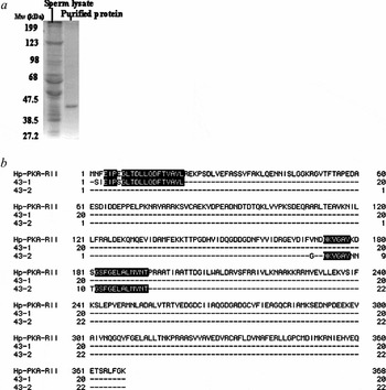
It has been demonstrated that, in starfish, asterosap specifically elevates cGMP concentration by 100-fold in approximately 400 ms (Matsumoto et al., Reference Matsumoto, Solzin, Helbig, Hagen, Ueno, Kawase, Maruyama, Ogiso, Godde, Minakata, Kaupp, Hoshi and Weyand2003). To identify the next signal for cGMP in starfish spermatozoa, we screened a cGMP-binding protein obtained from starfish spermatozoa using a cGMP-affinity column. The fraction contained a major 43 kDa protein, which showed a single band after being subjected to SDS-PAGE (Fig. 1a). The purified 43 kDa protein was chemically digested with hydroxylamine. Twenty residues of the N-terminal sequence of this 43 kDa protein (43–1) and 22 residues of the mid-sequence (43–2) were determined. The sequences showed high homology with sea urchin cAMP-dependent protein kinase type II regulatory subunit (Hoshino et al., Reference Hoshino, Nomura and Suzuki1997) (Fig. 1b).
(a) Analysis of cGMP-binding protein by SDS-PAGE. The purified protein was subjected to 10% SDS-PAGE and treated with CBB stain (Nacalai Tesque). Size markers are indicated on the left. (b) Comparison of amino acid sequences of the purified 43 kDa protein with deduced amino acid sequence of sea urchin spermatozoa PKA-R (Hp-PKA-RII). Purified 43 kDa protein was digested chemically and yielded two fragments (43-1 and 43-2). Homologous regions of the amino acid sequences are shaded black. Amino acid positions are on the right.

Cloning of the PKA-R from starfish testes
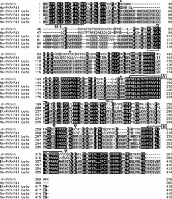
To identify the complete structure of the 43 kDa protein in starfish spermatozoa, RT-PCR was performed using degenerate primers from the N-terminal amino acid sequence of the 43 kDa protein. Furthermore, a cDNA library of starfish testes was screened using a fragment obtained by RT-PCR. The sequence of the full-length clone (approximately 2.3 kbp) includes an open reading frame of 1110 nucleotides that encodes a 370 amino acid peptide (Fig. 2). This full-length cDNA encodes the predicted 41.5 kDa protein, which is comparable with the size estimated by SDS-PAGE (Fig. 1a). A BLAST search revealed that the predicted protein displays sequence homology to the regulatory protein kinase A (PKA-R). This subunit contains a highly conserved region for the inhibitory site and two putative cAMP-binding sites. The inhibitory site retains two arginines that are essential for recognition by the catalytic subunit (Taylor et al., Reference Taylor, Buechler and Yonemoto1990; Hoshino et al., Reference Hoshino, Nomura and Suzuki1997). The alignment of PKA-R sequences from various species is shown in Fig. 2 and shows a 53–56% similarity among these species.
Homology of A. amurensis sf-PKA-R to proteins derived from sea urchin (Hp), bovine (Bt), human (Hs), rat (Rn) and mouse (Mm). Three N-linked glycosylation sites are marked by asterisk. The inhibitory site is boxed and the two cAMP-binding sites (sites A and B) are marked by a line. Disulphide bonds with the corresponding residues of the second protein are denoting by triangles. Identical amino acids are indicated by black backgrounds and conservative substitutions by grey backgrounds. Partial amino acid sequences of 43 kDa protein (43-1 and 43-2) are marked by a line. Gaps introduced to optimize alignments are marked by dashes. Amino acid positions are on the right.

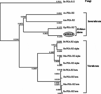
This sequence consists of one inhibitory site (residues 76–81), three N-linked glycosylation sites (residues 40–43, 95–98 and 178–181) and two cAMP-binding sites (site A: 183–200 and site B: 308–324) (Fig. 2). For the 18 cAMP-binding site A residues, 17 residues were found to be identical in all species, whereas only 12 out of 17 cAMP-binding site B residues are conserved. The putative structure of mammalian PKA-R is a homodimer linked by disulfide bonds. The cysteine residue at amino acid 81 is found in every species, but other cysteine residues (264, 275 and 326) are not found in every species. A phylogenetic tree, which was constructed based on full-length PKA-RII proteins, classified this clone as belonging to the echinoderm group (Fig. 3). Based on the homology to a wide range of species, it is concluded that the isolated clone represents the regulatory subunit of starfish PKA and is named sf-PKA-R.
Phylogeny of sf-PKA-R. A phylogenetic tree was constructed by the UPGMA method using PKA regulatory subunit of fungi as an outgroup. The numbers above each branch of the tree correspond to the p-values, which are measure of proportional differences between sequences. GenBank accession numbers of different animals PKA regulatory subunits are given in Materials and methods.

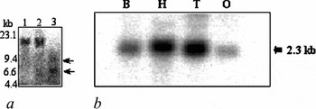
To determine the number of sf-PKA-R genes in the A. amurensis genome, we carried out a Southern blot analysis. As shown in Fig. 4a, restriction enzymes SalI (lane 1) and XhoI (lane 2), with no recognition site yielded one hybridization band. Conversely, NheI (lane 3), with one cutting site within the ORF, gave two bands. These results suggest that A. amurensis contains only one gene for sf-PKA-R.
(a) Southern blot analysis of the sfNCKX gene. Samples of A. amurensis DNA (10 µg/lane) were digested with SalI (lane 1), XhoI (lane 2) or NheI (lane 3) and processed as described in Materials and methods. Horizontal arrows show bands of genomic DNA digested with NheI (lane 3). Size markers in kb are indicated on the left. (b) Tissue distribution of sf-PKA-R transcripts. 7.5 µg of total RNA was isolated from testes (T), ovary (O), hepatopancreas (H) and body fluid (B) and analysed by northern blotting at higher stringency using a riboprobe from starfish PKA-R.

Northern blot analysis of sf-PKA-R distribution using total RNA isolated from various tissues from A. amurensis is shown in Fig. 4b. The hybridization signal was detected at 2.3 kb in length and was widely expressed in various tissues examined.
ARIS and asterosap increases the [cAMP]i
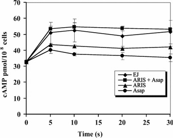
In sea urchins the sperm-activating peptide, speract, increases sperm cAMP concentrations (Garbers & Hardman, Reference Garbers and Hardman1976), but, in starfish, asterosap does not increase significantly (Matsumoto et al., Reference Matsumoto, Solzin, Helbig, Hagen, Ueno, Kawase, Maruyama, Ogiso, Godde, Minakata, Kaupp, Hoshi and Weyand2003). Therefore, an attempt was made to see the effects of another component of EJ, ARIS, on the increase in the [cAMP]i. By using a cAMP enzyme-immunoassay, the [cAMP]i in normal pH seawater was 30 pmol/108 cells (Fig. 5). When spermatozoa were stimulated with EJ or with ARIS plus asterosap, [cAMP]i was elevated to 51 pmol/108 cells or 54 pmol/108 cells respectively within 5 s. The [cAMP]i levels were sustained for more than 30 s after both treatments. ARIS alone could elevate [cAMP]i slightly. Recently it was found that PDE inhibitors suppress the hydrolysis of the asterosap-induced [cAMP]i increase (Kawase et al., Reference Kawase, Ueno, Minakata, Hoshi and Matsumoto2004).
Increase in the [cAMP]i induced by the EJ in normal seawater. Spermatozoa were incubated with EJ or with individual components of EJ as described in Materials and methods. The [cAMP]i was measured when spermatozoa were treated with EJ or individual components of EJ at different time points. Each measurement is the mean ± SD.

ARIS induces an increase in [cAMP]i in high pH seawater
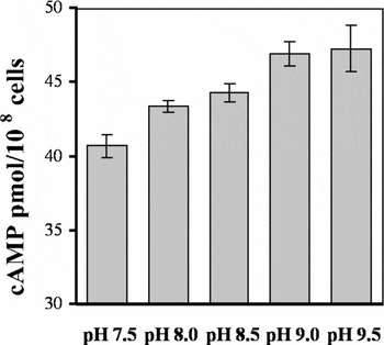
ARIS induces the AR in high pH seawater (Matsui et al., Reference Matsui, Nishiyama, Hino and Hoshi1986). Taking this result into account, we attempted to find if the increase in the [cAMP]i in ARIS-treated spermatozoa was pH dependent. When spermatozoa were treated with ARIS in ASW at pH 7.5, [cAMP]i rose to about 40 pmol/108 cells within 5 s. Conversely when external pH was stepped up gradually, the rate of increase also rose (Fig. 6). The [cAMP]i at pH 9.5 was 48 pmol/108 cells, which is similar to the value of EJ-stimulated spermatozoa in normal seawater.
Increase in the [cAMP]i induced by ARIS in seawater of differing pH. Spermatozoa were suspended in pH seawater of varying pH and incubated with ARIS as described in Materials and methods. The [cAMP]i was measured when spermatozoa were treated with ARIS for 5 s. Each measurement is the mean ± SD.

PKA participates in the AR
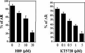
In the above experiment, we have found that EJ or ARIS plus asterosap can induce a rise in the cAMP concentration in starfish spermatozoa. To determine what were the subsequent signals in the PKA-regulated pathway that controlled the increase in cAMP levels and the induction of the AR, the spermatozoa were treated with PKA inhibitors KT5720 or H89 and the AR efficiency was examined. H89 or KT5720 suppressed the induction of AR in a concentration-dependent manner (Fig. 7). The inhibition of AR was especially evident when the spermatozoa were treated with H89 or KT5720 at 20 µM and 5 µM respectively.
Effect of PKA inhibitors in the AR. Spermatozoa were incubated in either the absence or presence of different concentrations of H89 and KT5720 for a period of 3 min then incubated with EJ (0.1 mg sugar/ml) for 5 min according to the procedure outlined in the Materials and methods. The data represent the means ± SD of at least four batches of spermatozoa.

cAMP-dependent kinase activity in starfish spermatozoa
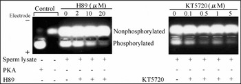
The kinase activity of starfish sperm lysate was measured in the presence of 1 µM cAMP using a fluorescent kemptide, which is a synthetic substrate for PKA. As shown in the control (Fig. 8), substrate that was completely phosphorylated by bovine PKA migrated towards the positive electrode in the agarose gel and non-phosphorylated substrates migrated towards the negative electrode, depending on the charge of the phosphate residues.
Kinase activities in sperm lysate. Phosphorylated kemptide was separated from non-phosphorylated by agarose gel electrophoresis and visualized on a UV transluminator. PepTag® A1 peptide (2 µg) were incubated as described in Materials and methods with or without varying amounts of bovine PKA (0 or/and 33 ng) provided by the kit in either the absence or presence of H89 and KT5720. About 10 µg protein of sperm lysate was used to test the kinase activity. This experiment was repeated three times, the gel photograph shows one of the three experiments.

When the substrate was incubated with starfish sperm lysate, it showed remarkable phosphorylation (Fig. 8). In the presence of different concentrations of PKA inhibitors, H89 (2–20 µM) and KT5720 (0.1–5 µM), the phosphorylated substrate decreased in a concentration-dependent manner.
Dependency of PKA on cyclic nucleotides
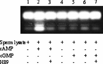
At the beginning of this experiment, we tried to isolate a cGMP-binding protein from starfish spermatozoa using a cGMP-affinity column, however the protein found was PKA. To confirm the existence of PKA in starfish spermatozoa we compared the dependency of kinase activity on cAMP and cGMP of starfish. In the presence of 1 µM cAMP, a larger amount of the phosphorylated substrate was observed (Fig. 9, lane 2). This phosphorylated substrate disappeared after treatment with 20 µM H89 (Fig. 9, lane 3), whereas in presence of 1 µM cGMP, the phosphorylated substrate was not observed (Fig. 9, lane 5). Only a lower level of phosphorylated substrate was observed when increasing the concentration of cGMP to 10 µM (Fig. 9, lane 6). This phosphorylated substrate was not observed after treatment with 20 µM H89 (Fig. 9, lane 7). These results indicate that PKA in starfish spermatozoa is specific to cAMP and is inhibited by its conventional inhibitors.
Ability of cyclic nucleotides to activate the kinase. Activity was measured as described in Fig. 8. Two concentrations of cGMP (1 µM and 10 µM in lane 5 and 6 respectively) were used, for cAMP only one concentration (1 µM) was used. A concentration of 20 µM of H89 inhibited the kinase activity (lane 3 and 7). The photograph shows one of three independent experiments.

Discussion
In starfish spermatozoa, the sperm-activating peptide, asterosap, evokes a rapid and transient increase in the cGMP concentration; however, the cAMP concentration does not change significantly (Matsumoto et al., Reference Matsumoto, Solzin, Helbig, Hagen, Ueno, Kawase, Maruyama, Ogiso, Godde, Minakata, Kaupp, Hoshi and Weyand2003). In this study, we attempted to isolate a cGMP-binding protein from starfish spermatozoa by cGMP-affinity chromatography. The extracted protein was not a cGMP-binding protein, but was a cAMP-binding protein. This protein encoded a regulatory subunit of PKA, referred to as sf-PKA-R, which is conserved in mammals, sea urchins and in various other animals.
The increase in cAMP concentration could regulate various channels. For example, EJ increases the cAMP concentration over a 100-fold and increases PKA activity from 4- to 8-fold in sea urchins (Garbers & Kopf, Reference Garbers and Kopf1980, Garbers et al., Reference Garbers, Tubb and Kopf1980). Intracellular levels of cAMP are also elevated during the AR by EJ (Ward & Kopf, Reference Ward and Kopf1993). Furthermore, PKA-mediated phosphorylation in sea urchin spermatozoa is also required for the EJ-induced AR (Su et al., Reference Su, Chen, Zhou and Vacquier2005). In mammalian spermatozoa, PKA induces the phosphorylation of several proteins required for capacitation and the AR (Harrison, Reference Harrison2004; O'Flaherty et al., Reference O'Flaherty, de Lamirande and Gagnon2004). Here, we observed that in starfish, EJ components evoke an AR, which might be mediated by the increase in the cAMP followed by the activation of PKA.
In starfish, EJ comprises three components, i.e. ARIS, Co-ARIS and a sperm-activating peptide named asterosap, and these are responsible for triggering the AR (Hoshi et al., Reference Hoshi, Nishigaki, Ushiyama, Okinaga, Chiba and Matsumoto1994). ARIS and asterosap function in concert to induce AR and asterosap facilitates the ARIS-triggered AR via a cGMP-dependent pathway (Matsumoto et al., Reference Matsumoto, Solzin, Helbig, Hagen, Ueno, Kawase, Maruyama, Ogiso, Godde, Minakata, Kaupp, Hoshi and Weyand2003). In sea urchins, while fucose sulphate polymer (FSP) is sufficient to induce the AR, another EJ component, sialoglycan, can serve to potentiate FSP induction (Hirohashi & Vacquier, Reference Hirohashi and Vacquier2003). The activation of adenylyl cyclase leads to an increase in [cAMP]i in sea urchin spermatozoa due to the effect of speract, which is a sperm-activating peptide in Strongylocentrotus purpuratus (for review see Darszon et al., Reference Darszon, Nishigaki, Wood, Trevino, Felix and Beltran2005). Here we observed that, in starfish, ARIS is a major component that could induce an increase in the level of cAMP in combination with asterosap for the initiation of PKA activity. In sea urchin, sialoglycan and speract in the EJ potentiate the FSP-induced AR in low pH seawater (Hirohashi & Vacquier, Reference Hirohashi and Vacquier2002b) and sialoglycan reduces the concentration of FSP required for the induction in normal seawater (Hirohashi & Vacquier, Reference Hirohashi and Vacquier2002a). Both sialoglycan and speract seem to facilitate the AR through a rise in pHi. In starfish, Matsui et al. (Reference Matsui, Nishiyama, Hino and Hoshi1986) and Kawase et al. (Reference Kawase, Minakata, Hoshi and Matsumoto2005) reported that, in high pH ASW, ARIS alone could induce the AR. We also found using high pH ASW, ARIS alone could increase the [cAMP]i of starfish spermatozoa. Therefore, we assumed that cAMP might participate in the induction of AR via a rise in the pHi. The increase in the cAMP-dependent PKA activity induced by EJ was inhibited when spermatozoa were preincubated with KT5720, which is a cell permeable inhibitor of PKA (Fig. 7). It was reported that cAMP could stimulate the AR in bovine spermatozoa; this is because AR is completely inhibited in mammals using KT5720 (Spungin & Breitbart, Reference Spungin and Breitbart1996). Here we also found that a complete inhibition of starfish spermatozoa AR when spermatozoa were preincubated with KT5720 or H89. The in vitro kinase activity is other evidence for the involvement of PKA in AR in starfish spermatozoa. A significant kinase activity was observed in starfish spermatozoa in presence of cAMP. In this case, cAMP could bind to the regulatory subunit of PKA of which cDNA was cloned from starfish testes and the activated form of PKA could subsequently phosphorylate various sperm proteins.
Crosstalk between cGMP and cAMP pathways has been described among vertebrates and invertebrates. While activation of mammalian PKA requires a 50- to 100-fold higher concentration of cGMP compared to cAMP (Reed et al., Reference Reed, Sandberg, Jahnsen, Lohmann, Francis and Corbin1996), in invertebrates the discrimination between cAMP and cGMP is not so clear. Bardales et al. (Reference Bardales, Diaz-Enrich, Ibarguren and Villamarin2004) found in the bivalve mollusc that both cAMP and cGMP can activate PKA, although PKA exhibits sensitivity approximately 100-fold higher for cAMP than for cGMP. In contrast, PKA holoenzymes from yeast (Cytrinska et al., Reference Cytrinska, Wojda, Frajnt and Jakubowicz1999) and from some invertebrates, such as insects (Vardanis, Reference Vardanis1980; Altfelder & Müller, Reference Altfelder and Müller1991) or the crab Chasmagnathus (Locatelli et al., Reference Locatelli, LaFourcade, Maldonado and Romano2001), have comparable affinities for cAMP and cGMP and are likely to be activated in vivo by either nucleotide. Our data suggest that PKA might have a higher affinity for cGMP, in fact it was initially purified on a cGMP–agarose column. As starfish spermatozoa PKA-R is extracted by cGMP affinity chromatography, it might be able to induce the activation of PKA with cGMP. However, in starfish spermatozoa, this PKA activity exhibited a higher sensitivity to cAMP than to cGMP (Fig. 9). This result shows that PKA could play a vital role in starfish spermatozoa.
Many of the genes and proteins involved in cAMP-mediated cell signaling have been identified in different animals. We found that starfish spermatozoa contained a single copy of the sf-PKA-R gene. The effects of the specific-PKA inhibitors H89 and KT5720 strongly suggest that PKA might participate in the signal transduction cascade leading to induction of AR through the involvement of EJ.
In various animals, PKA plays an important role in the later steps for induction of AR. Even in echinoderms such as sea urchins, the small molecules in EJ, including speract, can induce a rise in cAMP levels; however, molecules similar to asterosap in EJ cannot induce them. If the small molecules in sea urchin and starfish EJ diversified almost 500 million years ago (Matsumoto et al. Reference Matsumoto, Solzin, Helbig, Hagen, Ueno, Kawase, Maruyama, Ogiso, Godde, Minakata, Kaupp, Hoshi and Weyand2003), then this implies that the later steps for AR induction might utilise similar signalling systems in different animals, but that the first stages of these signalling systems differ among species. Species specificity could be provided by the triggering step in fertilization.
Acknowledgements
We thank the Director and staff members of the Otsuchi Marine Research Center and the Misaki Marine Biological Station, the University of Tokyo for their help in collecting the starfish. We also thank Dr H. Kawahara, Hokkaido University, Japan for the amino acid sequencing. M.S. Islam extends his thanks to the Jinnai International Student Scholarship Foundation for its generous financial assistance. This research was partially supported by the Ministry of Education, Culture, Sports, Science and Technology, Grants-in-Aid for Scientific Research on Priority Area (A) (to M.M.) and Grant-in-Aid for the 21st Century Center of Excellence (COE) Program entitled ‘Understanding and Control of Life's Function via Systems Biology (Keio University)’ (to M.S.I).