Introduction
The prognosis of congenital heart disease (CHD) has improved dramatically because of therapeutic advances in congenital cardiology and surgery; therefore, the number of patients with adult CHD successfully reaching adulthood has increased. Although numerous previous reports have mentioned the association of sleep apnea with heart failure, hypertension, and arrhythmia in the general population,Reference Shamsuzzaman, Gersh and Somers1–Reference Oldenburg, Lamp and Faber3 few studies have examined sleep apnea in patients with adult CHD. Studies have shown a high prevalence of sleep apnea in the children with tetralogy of FallotReference Herold, Young, Ge, Snieder and Lovrekovic4 as well as in patients with transposition of the great arteries after an atrial switch procedure.Reference Cotts, Smith, Lu, Dorfman and Norris5 The prevalence, risk factors, and effects on prognosis of sleep apnea in adult CHD have not been studied well.
The aim of this study was to investigate the prevalence and characteristics and to identify the risk factors associated with the development of sleep apnea in a population of hospitalised patients with adult CHD.
Materials and methods
Study design and participants
This was a single-centre retrospective study of sleep apnea in 104 consecutive patients older than 15 years (median age: 36 [interquartile range: 28–48] years) who had various adult CHDs with or without a medical history of cardiac surgery. The medical records of patients who attended Tokyo Women’s Medical University Hospital between May 2010 and June 2016 were reviewed. All participants underwent a sleep study in the hospital. Indications for admission were classified as heart failure, diagnostic catheterisation, interventional catheterisation, or arrhythmia. We excluded patients with unstable status, such as those with New York Heart Association (NYHA) class IV; those receiving any intravenous drip infusions, oxygen supplementation, or positive airway pressure treatment; and those with cyanosis or arterial oxygen saturation less than 90%.
First, we studied the prevalence of sleep apnea in patients with adult CHD. Second, we compared the characteristics based on the severity of sleep apnea. Third, we analysed the risk factors associated with respiratory disturbance index. Finally, we reviewed the outcome of patients with sleep apnea treated by continuous positive airway pressure or adaptive servo-ventilation.
Sleep study
All patients underwent type-3 portable overnight polygraphy: Pulsleep LS-120S (Fukuda Denshi, Tokyo, Japan)Reference Uesugi, Kobayashi and Hasebe6 in 57 patients and Morpheus R (Compumedics, Victoria, Australia)Reference Satake, Sugimura and Fukumoto7 in 47 patients. Episodes of apnea and hypopnea, the mean and lowest percutaneous arterial oxygen saturation levels, and the number of desaturation from baseline saturation episodes were assessed based on previously described methods.Reference Chesson, Berry and Pack8 Pulsleep LS-120S has a pressure sensor for nasal airflow and snoring, a pressure-sensitive sensor on the suprasternal notch to estimate oesophageal pressure, and a body position sensor. Heart rate and percutaneous arterial oxygen saturation were continuously monitored using a pulse oximeter. Morpheus R has a pressure sensor for nasal airflow and snoring, two stress-sensitive belts (one each for the rib cage and abdomen), a body position sensor, continuous pulse oximetry, and three-channel electrocardiograms.
Sleep duration, the estimated length of time between the time the participant went to bed and the time one got out of bed, was estimated from a sleep diary. Estimated sleep duration was used for portable monitor analyses.
Apnea was defined as a continuous cessation of breathing airflow for 10 s or more; hypopnea was defined as a reduction of 50% or greater in breathing airflow with percutaneous arterial oxygen desaturation ≥3%.9 Respiratory disturbance index, the number of apneas and hypopneas per hour of the estimated sleep duration, was calculated from the portable monitor data.10 Apnea was classified as obstructive sleep apnea or central sleep apnea based on the presence or absence of laboured breathing estimated using an oesophageal pressure sensor in Pulsleep LS-120S and by thoracoabdominal motion in Morpheus R during apnea. We also calculated the 3% oxygen desaturation index, which was the total number of 3% desaturation from baseline saturation episodes per hour of estimated sleep duration.9 The portable monitor records were manually analysed by an expert technician and medical doctor specialising in respiratory medicine.
Severity and type of sleep apnea
We defined a respiratory disturbance index ≥5 as sleep apnea. The severity of sleep apnea was classified into four categories: no sleep apnea, patients with a respiratory disturbance index <5; mild SDB, patients with a 5≤ respiratory disturbance index <15; moderate sleep apnea, patients with a 15≤ respiratory disturbance index <30; and severe sleep apnea, patients with a respiratory disturbance index ≥30.
We divided the participants into three groups: those without sleep apnea (respiratory disturbance index <5), those with mild sleep apnea (5≤ respiratory disturbance index <15), and those with moderate/severe sleep apnea (respiratory disturbance index ≥15). We also stratified the types of sleep apnea into two categories as described above: obstructive sleep apnea and central sleep apnea.
Demographic data collection
We obtained demographic data within 1 week before or after the sleep study from the patients’ medical records, such as NYHA functional class, history of cardiac surgery (particularly the performance of the Fontan procedure), blood test results for brain natriuretic peptide (normal: <20 pg/mL) measured using a chemiluminescence enzyme immunoassay (Lumipulse Presto BNP; Fujirebio, Tokyo, Japan) and noradrenaline (normal: <5 pg/mL) measured using high-performance liquid chromatography (CA test; TOSHO, Tokyo, Japan), echocardiographic parameters of the shortening fraction of the systemic ventricle, catheterisation parameters, drug use, and treatment using pacemaker and implantable cardioverter defibrillators. Arrhythmia was defined as sinus rhythm, atrial tachyarrhythmia, atrial fibrillation, ventricular tachyarrhythmia, and ventricular fibrillation detected by 12-lead and Holter electrocardiogram. Hypertension was determined when patients had systolic and diastolic blood pressures of >140 mmHg or >90 mmHg, respectively. Dyslipidaemia was defined as low-density lipoprotein cholesterol ≥140 mg/dl, high-density lipoprotein cholesterol <40 mg/dl, or triglycerides ≥150 mg/dl. Patients with glucose ≥126 mg/dl or HbA1c ≥6.5% were defined as having diabetes mellitus. Chronic kidney disease was defined as an estimated glomerular filtration rate <60 mL/min/1.73 m2. Thyroid dysfunction, including hyper- and hypothyroidism, was defined as a previous diagnosis of thyroid dysfunction by endocrinologists. Protein-losing enteropathy was diagnosed by documenting enteric protein loss using nuclear scintigraphy. We obtained central venous pressure, mean pulmonary artery pressure, pulmonary capillary wedge pressure, systemic end-diastolic pressure, ascending aortic pressure, pulmonary vascular resistance, and cardiac index from cardiac catheterisation (n = 78). From 78 patients, 60 (77%) underwent cardiac catheterisation within 1 month after the sleep study, whereas 3 (4%), 6 (7%), and 9 (12%) patients underwent the procedure after 4–6 months, 7–11 months, and 12 months, respectively.
Complexity of congenital heart disease
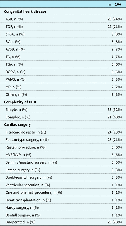
According to the complexity of CHD, based on the ICD-9 codes,Reference Warnes, Williams and Bashore11–Reference Khairy, Van Hare and Balaji13 we divided the patients’ CHD into simple or complex CHD, including moderate/severe CHD. Simple CHD included atrial septal defects, ventricular septal defects, mitral valve regurgitation, and patent ductus arteriosus. Other CHDs such as tetralogy of Fallot, congenitally corrected transposition of the great arteries, single ventricle, and double-outlet right ventricle were considered complex CHDs.
Statistical analysis
Data regarding patient characteristics and various biomarker levels were expressed as number (percentage) or median (interquartile range: 25th–75th percentile).
We used the non-parametric test (Mann–Whitney U test or Kruskal–Wallis one-way analysis) to compare continuous variables (e.g. various biomarker levels) and the chi-square test to compare the proportion of categorical variables (e.g. atrial arrhythmia) among our study groups. We used multivariate logistic regression analysis to analyse risk factors associated with a respiratory disturbance index ≥15. This model included body mass index, NYHA≥II, shortening fraction <0.25, atrial tachycardia/atrial fibrillation, complex CHD, and Fontan-type surgery. Body mass index and atrial tachycardia/atrial fibrillation were previously reported as factors associated with sleep apnea in patients with congestive heart failure.Reference Sin, Fitzgerald and Parker14 Relationships were summarised as an odds ratio with 95% confidence interval. JMP Pro version 12 (SAS Institute, Cary, NC, USA) was used for all analyses. A p-value of <0.05 was considered statistically significant.
Results
Participants’ characteristics
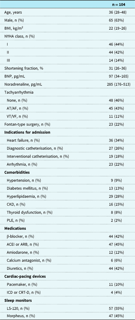
The baseline patient characteristics of these patients are summarised in Tables 1 and 2. The median age of the patients was 36 (28–48) years, the body mass index was 22 (19–26) kg/m2, and the indications for admission were heart failure (34%, n = 36), diagnostic catheterisation (26%, n = 27), interventional catheterisation (18%, n = 19), or arrhythmia (22%, n = 23).
The number of prior Fontan-type surgeries was 23 (22%). The number of patients with simple and complex CHDs was 33 (32%) and 71 (68%), respectively.
The numbers of patients with NYHA classes I and II+III were 46 (44%) and 58 (56%), respectively, and the shortening fraction and brain natriuretic peptide concentrations were 31% (26%–36%) and 97 pg/mL (34–165 pg/mL), respectively.
The type of atrial tachyarrhythmia/atrial fibrillation included 37 paroxysmal (stopping within a week of onset) and 8 permanent types (normal heart rhythm could not be restored with treatment).
Patient characteristics.

The values presented are in median (interquartile range: 25th–75th percentile) or n (%).
ACEI = angiotensin-converting enzyme inhibitor; AF = atrial fibrillation; ARB = angiotensin receptor blocker; AT = atrial tachyarrhythmia; BMI = body mass index; CKD = chronic kidney disease; CRT-D = cardiac resynchronisation therapy defibrillator; ICD = implantable cardioverter defibrillators; NYHA = New York Heart Association; PLE = protein-losing gastrointestinal disease; VF = ventricular fibrillation; VT = ventricular tachyarrhythmia.
Cardiac diagnosis and previous cardiac surgeries.

The values presented are in n (%).
ASD = atrial septal defect; AVSD = atrioventricular septal defect; CHD = congenital heart disease; cTGA = corrected transposition of the great arteries; DORV = double-outlet right ventricle; MR = mitral valve regurgitation; MVP = mitral valve plasty; MVR = mitral valve replacement; PAIVS = pulmonary atresia with intact ventricular septum; SV = single ventricle; TA = tricuspid atresia; TGA = transposition of the great arteries; TOF = tetralogy of Fallot.
Prevalence of sleep apnea in adult CHD
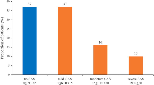
The prevalence of sleep apnea is shown in Figure 1. The prevalence of sleep apnea, defined as respiratory disturbance index ≥5, was 63% (n = 66). The distribution of mild, moderate, and severe sleep apnea was 37%, 16%, and 10%, respectively. Moderate/severe sleep apnea was found in 26% of participants. The type of sleep apnea was 92% (n = 58) for obstructive sleep apnea, and 8% (n = 5) for central sleep apnea.
Prevalence of sleep apnea based on the degree of RDI. RDI=respiratory disturbance index; SAS = sleep apnea syndrome.

Comparison of characteristics based on the degree of sleep apnea
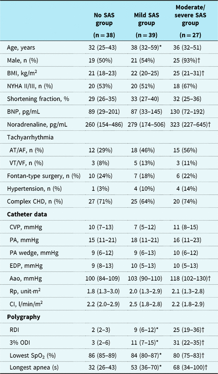
Comparison of the characteristics between patients without sleep apnea and those with mild or moderate/severe sleep apnea is shown in Table 3. In brief, the group with moderate/severe sleep apnea had a significantly higher proportion of male patients and higher body mass index, noradrenaline level, and ascending aortic pressure than the group without sleep apnea. In contrast, the group with mild sleep apnea showed no significant difference in various parameters, except age, compared with the group without sleep apnea. All patients with suspected central sleep apnea had a high NYHA class (NYHA class III).
Characteristics of patients based on the degree of sleep apnea.

The values presented are in median (interquartile range: 25th–75th percentile) or n (%).
Aao = ascending aorta; CI = cardiac index; CVP = central venous pressure; EDP = end-diastolic pressure; ODI = oxygen desaturation index; PA = pulmonary artery; RDI = respiratory disturbance index; Rp = resistance of pulmonary; SAS = sleep apnea syndrome.
* p<0.05, no SAS vs. mild SAS.
† p<0.05, no SAS vs. moderate–severe SAS.
Risk factors associated with respiratory disturbance index ≥15
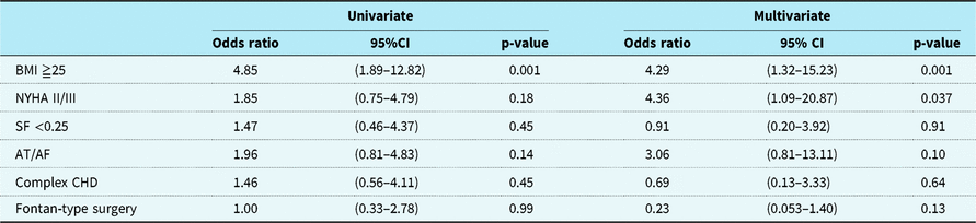
Multivariable analysis showed that NYHA ≥II (odds ratio, 4.36; 95% confidence interval, 1.09–20.87) and body mass index ≥25 (odds ratio, 4.29; 95% confidence interval, 1.32–15.23) were independent risk factors for respiratory disturbance index ≥15 in patients with adult CHD (Table 4).
Multivariable odds ratios for RDI.

AF = atrial fibrillation; AT = atrial tachyarrhythmia; BMI = body mass index; CHD = congenital heart disease; CI = confidence intervals; NYHA = New York Heart Association; RDI = respiratory disturbance index; SF = shortening fraction.
Treatment of sleep apnea
We treated eight patients with severe sleep apnea using continuous positive airway pressure in five patients and adaptive servo-ventilation in three patients. After continuous positive airway pressure or adaptive servo-ventilator therapy, symptoms associated with sleep apnea, such as sleepiness, arousal during sleep, and headache, improved in 88% of patients (7/8 patients). Although the respiratory disturbance index after respiratory therapy showed a significant decrease compared with the respiratory disturbance index prior to respiratory therapy (3.7±1.9 vs 55.4±19.7, p = 0.002) in six patients who underwent sleep study before and after respiratory therapy, heart failure worsened in one patient with repaired tetralogy of Fallot who experienced severe right ventricle dysfunction after automatically adjusted continuous positive airway pressure with the pressure level ranged 4–16 mmH2O.
Discussion
This study showed a high prevalence of sleep apnea in adult CHD, with most cases being obstructive sleep apnea, and that NYHA ≥II and higher body mass index were risk factors for developing respiratory disturbance index ≥15 in patients with adult CHD.
A high prevalence of sleep apnea in patients with adult CHD was observed in this study compared with a previously reported prevalence of sleep apnea in a healthy community sample (16.5% and 6.5% for apnea–hypopnea index ≥5 and apnea–hypopnea index ≥15, respectively).Reference Young, Palta and Dempsey15 In a previous study that screened sleep apnea using the Paediatric Sleep Questionnaire, which defined sleep apnea as having Paediatric Sleep Questionnaire scores ≥8, the reported prevalence of sleep apnea in the children with tetralogy of Fallot was 38% higher than that in the healthy general population (5%),Reference Herold, Young, Ge, Snieder and Lovrekovic4 and screening of sleep apnea in CHD using type-3 polysomnography has not been reported yet. Several factors may contribute to this high prevalence of sleep apnea in adult CHD. In this study, the median central venous pressure of all patients and even in the group without sleep apnea was high at 10 mmHg. The underlying adult CHD itself may be associated with abnormal haemodynamics and congestive state even without clinical heart failure. The high venous pressure and venous blood flow congestion in pharyngeal and neck tissues may decrease upper airway size,Reference Shepard, Pevernagie, Stanson, Daniels and Sheedy16 and fluid displacement from the oedematous leg to the peripharyngeal soft tissues when lying supine may narrow the pharynx during sleep.Reference Yumino, Redolfi and Ruttanaumpawan17 There was a high rate of complex CHD, heart failure that required hospitalisation, cardiac conditions that required catheter intervention, and arrhythmia in the present study. Such findings may reflect a more unhealthy patient population within our institute. Together, these mechanisms and factors may be associated with the high prevalence of sleep apnea in adult CHD.
Although adult patients with CHD have unique haemodynamics such as complex CHD with chronic right or left heart failure and repaired Fontan-type surgery with single ventricle physiology, these conditions involved in the congestive state with high central venous pressure may contribute to the high prevalence of sleep apnea in adult CHD; however, the presence of complex CHD and history of Fontan-type surgery were not independent risk factors of respiratory disturbance index ≥15 after multivariate analysis. Because this study included various heterogenous CHDs and haemodynamics, the risk of sleep apnea in each specific condition should be studied in the future.
The findings of heart failure (high NYHA class) and obesity as risk factors for sleep apnea were compatible with previous reports in patients without CHD.Reference Yumino, Redolfi and Ruttanaumpawan17–Reference Tkacova, Niroumand, Lorenzi-Filho and Bradley21 Obstructive sleep apnea and central sleep apnea are common in patients with heart failure, and the predominant type of sleep apnea can shift from obstructive to central based on the severity of heart failure.Reference Vazir, Hastings and Dayer20, Reference Tkacova, Niroumand, Lorenzi-Filho and Bradley21 The incidence of central sleep apnea was higher in patients with low cardiac index and increased pulmonary wedge pressure.Reference Mortara, Sleight and Pinna22 In patients with heart failure, fluid displacement from the leg to the neck narrows the pharynx, contributes to obstructive sleep apnea, and increases venous blood flow; consequently, pulmonary congestion with increasing pulmonary wedge pressure leads to central sleep apnea.Reference Yumino, Redolfi and Ruttanaumpawan17 Obesity is also a risk factor for sleep apnea due to airway size affected by fat deposition.Reference Horner, Mohiaddin and Lowell18, Reference Mortimore, Marshall, Wraith, Sellar and Douglas19 Although congestive heart failure and obesity were already known as risk factors of sleep apnea, this was the first study to document these findings in adult CHD. Recently, there has been a focus on the management of cardiovascular risk factors such as obesity, hypertension, diabetes, and hyperlipidaemia in adult CHD.Reference Lui, Fernandes and McElhinney23 First, the management of obesity and lifestyle in adult CHD patients may be important to reduce sleep apnea and improve long-term outcomes similar to what is seen in the general population.Reference Nishihata, Takata and Usui24
This study showed high noradrenaline level and ascending aortic pressure in the group with moderate/severe sleep apnea compared with the group without sleep apnea and with mild sleep apnea. Sleep apnea contributed to the autonomic and hemodynamic changes by hypoxia, hypercapnia, negative intrathoracic pressure, and arousal to increase sympathetic activity and noradrenaline level.Reference Kapa, Sert Kuniyoshi and Somers25–Reference Somers, Dyken, Clary and Abboud27
Most of the sleep apnea types were obstructive sleep apnea (92%). A small percentage of central sleep apnea may be associated with a relatively young age and low percentage of severe heart failure (NYHA class III in 14%) of the participants in this study. The Japanese Circulation Society recommended using portable sleep monitoring for screening of sleep apnea.10 More detailed tests for sleep apnea using detailed polysomnography are needed to show the exact sleep apnea type and apnea–hypopnea index in patients with adult CHD whose symptoms are highly suggestive of sleep apnea.
The effectiveness of sleep apnea treatments, such as continuous positive airway pressure and use of an adaptive servo-ventilator, in the CHD population is still unknown. In our limited experience, although continuous positive airway pressure or an adaptive servo-ventilator seemed to be effective to improve sleep apnea-associated symptoms for most patients treated, right heart failure worsened in one patient with tetralogy of Fallot and severe right ventricular dysfunction after starting continuous positive airway pressure. High positive airway pressure was suggested to increase intrathoracic pressure, resulting in disturbed venous return into the right ventricle in this case. Some CHD patients have unique haemodynamics with chronic right heart failure, such as the Fontan circulation. Because high positive air pressure may exacerbate haemodynamics in patients with predominant right heart failure, appropriate identification of positive air pressure level for those patients with sleep apnea and right heart failure may be necessary to start continuous positive airway pressure therapy. Positive pressure should be used with caution in patients with Fontan physiology, as well as in those with biventricular circulation and right ventricular dysfunction.
Limitations of the study
Our study has several limitations. Because the number of study patients was small, the participants in this study included heterogeneous CHD, and each hemodynamic condition was different such as single or biventricular ventricle physiology. The role of haemodynamics in and the risk factors for developing sleep apnea may be different among various conditions. Because the participants in this study had a high rate of complex CHD (68%) and heart failure (34%) requiring hospitalisation as mentioned above, we cannot exclude the possibility that the results (particularly with regard to the prevalence of sleep apnea) may not be as applicable to an outpatient population of adults with simple forms of CHD.
We used type-3 sleep monitoring for screening sleep apnea and did not use detailed polysomnography. Type-3 portable monitoring may somewhat over- or underestimate the actual prevalence of sleep apnea in the adult CHD population. Identifying the accurate sleep apnea type using type-3 portable sleep monitoring may be difficult. Although type-1 monitoring polysomnography remains the gold standard for the diagnosis of sleep apnea, a recent report showed the usefulness of portable device compared to home-based polysomnography for screening the presence of sleep apnea in patients with heart failure.Reference El Shayeb, Topfer, Stafinski, Pawluk and Menon28, Reference de Vries, van der Wal and Kerstjens29 Because more than 90% of sleep apnea was obstructive sleep apnea in this study, we believe using type-3 monitoring in our study did not affect our results largely.
Conclusion
Our results indicated a high prevalence of sleep apnea in patients with adult CHD, and that congestive heart failure (NYHA ≥II) and obesity (body mass index ≥25) were risk factors for sleep apnea in adult CHD, which were also recognised as risk factors for SDB in the non-adult CHD population. These results indicate that the unique haemodynamics and congestive state of CHD may be associated with the high prevalence of sleep apnea, and lifestyle improvement and heart failure control in adult CHD patients is critical. The role of screening, prevention, and treatment for sleep apnea in adult CHD should be clarified in future studies.
Conflicts of Interest
The authors declare that they have no conflicts of interest.
Ethical Standard
All procedures performed in studies involving human participants were conducted in accordance with the ethical standards of the institutional and/or national research committee and with the Declaration of Helsinki (1975, as revised in 2008) or comparable ethical standards. This study was approved by the Institutional Review Board of Tokyo Women’s Medical University Hospital. Informed consent was obtained from patients in accordance with the university hospital policies.