Introduction
Laryngeal haemangioma is a benign vascular tumour of the larynx, which can be categorised into adult and infantile forms.Reference Sweetser 1 The infantile form typically occurs in the first year of life during a period of rapid growth, followed by spontaneous involution (by five years of age in 50 per cent of cases). There is a female preponderance, with the majority of lesions arising from the subglottis. In contrast, the rare adult form has a male preponderance, with the majority of lesions arising from the glottis or supraglottic region.Reference Berkes and Sente 2 Importantly, they do not readily undergo involution without intervention, and can occasionally result in fatal acute airway compromise.Reference Kiho and Byard 3 , Reference Lin and Ho 4
Surgical excision has therefore become the principal management strategy in symptomatic patients. Various factors influence the choice of surgical approach, including patient age, lesion size and location.Reference Wang and Tsai 5 Previously documented approaches include: micro-scissor excision, carbon dioxide (CO2) or potassium titanyl phosphate laser excision,Reference Lucioni, Marioni, Libera and Rizzotto 6 sclerotherapy,Reference Orlando, Caldas, Campos, Nishinari, Krutman and Wolosker 7 and cryotherapy.Reference O'Neill, Snyder and Toomey 8 Wang and Tsai recently described transoral robotic resection using the da Vinci System.Reference Wang and Tsai 5
The above approaches are often effective for the excision of small to moderate lesions. However, large laryngeal haemangiomas are less amenable to micro-scissor or laser resection. We describe the use of a laparoscopic bipolar tissue-sealing device to resect a laryngeal haemangioma originating from the right arytenoid.
Case report
A 34-year-old male presented with a 6-hour history of acute onset haemoptysis and dyspnoea, on a background of a foreign body sensation in the throat for 8 months.
Flexible nasendoscopy revealed a large vascular mass arising from the right arytenoid and causing intermittent airway obstruction. Acute obstruction occurred during the examination, necessitating surgical cricothyroidotomy and emergent tracheostomy. An initial debulking procedure was attempted at this time using suction monopolar electrocautery; however, excessive bleeding from the lesion hindered progress. The pharynx was packed and the patient nursed in an intensive treatment unit.
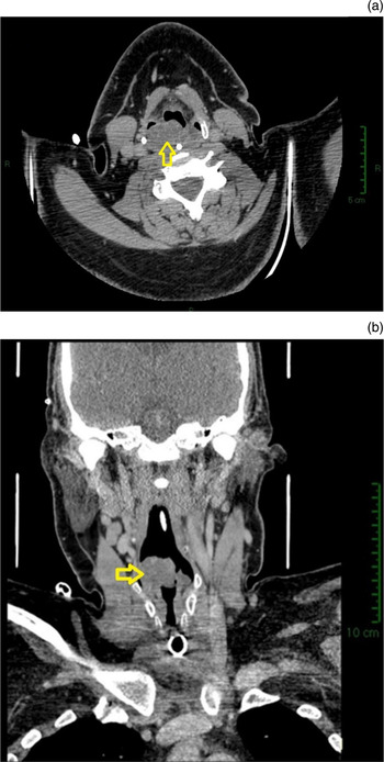
Computed tomography of the larynx revealed a 3.7 cm enhancing mass originating from the right arytenoid with extension into both piriform fossae (Figure 1). Histological analysis of tissues obtained intra-operatively was inconclusive; however, there was a strong suspicion of haemangioma given the appearance of the lesion on direct laryngoscopy.
Computed tomography images of the neck, with contrast. The 3.7 cm enhancing mass (arrows) is seen on both axial (a) and coronal (b) views.

Radiological embolisation was attempted, but no definitive feeding vessel could be visualised. As a result, the decision was made to attempt surgical resection using a laparoscopic bipolar tissue-sealing device (Enseal Tissue Sealer; Ethicon, Wokingham, UK).
Operative technique
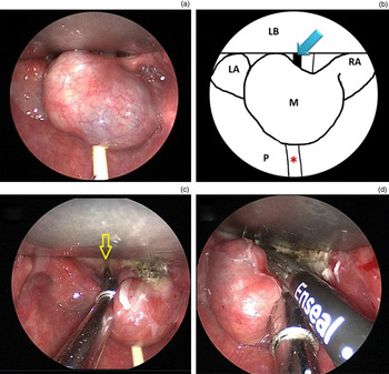
General anaesthesia was achieved with ventilation via the previous tracheostomy. Direct laryngoscopy was performed using a Dedo laryngoscope with the patient in the supine position. Lateral retraction revealed the mass to be pedunculated, with its base attached to the arytenoid, aryepiglottic fold and lateral pharyngeal wall as a linear pedicle (Figure 2).
Intra-operative images. (a) Exophytic pedunculated mass arising from the right arytenoid. (b) Diagrammatic representation of (a). (c) The mass is retracted laterally, revealing the rima glottidis (arrow). (d) Excision of haemangioma using the laparoscopic bipolar tissue-sealing device (Enseal Tissue Sealer). LB = laryngoscope blade; LA = left arytenoid; RA = right arytenoid; M = mass; P = posterior pharyngeal wall; arrows = rima glottidis; asterisk = nasogastric tube

The laparoscopic bipolar tissue-sealing device was introduced through the laryngoscope, with satisfactory excision of the lesion at its pedicle. The lesion was successfully removed with minimal blood loss.
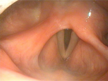
The patient subsequently returned to the intensive treatment unit for further monitoring. The patient was discharged following a step down to ward-based care from the intensive treatment unit. Four months following the procedure, he underwent decannulation of the tracheostomy tube. Flexible nasendoscopy was performed during the clinic review one year after the resection; no evidence of recurrence was seen (Figure 3).
No recurrence of the lesion was visible on flexible nasendoscopy at one-year follow up.

Discussion
Adult laryngeal haemangiomas are typically slow-growing, benign, vascular tumours that can be diagnosed on visualisation via nasendoscopy or direct laryngoscopy.Reference Wang and Tsai 5 Hence, a conservative strategy can be adopted for small lesions that are asymptomatic, with periodic clinic review including nasendoscopy to monitor progression. Symptomatic lesions require intervention, the choice of which is dependent upon the size and extent of the lesion.
Given the rarity of the condition, there are no randomised studies comparing the efficacy of the available options for intervention. However, successful CO2 laser excision has been documented in 2 case series, with low recurrence rates: of 10 patients with either a glottic or supraglottic haemangioma, only 1 developed partial recurrence.Reference Lucioni, Marioni, Libera and Rizzotto 6 , Reference Lomeo, McDonald and Finneman 9 Laser excision is unsuitable for larger lesions because of the risk of uncontrollable haemorrhaging and extensive scarring.Reference Wang and Tsai 5 , Reference Kawakami, Hayashi, Yoshimura, Ichihara, Nishikawa and Ichihara 10 Transoral robotic resection has been recommended by Wang and Tsai for small to medium haemangiomas of the larynx, as the device is more precise and thus superior in achieving normal tissue preservation. Additionally, the procedure is minimally invasive and the bleeding risk is lower.Reference Wang and Tsai 5 At present, transoral robotic surgery is expensive, available in few centres and not recommended for large lesions.
Interventions for large lesions include embolisation,Reference Kawakami, Hayashi, Yoshimura, Ichihara, Nishikawa and Ichihara 10 radiotherapy or open surgery (requiring a tracheostomy).Reference Wang and Tsai 5 In the case of our patient, no feeding vessel from the carotid or vertebral arterial systems could be identified on angiography, precluding embolisation. Radiotherapy led to the partial regression of a laryngeal haemangioma, as described by Huang et al.;Reference Huang, Lee and Huang 11 however, the risk of secondary malignancy and other radiation-induced side effects must be taken into account.
Laparoscopic bipolar tissue-sealing devices (such as Enseal) are routinely used in laparoscopic abdominal surgery to coagulate and divide tissues containing vessels or lymphatics. They utilise pulsed radiofrequency current applied to tissue held between ratcheted bipolar scissors. A microprocessor tied to a voltage sensor determines the initial power based on tissue impedance, with power adjusted throughout the coagulating process via a feedback circuit. By this mechanism, tissues are sealed within 1–6 seconds and the maximum temperature does not exceed 100 °C at the active surface, limiting damage to surrounding tissues. An integrated cutting blade incises the coagulated tissue on releasing the bipolar scissors.Reference Campbell, Cresswell, Frank and Cuschieri 12 , Reference Seehofer, Mogl, Boas-Knoop, Unger, Schirmeier and Chopra 13 The instrument design allows a large piece of tissue to be grasped, and the combined coagulating and cutting mechanism prevents excessive bleeding. These properties proved particularly useful when resecting this large pedunculated vascular lesion – use of the bipolar scissors allowed us to grasp the stalk at its base, whilst simultaneous coagulation prevented haemorrhage. Our approach therefore enabled successful excision, with no recurrence at one year, after conventional methods had failed.
Conclusion
Laparoscopic bipolar tissue-sealing devices can be used to safely excise moderate to large laryngeal haemangiomas via direct laryngoscopy. This novel technique offers an alternative to laser excision or transoral robotic resection for complex cases.