Introduction
Larval sea urchins have become ‘model organisms’ for conducting developmental and evolutionary research on skeletogenesis (e.g., Hinegardner Reference Hinegardner1969; Mazur & Miller Reference Mazur and Miller1971; Vaїtilingon et al. Reference Vaїtilingon, Morgan, Grosjean, Gosselin and Jangoux2001; George et al. Reference George, Lawrence and Lawrence2004; Sewell et al. Reference Sewell, Cameron and McArdle2004; Smith et al. Reference Smith, Smith, Cameron and Urry2008) and even studying effects imparted by spaceflight conditions on fertilization and embryogenesis (Schatten et al. Reference Schatten, Chakrabarti, Taylor, Sommer, Levine, Anderson, Runco and Kemp1999). Adult sea urchins, however, also constitute systems that are amenable to ontogenetic and phylogenetic studies on skeletons, which may provide data that could be utilized to conduct extraterrestrial biology research, specifically how organisms on Earth would respond to gravitational conditions during space travel or on other planets.
Adult sea urchin skeletons, or tests, are endoskeletons, each situated between an overlying, external epidermis and an underlying, internal coelomic lining (Hyman Reference Hyman1955). Formally, each test may be divided structurally into three parts (from top to bottom in supra-sediment orientation): the apical system, comprising ocular and genital plates (and, in regular echinoids and early irregular echinoids, the periproct; Smith Reference Smith1984); the corona, containing ambulacral and interambulacral plates; and the peristome, containing buccal (mouth) plates and sometimes primordial ambulacral plates (Hyman Reference Hyman1955; Smith Reference Smith1984), which surround the mouth, or Aristotle's lantern.
The plates constituting adult sea urchin tests comprise magnesium calcite associated with protein and are interlocked and sutured together by collagen fibres (Telford Reference Telford1985; Johnson et al. Reference Johnson, Ellers, Limire, Minor and Leddy2002). Skeleton growth involves plate addition at the apical system; plate gapping as collagen fibres loosen; plate accretion at peripheries; plate interactions at boundaries; and integrated effects imparted by somatic growth onto the body wall (e.g., visceral growth; Abou Chakra & Stone Reference Abou Chakra and Stone2011). These processes occur in sections called ‘growth zones,’ biserial ambulacral columns flanked by two interambulacral columns, one on either side (Jackson Reference Jackson1912; Mooi et al. Reference Mooi, David, Marchant, David, Guille, Feral and Roux1994; David & Mooi Reference David and Mooi1996, Reference David, Mooi, Mooi and Telford1998; Mooi & David Reference Mooi and David1997).
New plate addition adjacent to ocular plates results in older plates within a growth zone becoming situated closer to the peristome (Ellers Reference Ellers1993). This repositioning is made possible by lengthening collagen fibres, which enable plate gapping and, consequently, plate accretion along peripheries (Telford Reference Telford1985; Johnson et al. Reference Johnson, Ellers, Limire, Minor and Leddy2002). Test growth initiates during active feeding; test diameters increase when adult sea urchins are fed (McCarron et al. Reference McCarron, Burnell and Mouzakitis2009). Unfed adult sea urchins maintain rigid tests; tests may shrink due to contracting collagen fibres, which narrows plate gaps (Ellers et al. Reference Ellers, Johnson and Moberg1998; Johnson et al. Reference Johnson, Ellers, Limire, Minor and Leddy2002). Adult sea urchin ontogeny, therefore, is effected and affected by a variety of factors relating to food availability (Russell Reference Russell1998), including environmental conditions (Fernandez & Boudouresque Reference Fernandez and Boudouresque1997).
The relatively minimal musculature contained within adult sea urchin bodies entails low ‘maintenance costs,’ resulting in resilient organisms with high tolerance for starvation (Lau et al. Reference Lau, Lau, Quian and Qiu2009). The high tolerance for starvation combined with the immediate and episodic dependence on feeding for growth renders adult sea urchins as excellent candidates for studying skeletogenesis and its malleability under controlled experimental conditions. We utilized this combination to conduct the extraterrestrial simulation, altered-gravity research described herein.
-
1. Biomechanical growth model: Among the models that have been proposed to explain adult sea urchin test shapes (reviewed in Abou Chakra & Stone Reference Abou Chakra and Stone2011), the idea that sea urchin tests may be analogized with liquid drops particularly is elegant. The analogy was published originally by Thompson (Reference Thompson1917) and since has been applied by researchers for a variety of purposes, practically for instance, in engineering underwater storage vessels that minimize buckling stresses and resemble tests in echinoids (e.g., sea urchins), the so-called ‘echinodomes’ (Royles et al. Reference Royles, Sofoluwe, Baig and Currie1980).
One theoretical application has become known as the ‘pneu model’ for test growth (Moss & Meehan Reference Moss and Meehan1968; Seilacher Reference Seilacher1979; Dafni Reference Dafni1983, Reference Dafni, Keegan and O'Connor1985, Reference Dafni1986, Reference Dafni2011; Dafni & Erez Reference Dafni and Erez1987; Baron Reference Baron and Burke1988). According to the pneu model, adult sea urchin tests behave like water-filled balloons resting on surfaces; the resulting dome shapes are determined primarily by internal pressures, and tests grow when internal pressures exceed external surface tensions (Dafni Reference Dafni1986). Consequently, expansive forces within sea urchins, such as visceral growth (e.g., gut and gonad development), induce test expansion, which may be relieved or balanced by tensional forces (e.g., along collagen sutures or in the peristomial membrane surrounding the mouth; Russell Reference Russell1998). Positive pressures predicted on the basis of the pneu model failed to be measured or observed in empirical investigations and experimental tests; instead, internal pressures frequently were negative (70% the time, with mean value −8 Pa), suggesting that the pneu model in its original form required modification at least (Ellers & Telford Reference Ellers and Telford1992).
Indeed, other pressure-based models have been published. Baron (Reference Baron and Dudley1990, Reference Baron1991) developed a ‘tensile stress model,’ involving finite-element analysis to integrate self-weight, Aristotle's lantern forces, fluctuating coelomic pressures and tube feet forces to describe test shapes (tube feet are small projections connected to the hydraulic, water-vascular system and function in respiration, ingestion and locomotion). Ellers (Reference Ellers1993) developed a ‘membrane model,’ involving thin-shell theory to integrate apical curvature and vertical pressure gradients (that could be generated by forces caused by activities such as tube feet movements) to describe curvatures exhibited by regular echinoid tests. One prediction common to and derivable from the pneu model and other pressure-based models is that sea urchins grown under zero-gravity conditions would develop spherical tests (Dafni & Erez Reference Dafni and Erez1987; Dafni Reference Dafni2011).
-
2. Height/diameter (h/d) for sea urchin tests: The dome shapes for sea urchin tests are produced partially from reactive forces resulting when individuals adhere to surfaces with tube feet and the aforementioned flexible, collagen-sutured tests. Sea urchin species are constrained to characteristic h/d ratios; low-domed juvenile sea urchins tend to increase h/d during ontogeny, whereas high-domed juvenile sea urchins tend to decrease h/d; once a species-specific constant is achieved, subsequent growth tends to be isometric (Ebert Reference Ebert1988; Ellers Reference Ellers1993). Environmental factors can influence h/d. For instance, Dafni (Reference Dafni1980, Reference Dafni1983) examined adult sea urchins in populations that were exposed to thermohaline and heavy metal ion and laundry wastewater pollutants; tests were deformed and flattened from pollution-induced calcification inhibition. Dafni (Reference Dafni1986) and Dafni & Erez (Reference Dafni and Erez1987) also studied experimentally effects resulting from different substrates: adult sea urchins that were maintained on soft-bottom substrates, such as sand, achieved greater h/d relative to sea urchins that were maintained on hard-bottom substrates, such as glass. In natural, soft-substrate habitats, such as seaweed beds, tube feet adhere relatively ineffectively and, thus, impart smaller forces on tests (i.e., towards the substrate surface).
In the experiment described subsequently herein, effects on test shape from tube feet were diminished, effectively ‘cancelled-out’ by balancing ‘reactionary gravitational forces’ to produce approximately equal tensile forces in collagen fibres over test surfaces. This resulted in tests experiencing forces acting approximately equally in all directions, including the apical system and peristome. The high tolerance for starvation combined with the immediate and episodic dependence on feeding for growth allowed changes to h/d to accumulate rapidly when food became available and feeding commenced after a brief starvation period. The malleability afforded by the collagen fibres deconstrained tests from their characteristic, species-specific h/d, producing more-spherical shapes. By developing an experimental design that prevented sea urchins from attaching to substrates and generating unbalanced reactionary gravitational forces, we observed that morphological changes to endoskeletons, manifested as increases in h/d, can be effected during growth under uniformly distributed force conditions.
Materials and methods
We chose the species Arbacia punctulata for its availability and being reproductively inactive at the time that the experiment was performed ( i.e., so that all energy input through feeding would be allotted to somatic growth). We acquired adult specimens from Gulf Specimen Marine Laboratory (Panacea, FL, USA) and, after a brief acclimatization period in which no food was provided ( i.e., the aforementioned starvation period), measured with Somona digital callipers (Ottawa, ON, Canada) on each specimen the variables height (h) and diameter (d). Due to the extensive logistics involved with the growth protocol (described subsequently), sample sizes for the control and treatment groups were limited to n = 3 specimens.
We maintained specimens in filtered, artificial sea water (Oceanic Natural Sea Salt Mix, Central Garden & Pet Company, Walnut Creek, CA, USA) in glass aquaria (TopFin, PetSmart, Pheonix, AZ, USA; 6 litres maximum capacity, filled to 5 litres) at room temperature ( i.e., T = 297.15 K). We provided ad libitum to control and treatment groups carrots sliced transversely into discs and replenished the food supply whenever it became depleted. We cleaned aquaria and replenished water whenever necessary (approximately once per week).
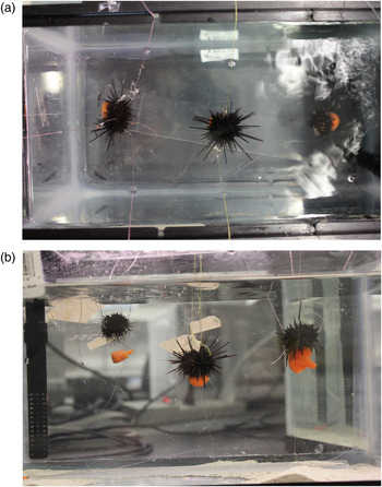
We suspended specimens in the treatment group aquarium by adhering threads to spines with glue (Krazy Glue, Columbus, OH, USA) and affixing the threads to the aquarium outer walls with tape (3 M Scotch Adhesive Tape, London, ON, Canada) (Fig. 1). To assist suspended specimens in obtaining food, we designed a feeding apparatus. We strung fishing line through centres in carrot discs and tied the fishing line to Plexiglas strips, which served as platforms and allowed specimens to access the carrot discs. We fortuitously observed that specimens in the treatment group were able to hold the carrot discs next to their mouths indefinitely and, so, were able to feed continuously throughout the entire experimental time period (including daily observations and occasional maintenance, Fig. 1(b)), as were their counterparts in the control aquarium (who could access carrot discs on the aquarium floor). We maintained specimens in the control group aquarium under identical conditions (i.e., filtered artificial seawater ρspecific = 1.020–1.023, T = 297.15 K) but for the immobilization and feeding apparatus.
Suspended individuals in the treatment group: (a) top view and (b) side view (the individual at left temporarily and atypically has released its food; such situations were corrected by individuals, themselves, or through daily maintenance). Threads were glued to spines and affixed to aquarium outer walls. Fishing line threaded through centres in carrot discs and tied to Plexiglas strips, which served as platforms on aquarium floors, allowed individuals to access food (carrots).

The elaborate design in the treatment group aquarium eliminated reactionary gravitational forces in the sea urchins that were contained therein. These reactionary gravitational forces were redistributed in the threads that were used to immobilize specimens and maintain them in suspension.
We measured and recorded h and d for the control and treatment group specimens, once before any feeding had started (i.e., immediately following the starvation period) and again after feeding had finished (i.e., after a 9-week feeding period) and compared groups by calculating effect sizes (as the raw difference between means and using Cohen's d (Cohen Reference Cohen1992); we also analysed the data using conventional quantitative protocols: as we expected specimens in both groups generally to grow and in the treatment group specifically to grow tests that were more-spherical and, so, characterized by greater mean h/d than the treatment group, we established the null hypothesis ‘no difference between groups’ and tested the related predictions mean (h/d)after = mean (h/d)before, mean d after = mean d before, and mean (h/d)control = mean (h/d)treatment by applying paired, one-tailed t-tests – insufficient degrees of freedom were available to conduct the nonparametric paired, one-tailed Wilcoxon signed-rank test; Sokal & Rohlf Reference Sokal and Rohlf1995).
Results
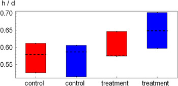
Mean h/d was greater after the experiment than before for only the treatment group (Fig. 2, effect sizes, determined as mean changes in h/d, were −0.003 and 0.051 for the control and treatment group, respectively; Table 1); mean d was greater after than before the experiment for both groups (Table 1); h/d values between control and treatment groups differed insignificantly before but significantly after the experiment.
Values for test h/d ratio in control and treatment groups for Arbacia punctulata specimens at 0 and 9 weeks (i.e., before and after feeding, red and blue, respectively); each rectangular box contains a median value (dashed line) with a top and bottom (i.e., ‘whiskers’) that span the range observed.

Values for test diameter (d) and height-to-diameter ratio (h/d) (mean ± SD) in control and treatment groups for Arbacia punctulata specimens at 0 and 9 weeks

Differences in means after and before the experiment were tested with paired, one-tail t-tests: d for control group: t = 3.73, P = 0.0325 (power, 1−β estimated as 0.111; Cohen's d = −0.246); d for treatment group: t = 0.173, P = 0.440 (power, 1−β estimated as 0.0550; Cohen's d = 0.0252); h/d for control group: t = 0.210, P = 0.426 (power, 1−β estimated as 0.0660; Cohen's d = 0.0797); h/d for treatment group: t = 3.08, P = 0.0373 (power, 1−β estimated as 0.738; Cohen's d = −1.36). Power was estimated (post hoc) with the Researcher's Toolkit provided by DSS Research (2012), using initial values for the sample average and final values for the test average; pooled standard deviations (Tabachnick and Fidell Reference Tabachnick and Fiddel2007, 55); and α = 0.05. Cohen's (Reference Cohen1992) d was calculated as the difference between means divided by one standard deviation.
Discussion
The h/d characterizing tests for sea urchins that were grown without reactionary gravitational forces increased relative to the h/d characterizing tests for sea urchins that were grown in a glass aquarium that allowed strong adherence to surfaces and, so, with reactionary gravitational forces (even according to conventional quantitative protocols and with the small sample sizes that were necessitated by the labour-intensive logistics involved with the experimental design). The h/d for all individuals in the treatment group changed positively over the 9-week period, whereas the h/d for all individuals in the control group changed non-uniformly, one positively and two negatively.
That d for individuals in the control group increased, but their collective h/d remained unchanged (Table 1), which indicates that they grew in an isometric manner (i.e., they increased in size but maintained constant proportions, as expected in echinoids; Ebert Reference Ebert1988; Ellers Reference Ellers1993) because they were subjected to reactionary gravitational forces akin to those that are experienced by individuals in natural populations; individuals in the treatment group grew in an allometric manner (i.e., they increased in size and changed proportions) because they were grown in altered-gravity conditions (Table 1).
In biomechanical models for growth, adult sea urchin test shapes are governed by a variety of forces, including self-weight, which generates compressive forces; Aristotle's lantern movements caused by muscular contraction during feeding, which generate forces that pull together plates; expanding, internal, visceral tissues such as guts and gonads, which generate tensional forces against test plates; and tube feet retracting and adhering to substrates, which generate tensional forces approximately normal to plate surfaces (Dafni Reference Dafni1986). Effectively eliminating the effects that are imparted by tube feet, by suspending adult sea urchins, resulted in tests deforming from their characteristics and species-specific h/d ratio during growth. Redistributing reactionary gravitational forces in the tethering threads enabled plates to accrete and interact uniformly over entire test curvatures, unconstrained by unbalanced reactionary forces or the previously generated dome shapes such forces caused, which otherwise would have constrained tests to remain more flattened.
Sea urchins alter their test morphologies when grown under balanced reactionary gravitational forces. This finding indicates that physical factors such as substrates may have played a substantial role in sea urchin phenotypic evolution (Dafni Reference Dafni1986) and effected the disparate test morphologies that are observed among echinoids (i.e., sea urchins, sand dollars, sea biscuits, heart urchins and their relatives, which are related evolutionarily).
Implications
In addition to providing a mechanistic factor that may be integrated into the evolutionary framework for explaining echinoid morphological evolution, the findings have application in at least two other, practical areas: sea urchin aquaculture (discussed only briefly in the next paragraph, as this topic is peripheral to the experiment described herein) and studies on the effects imparted to skeletons during space travel or on other planets.
Sea urchin fishing outside Japan has gained popularity and become a lucrative industry since Japanese stocks have declined and availability within Japan has become reliant on foreign supplies (Russell Reference Russell1998), especially for gonads, which constitute the delicacy ‘uni’ (Pfister & Bradbury Reference Pfister and Bradbury1996). A paramount objective is to determine feeding regimes and environmental conditions to optimize growth. Growth could be optimized economically by growing more-spherical sea urchins, maximizing gonad size and yield for a given test radius (i.e., size).
The observations that are documented herein support the hypothesis that sea urchins that were grown completely in a zero-gravity environment would become spherical (Dafni & Erez Reference Dafni and Erez1987; Dafni Reference Dafni2011). An experiment to test this hypothesis could be setup for performance during a lengthy voyage, as would be expected for a vessel journeying to Mars. Results obtained in such an experiment could be extrapolated to provide insight into how space travel or living on other planets over extended time periods might affect skeletons in other animals, such as vertebrates (albeit with caution, as echinoid test comprise calcium carbonate and vertebrate skeletons comprise hydroxyapatite, i.e. calcium phosphate).
The treatment that we imposed on growing sea urchins differs from microgravity, defined formally as reduced acceleration due to gravity or g-force, and described informally as ‘effective weightlessness’. Bona fide microgravity conditions could be achieved in deep space (i.e., in locations remote from any large bodies), requiring transport by spacecraft (as described in the preceding paragraph). Generating free-fall-like conditions approximating microgravity can be achieved by flying reduced gravity aircraft in approximately parabolic arcs (e.g., C131 Samaritan – affectionately dubbed the ‘vomet comet,’ KC135 Stratotankers, McDonnell Douglas C-9B Skytrain II and the Zero Gravity Corporation G-FORCE ONE modified Boeing 727–200) or using ‘drop tubes’ or ‘drop towers’ (e.g., NASA Glenn Zero Gravity Research Facility 155 m subterranean shaft). Both methods provide microgravity conditions for very short time periods and would be impractical for studying skeletogenesis.
In addition to the aforementioned practical applications, future directions for related research include repeating the experiment with larger sample sizes and additional species to explore other modes for expressing phenotypic plasticity or accommodation (West-Eberhard Reference West-Eberhard2003, Reference West-Eberhard2005) like modified skeletons. An interesting instance for this would be to repeat the experiment with sand dollars, sea biscuits and heart urchins to examine whether altered reactionary gravitational forces impart effects on their disc-, ellipsoid- and cardioid-shaped tests similar to those described herein for dome-shaped sea urchin tests.
Acknowledgements
Resources to develop ideas in an unconstrained manner were provided by the Shared Hierarchical Academic Research Computing Network, Department of Biology and Origins Institute at McMaster University; and funds to produce results were provided by the Natural Sciences and Engineering Research Council of Canada (Discovery Grant 261590).