Introduction
Many studies have reported how normal aging influences chromatic and luminance contrast sensitivity (Fiorentini et al., Reference Fiorentini, Porciatti, Morrone and Burr1996; Page & Crognale, Reference Page and Crognale2005; Mateus et al., Reference Mateus, Lemos, Silva, Reis, Fonseca, Oliveiros and Castelo-Branco2013; Tamura & Sato, Reference Tamura and Sato2020). Both optical and neural factors affect luminance and color processing. However, blue–yellow color discrimination seems to be more affected by senescence than red–green color and luminance discrimination (Werner & Steele, Reference Werner and Steele1988; Knoblauch et al., Reference Knoblauch, Vital-Durand and Barbur2001; Mateus et al., Reference Mateus, Lemos, Silva, Reis, Fonseca, Oliveiros and Castelo-Branco2013; Barbur & Rodriguez-Carmona, Reference Barbur, Rodriguez-Carmona, Elliott, Fairchild and Franklin2016).
Luminance and color information are processed relatively independently in the early stages of the visual system but interact in later stages of the visual system (Kaplan & Shapley, Reference Kaplan and Shapley1986; Lee et al., Reference Lee, Martin and Valberg1989; Dacey & Lee, Reference Dacey and Lee1994; Johnson et al., Reference Johnson, Hawken and Shapley2001; Shapley & Hawken, Reference Shapley and Hawken2002; Ventura et al., Reference Ventura, Silveira, Rodrigues, de Souza, Gualtieri, Bonci, Costa, Mollon, Pokorny and Knoblauch2003; Lee et al., Reference Lee, Sun and Valberg2011; Li et al., Reference Li, Chen, Lashgari, Bereshpolova, Swadlow, Lee and Alonso2015; Xing et al., Reference Xing, Ouni, Chen, Sahmoud, Gordon and Shapley2015).
Recently, we introduced a novel paradigm using a mosaic with chromatic noise to mask luminance contrast in order to quantify the interaction between color and luminance discrimination (Miquilini et al., Reference Miquilini, Walker, Odigie, Guimarães, Salomão, Lacerda, Cortes, de Lima Silveira, Fitzgerald, Ventura and Souza2017; Sousa et al., Reference Sousa, Loureiro, Goulart, Cortes, Costa, Bonci, Baran, Hauzman, Ventura, Miquilini and Souza2020). Miquilini et al. (Reference Miquilini, Walker, Odigie, Guimarães, Salomão, Lacerda, Cortes, de Lima Silveira, Fitzgerald, Ventura and Souza2017) introduced a method that measures changes in luminance contrast threshold as a function of hue saturation of chromatic noise. We also showed that luminance thresholds increased linearly with chromatic noise saturation over the range between low-saturated chromatic noise and medium-to-high saturated chromatic noise. The magnitude of chromatic noise suppression of luminance contrast thresholds can be estimated from the difference between the thresholds measured without chromatic noise and those measured in the presence of medium-to-high saturated chromatic noise (Sousa et al., Reference Sousa, Loureiro, Goulart, Cortes, Costa, Bonci, Baran, Hauzman, Ventura, Miquilini and Souza2020).
We hypothesized that interactions between luminance and color processing would be modified in elderly subjects compared to young people, especially under stimulus conditions that used chromaticities targeting the blue–yellow channel. Therefore, the present study evaluates luminance threshold discrimination masked by chromatic noise in elderly and young people.
Materials and methods
Ethical considerations
The Ethical Committee of the Federal University of Amapá approved the present investigation (report #4557848) which adhered to the Declaration of Helsinki of 1964 and all its amendments. All participants were informed about the purpose and methods used in the purpose of the experiments and gave written consent for their participation.
Participants
Forty subjects participated in the experiments: 20 participants in the Young Group (29.6 ± 6.3-year-old, 10 females and 10 males) and 20 in the Elderly Group (57.8 ± 6.6-year-old, 10 females and 10 males). All participants had best-corrected visual acuity of 20/20 or better determined with the ETDRS acuity chart, and normal color vision based on the Ishihara test (Ishihara, Reference Ishihara and Ishihara1997). In addition, no participant had a history of ocular or neurological diseases, degenerative diseases, or color vision deficits in the family.
Luminance contrast discrimination
The experimental set-up has been described previously (Miquilini et al., Reference Miquilini, Walker, Odigie, Guimarães, Salomão, Lacerda, Cortes, de Lima Silveira, Fitzgerald, Ventura and Souza2017; Sousa et al., Reference Sousa, Loureiro, Goulart, Cortes, Costa, Bonci, Baran, Hauzman, Ventura, Miquilini and Souza2020). Briefly, the experiment was carried out using MATLAB software (MATLAB R2017b, Mathworks, Natick, MA, USA).
The stimulus was displayed on a 17″ LCD MacBook Pro monitor (1690 × 1050 pixels, 75 Hz frame rate, 10 bits color resolution). A CS-100A chromameter (Konica Minolta, Osaka, Japan) was used to calibrate the color and luminance of the display.
The stimulus was a mosaic comprised of 428 circles with diameters varying between 0.12° and 0.37° viewed 234 cm from the monitor. A subset of the circles, each with a starting luminance of 4 cd/m2, were perceptually integrated to form a Landolt-C shaped target (outer diameter: 4.4°, inner diameter: 2.2°, gap: 1°) within a background of 40 cd/m2 circles. The C-gap was defined by the luminance contrast between the target circles and background circles. The observer’s task on each forced-choice trial was to identify the location of the gap in the Landolt-C (up, down, left, or right). Each stimulus was presented for 3 s. Thus, stimuli were randomly selected from 16 possible Landolt-C mosaic arrangements: four possible locations for each of four test protocols described below.
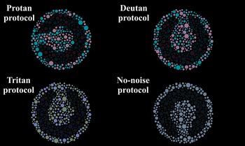
The participants performed four test protocols, in which a static chromatic noise was applied to the entire mosaic. The noise was comprised of 10 chromaticities with a vector size of 0.04 u′v′ units projected radially from a reference color (CIE 1976: u′ = 0.1947; v′ = 0.4639) close to the protan confusion line (protan protocol), deutan confusion line (deutan protocol), tritan confusion line (tritan protocol), and a condition without chromatic noise (no-noise protocol) with only the reference chromaticity across the mosaic.
Fig. 1 shows an example of the stimuli used in the protan, deutan, tritan, and no-noise protocols. The sequence of protocols tested was randomly chosen for each participant. The participant’s task was to indicate the orientation of the C-gap of the target (up, bottom, left, or right). The luminance of the target circles changed by a two down/one up adaptive staircase procedure in each presentation. Hits increased the luminance of the target circles and errors decreased the luminance of the target circles. The luminance of the background circles was kept at 40 cd/m2 during the experiments. The background circles remained fixed at 40 cd/m2. The luminance contrast thresholds thus obtained corresponded to 70.7% correct responses (Levitt, Reference Levitt1971). The threshold was taken as the mean of the contrasts in the final six reversals. We used the Weber contrast to quantify the luminance contrast between the target circles and background circles (1)).
 $$ \mathrm{Contrast}\hskip0.35em =\hskip0.35em \left({L}_t-{L}_b\right)/{L}_b, $$
where
 $ {L}_t $
is the luminance of the circles of the stimulus target, and
 $ {L}_b $
is the luminance of the circles in the stimulus background.
Example of stimulus from each of the four test protocols: protan, deutan, tritan, and no chromatic noise. Observers’ contrast thresholds were measured using a 4AFC staircase (choose the direction of the Landolt-C gap on each trial).

Data analysis
For each age group, we conducted a one-way analysis of variance to compare the luminance contrast thresholds estimated from each stimulus protocol followed by Tukey’s test for multiple comparisons. For each protocol, an independent-samples t-test with Welch’s correction was performed comparing the mean contrast thresholds estimated from the Young and Elderly groups. We chose a confidence level of 5% for all statistical tests.
Results
Comparison of contrast thresholds across age groups
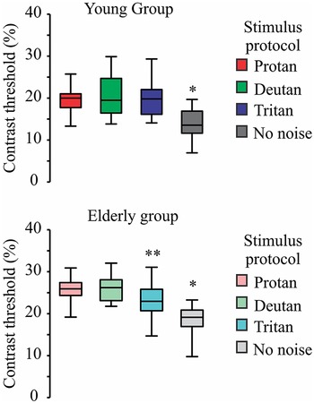
To evaluate the effect of each of the four chromatic noise protocols on luminance contrast thresholds in the Young age group, we conducted a one-way ANOVA with repeated measures. A significant effect was observed (F[2.562, 41] = 39.67, P < 0.0001; η 2 = 0.31). Analyses comparing across protocols showed that the luminance contrast thresholds in the no-noise protocol were significantly lower than those in the other three protocols (P < 0.001). No differences in luminance contrast thresholds were found between the three chromatic noise protocols (P > 0.05).
For the Elderly Group, the one-way ANOVA with repeated measures showed similar results compared with those observed in the Young Group. There was a significant effect (F[1.855, 29.69] = 8.039, P < 0.002; η 2 = 0.47) of the stimulus protocols on the luminance thresholds in the Elderly Group: contrast thresholds estimated in the no-noise protocol also were lower than any of the thresholds measured with the chromatic noise protocols (P < 0.001). In addition, thresholds from tritan protocol were lower than those measured in the protan and deutan protocols. (P < 0.05). Thresholds from the protan and deutan protocols were not significantly different (P > 0.05). Fig. 2 shows the contrast thresholds from the four test protocols for the Young and Elderly Groups.
Comparison of the luminance contrast thresholds in each age group. Box plots are represented by first and third quartiles (lower and upper horizontal lines of the box), median (horizontal line in the box), and minimum and maximum values (lower and upper bars). *Luminance contrast thresholds are lower than thresholds estimated from all three protocols with chromatic noise (P < 0.05). **Luminance contrast thresholds in the tritan protocol were lower than those measured in the protan and deutan protocols (P < 0.05).

Comparing contrast thresholds between young and elderly participants
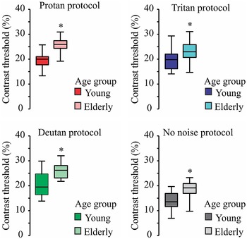
A t-test with Welch’s correction was used to compare the luminance contrast thresholds estimated from young and elderly people for each test protocol. For all test protocols, the elderly participants had higher contrast thresholds than obtained from the young participants: protan protocol (t[32.95] = 5.494, P < 0.0001), deutan protocol (t[29.67] = 4.365, P = 0.0001), tritan protocol (t[33] = 2.414, P < 0.0001), no-noise protocol (t[33] = 3.713, P = 0.0008). Fig. 3 shows the intergroup comparisons of the luminance contrast thresholds for each test protocol.
Intergroup comparison of the luminance contrast threshold obtained from each protocol. Box plot coding is the same as in Fig. 2. *Luminance contrast thresholds from older participants were higher than the thresholds from younger participants (P < 0.001).

Discussion
Using the same paradigm adopted in the present investigation, Miquilini et al. (Reference Miquilini, Walker, Odigie, Guimarães, Salomão, Lacerda, Cortes, de Lima Silveira, Fitzgerald, Ventura and Souza2017) and Sousa et al. (Reference Sousa, Loureiro, Goulart, Cortes, Costa, Bonci, Baran, Hauzman, Ventura, Miquilini and Souza2020) showed that the suppression even occurs in the presence of low-saturated chromatic noise and increases, reaching a maximum in the presence of medium-to-high saturated chromatic noise. In the present study, we expanded the chromatic noise range by including a condition without any chromatic noise (no-noise protocol, which optimized luminance contrast sensitivity), and extending the chromatic noise to include highly saturated colors to induce maximum suppression of the luminance discrimination. Some prior investigations found evidence for the suppression of luminance discrimination under chromatic masking (Switkes et al., Reference Switkes, Bradley and De Valois1988; Gegenfurtner & Kiper, Reference Gegenfurtner and Kiper1992; Chen et al., Reference Chen, Foley and Brainard2000; Kingdom et al., Reference Kingdom, Bell, Gheorghiu and Malkoc2010; Miquilini et al., Reference Miquilini, Walker, Odigie, Guimarães, Salomão, Lacerda, Cortes, de Lima Silveira, Fitzgerald, Ventura and Souza2017; Sousa et al., Reference Sousa, Loureiro, Goulart, Cortes, Costa, Bonci, Baran, Hauzman, Ventura, Miquilini and Souza2020; Negishi & Shinomori, Reference Negishi and Shinomori2021).
Neural substrate for color–luminance interactions
Along the visual pathway, there are cells responsive to luminance and chromatic signals (Johnson et al., Reference Johnson, Hawken and Shapley2001) that could subserve color and luminance interaction. Previous studies supported a hypothesis that cortical mechanisms for color and luminance perception are mutually inhibitory (Gordon & Shapley, Reference Gordon and Shapley2006; Xing et al., Reference Xing, Ouni, Chen, Sahmoud, Gordon and Shapley2015). Xing et al. (Reference Xing, Ouni, Chen, Sahmoud, Gordon and Shapley2015) found that luminance responses in V1 were suppressed by chromatic content of the stimuli. This suppression was maximal at the boundary between target and background and correlated more with local brightness contrast than with the measured luminance differences across the target-background boundary. An fMRI study by Negishi and Shinomori (Reference Negishi and Shinomori2021) also found suppression of luminance contrast sensitivity by chromatic content of stimuli. They suggested that the influence of chromatic information on luminance contrast sensitivity is initiated in the early stages of visual processing, then propagating to higher cortical stages. Attentional mechanisms can also be involved in chromatic suppression of luminance discrimination (Frey et al., Reference Frey, Honey and König2008). For example, dots with similar chromatic content can be perceptually grouped into multiple forms which would act as chromatic distractors to varying degrees, thus impacting luminance contrast discrimination differentially.
Age effects
Our results are consistent with the hypothesis that age influences the interaction between color and luminance, with an asymmetric degree of interaction according to the chromaticity space evaluated. Three findings support our conclusions: (1) elderly participants had higher luminance contrast thresholds than younger participants, (2) suppression of luminance discrimination under masking with protan or deutan chromatic noise was not influenced by age, and (3) suppression of luminance discrimination by tritan chromatic noise was less robust in the elderly group.
The latter finding likely reflects a generalized degradation of blue–yellow discrimination during aging. We know that senescence impacts the visual system due to both optical and neural factors (Weale, Reference Weale1973; Knoblauch et al., Reference Knoblauch, Saunders, Kusuda, Hynes, Podgor, Higgins and de Monasterio1987; Burton et al., Reference Burton, Owsley and Sloane1993; Paramei & Oakley, Reference Paramei and Oakley2014). The lens of eye progressively loses its malleability, and tends to scatter light more as we age (Rozema et al., Reference Rozema, Van den Berg and Tassignon2010). Moreover, at least two other nonneural changes associated with aging can impact contrast discrimination: (1) an increase in the optical density of the macular pigment (Werner et al., Reference Werner, Donnelly and Kliegl1987) and (2) the progressive yellowing of the crystalline lens (Pokorny et al., Reference Pokorny, Smith and Lutze1987). The latter (optical) factor is a candidate for explaining why tritan noise would be less effective in masking luminance discrimination in the older group.
Neural factors can also contribute to age-related changes in chromatic masking. For example, there is evidence that processing in the S-cone pathway progressively slows with age but not loss of the number of neurons (Shinomori & Werner, Reference Shinomori and Werner2012; Weinrich et al., Reference Weinrich, Powner, Lynch, Jonnal, Werner and Jeffery2017).
Preciado et al. (Reference Preciado, Martin, Manzano, Smet and Hanselaer2021) found that age affects brightness perception of blue and red stimuli: brightness perception for blue stimuli was decreased in older people compared with younger people. The effect of this would be to attenuate the impact of chromatic blue–yellow noise on luminance discrimination in older people. This may explain why the measured suppression in the tritan protocol was significantly less than in the protan and deutan protocols in older adults.
Limitations of the present study
The presence of different colors with same luminance in the target or in the background could elicit perceptual brightness differences simply due to the Helmholtz–Kohlrausch effect (Donofrio, Reference Donofrio2011) whereby an increase in saturation per se is perceived as a brightness increase. This effect could potentially counteract the masking effect of chromatic noise. This factor merits future investigation.
Other important issue to point out as limitation is that the assignment to a “young” or “old” age group is arbitrary. In the present study, we used the biological age to conclude about luminance contrast discrimination, but the individuals get older differently. Future studies could investigate a continuum of ages across the life spans. Finally, while no participant reported a history of eye disease, color vision deficits in the family, or visual complaints, conditions like glaucoma or cataract can go undetected in their early stages. Participants did not receive full ophthalmological examinations, and so it is possible that some elderly participants had subclinical ophthalmological factors that could affect their data in the current experimental protocols.
Future studies
Electrophysiological studies using chromatic pattern-ERG and multifocal ERG could reveal differences in inner retinal responses that might help explain the reduced effect of tritan chromatic noise on luminance discrimination along the tritan axis. Such studies are relevant to general question of the degree to which early visual processing can account for chromatic-luminance interactions. Future studies ought to explore the effect of aging using a continuum of ages across the lifespan.
Conclusion
Perceptual interaction between color and luminance changes with age. Specifically, in the elderly, the effectiveness of tritan, but not protan or deutan noise, in suppressing luminance contrast discrimination is diminished. The evaluation of color and luminance interactions is a promising candidate as an additional assay of the impact of neural and optical changes in contrast processing during the aging process.
Funding statement
This work was supported by research grants from the Brazilian funding agencies: CNPq Edital Universal (#431748/2016-0). L.M. is a CAPES postdoctoral fellow (CAPES-PROCAD #88887.200446/2018-00). D.F.V., M.F.C., and G.S.S. are CNPq Fellows, Productivity Grants 309409/2015-2, 302552/2017-0, and 310845/2018-1, respectively. The funders had no role in the study design.