INTRODUCTION
The iconic Regent Honeyeater, Anthochaera phrygia (Shaw, 1794) syn. Xanthomyza phrygia (Aves: Passeriformes) is a critically endangered passerine endemic to South-Eastern Australia. Historically, this bird could be seen in flocks of hundreds, ranging from Queensland to South Australia (Driskell and Christidis, Reference Driskell and Christidis2004; Gardner et al. Reference Gardner, Trueman, Ebert, Joseph and Magrath2010; Liu et al. Reference Liu, Gillespie, Atchison and Andrew2014). The current estimated wild population of less than 400 birds is distributed across four key sites in Victoria and New South Wales (Franklin et al. Reference Franklin, Menkhorst and Robinson1989; Thomas, Reference Thomas2009). Extensive loss of box-ironbark eucalyptus forests is attributed as the primary threatening process for this species throughout its range. The Regent Honeyeater is a highly mobile species that covers extensive distances to feed on sporadically available nectar and insects within box-ironbark eucalypt forests. In an effort to prevent extinction, the species has been the subject of an intensive recovery programme for the past 20 years. In 1995, nine male and nine female founders were collected from Chiltern (Victoria), and Capertee Valley (New South Wales) in a captive trial to provide some level of insurance from extinction. A small captive breed-and-release strategy began in 2000 and implemented into the programme in 2008. From the original founders, 312 chicks had been produced by 2013 and of these, 117 have been released (Liu et al. Reference Liu, Gillespie, Atchison and Andrew2014).
Identification and knowledge of the life history of infectious agents in wildlife is imperative for the implementation of satisfactory recovery programs. In a previous parasitological survey of a cohort of the Regent Honeyeaters at Taronga Zoo, Australia, we described a coccidian (Isospora lesouefi) present in essentially all captive birds, displaying diurnal periodicity of oocyst shedding (Morin-Adeline et al. Reference Morin-Adeline, Vogelnest, Dhand, Shiels, Angus and Šlapeta2011). The ecological significance of an ongoing I. lesouefi infection in the captive and the wild birds is unknown, nevertheless, recent observations on released birds showed that they thrive and pair with wild birds (Dean Ingwersen, personal observations). Besides coccidian and other particularly monoxenous intestinal parasitic organisms, birds are host to several groups of dixenous blood parasites including protists such as haemosporidia (Haemospororida) and trypanosomes (Kinetoplastida, Trypanosomatidae) (Baker, Reference Baker, Lumsden and .1976; Valkiūnas, Reference Valkiūnas2005).
In contrast to their mammalian relatives, avian trypanosomes are in most cases harmless to their hosts and remain understudied. Avian trypanosomes rarely cause clinical disease in domestic birds (Baker, Reference Baker, Lumsden and .1976), while the impact of Trypanosoma spp. on wild birds are scarcely documented (Molyneux and Gordon, Reference Molyneux and Gordon1975; Molyneux et al. Reference Molyneux, Cooper and Smith1983). The increasing avian trypanosome diversity, however, along with new species descriptions now attracts more attention on this rather neglected group of avian parasites (Votýpka et al. Reference Votýpka, Oborník, Volf, Svobodová and Lukeš2002; Votýpka and Svobodová, Reference Votýpka and Svobodová2004; Zídková et al. Reference Zídková, Cepicka, Szabová and Svobodová2012). The majority of known trypanosome species infect mammals, and only about one-fifth are described from birds (Podlipaev, Reference Podlipaev1990; Sehgal et al. Reference Sehgal, Valkiūnas, Iezhova and Smith2006). Presumed host specificity has been used as a criterion for species designation, although host–parasite relationships among avian trypanosomes are poorly understood (Zídková et al. Reference Zídková, Cepicka, Szabová and Svobodová2012). One bird species can harbour several trypanosome species (Molyneux and Gordon, Reference Molyneux and Gordon1975; Votýpka and Svobodová, Reference Votýpka and Svobodová2004; Zídková et al. Reference Zídková, Cepicka, Szabová and Svobodová2012). Such findings support the hypothesis of a high trypanosome specificity towards its vector (Apanius, Reference Apanius1991; Votýpka et al. Reference Votýpka, Szabová, Radrová, Zídková and Svobodová2012). Various blood-sucking insect groups such as flat flies (hippoboscids), black flies (simuliids) and mosquitoes are suggested as possible vectors, yet transmission of the majority of avian trypanosomes still remains unclear. Local transmission and distribution is suggested for many avian trypanosome lineages, while on the other hand, some species defined by gene sequence data are probably widespread and distributed intercontinentally (Sehgal et al. Reference Sehgal, Jones and Smith2001; Votýpka and Svobodová, Reference Votýpka and Svobodová2004; Zídková et al. Reference Zídková, Cepicka, Szabová and Svobodová2012).
The aim of this study was to undertake a parasitological survey focusing on the presence of trypanosomes in a cohort of Regent Honeyeaters that were part of a breeding and reintroduction programme at Taronga Zoo, Australia. We describe a novel trypanosome species, Trypanosoma thomasbancrofti sp. n., parasitizing an Australian passerine bird. The SSU rDNA of this new Trypanosoma species from the Regent Honeyeater is almost identical (>99·5%) to trypanosomes of the Common Chiffchaff (Phylloscopus collybita) and the common house mosquito (Culex pipiens complex) from Central Europe. The results suggest an intercontinental distribution of the newly described trypanosome and Culex mosquitoes as a suspected vector. This information is useful in establishing appropriate health screening protocols for the Regent Honeyeater, particularly prior to release into the wild.
MATERIAL AND METHODS
Blood collection, blood smears and cultivation of trypanosomes
Blood samples were collected from Regent Honeyeaters as part of the pre-release health screening protocol at Taronga Zoo, Mosman, Australia in 2013–2015. In June 2015, blood samples were also collected from one wild bird, one 2013 released captive bred bird and from 16 of the 2015 captive bred birds that were released in April 2015 when they were recaptured for refitting of transmitters as part of ongoing post-release monitoring in Chiltern, Victoria, Australia. Blood samples were collected post-mortem from the hearts of wild Noisy Miners (Manorina melanocephala), trapped and euthanased at Taronga Zoo, Mosman, Australia in May 2013. Noisy Miners are considered a pest bird species that lead to disruptive consequences to other birds (MacNally et al. Reference MacNally, Bowen, Howes, McAlpine and Maron2012). Periodic reduction of Noisy Miner numbers is undertaken under license MWL000100542 issued by the NSW Government Office of Environment and Heritage (NSW Parks and Wildlife Service).
Blood (10 µL) was taken directly from the birds and used for standard blood smear, methanol fixed and stained with Diff-Quick stain. All blood smears were inspected (5 min each) by a single observer (PT) using an upright microscope at 40× and 100× objective under immersion oil for the presence of trypanosomes and other blood parasites. Blood (50 µL) was taken directly from the birds and placed immediately into a culture flask. Samples were cultured in vitro in 15 mL glass cultivation flasks containing a biphasic medium (SNB-9) consisting of a sloping sheep blood agar with premixed vitamins (58980C, Sigma, Australia), then overlaid with a liquid phase supplemented with neomycin (80 µg mL−1). Cultivation was performed at 25–30 °C in a horizontal position with 10° slope. The flasks were checked every 2–6 days for the presence of trypanosomes. From positive samples, an aliquot (200 µL) was taken, and then pelleted by centrifuging and stored at −20 °C. Results from three repeated cultures over 3 weeks were compared with a single culture and used for the 2-way contingency table to evaluate a variety of standard statistical measures for diagnostics tests accompanied by 95% confidence interval (95% CI) (Fleiss et al. Reference Fleiss, Levin and Paik2013). Positive samples with trypanosomes were subcultured on a fresh biphasic medium. Non-contaminated isolates were stored in the cryobank of the Parasitology Laboratory, Faculty of Veterinary Science, The University of Sydney, Sydney, Australia.
Transmission electron microscope
Mid-exponential phase parasites were collected from culture and fixed in 2·5% glutaraldehyde (v/v) (PST, Queensland, Australia) in 0·1 m sodium cacodylate buffer (pH = 7·2) at 25 °C for 1 h. Fixed cells were washed in 0·1 m cacodylate buffer (pH = 7·2) and post-fixed in 1% (w/v) OsO4 in 0·1 m sodium cacodylate buffer (pH = 7·2). The cell pellet was further processed through an ethanol dehydration series carried out at 25 °C and samples were infiltrated and embedded in Epon resin (PST, Queensland, Australia). Ultrathins were obtained using a diamond knife and mounted on grids. Subsequently, grids were stained with 2% uranyl acetate followed by 1% lead citrate. The grids were viewed and imaged on a JOEL JEM 1400 TEM, using 120 kV, equipped with an ES500W Erlangshen CCD camera (Gatan, California, USA) and the kinetoplast ultrastructure analysed as described by (Lukeš and Votýpka, Reference Lukeš and Votýpka2000).
Amplification of the small subunit rRNA and glycosomal glyceraldehyde phosphate dehydrogenase genes
Total DNA was extracted from Trypanosoma culture pellets using Isolate II Genomic DNA kit (BioLine, Australia). DNA was eluted into 100 µL of Tris buffer (pH = 8·5) and stored at −20 °C. Small subunit rRNA gene (SSU rDNA, ~2100 bp) was PCR amplified using the Trypanosoma-specific primers: [S0359] S762 (5′-GAC TTT TGC TTC CTC TAW TG-3′)/[S0360] S763 (5′-CAT ATG CTT GTT TCA AGG AC-3′) (Sehgal et al. Reference Sehgal, Jones and Smith2001). Reactions of 30 µl contained MyTaq Red Mix (BioLine, Australia), and approximately 5 ng of genomic DNA template (2 µl). For selected SSU rDNA genotypes we PCR amplified partial glycosomal glyceraldehyde phosphate dehydrogenase (gGAPDH, ~900 bp). The gGAPDH was amplified using seminested PCR with Trypanosoma-specific primers: primary PCR [S0562] GAPDHF (5′-CTY MTC GGN AMK GAG ATY GAY G-3′)/[S0563] GAPDHR (5′-GRT KSG ART ADC CCC ACT CG-3′), followed by a secondary PCR [S0562] GAPDHF/[S0565] G4a (5′-GTT YTG CAG SGT CGC CTT GG-3′) as previously described (Hamilton et al. Reference Hamilton, Stevens, Gaunt, Gidley and Gibson2004; McInnes et al. Reference McInnes, Gillett, Ryan, Austen, Campbell, Hanger and Reid2009). The cycling for SSU rDNA was as follows: denaturing at 95 °C for 3 min followed by 35 cycles of 95 °C for 15 s, 53 °C for 15 s, 72 °C for 60 s, and a final elongation for 5 min at 72 °C. The gGAPDH primary cycling included denaturing at 95 °C for 3 min followed by 35 cycles of 95 °C for 15 s, 50 °C for 15 s, 72 °C for 30 s and a final elongation for 5 min at 72 °C. The gGAPDH secondary cycling included denaturing at 95 °C for 3 min followed by 35 cycles of 95 °C for 15 s, 52 °C for 15 s, 72 °C for 15 s and a final elongation for 5 min at 72 °C; 0·5 µL of the primary reaction was used as template for the secondary PCR. The PCRs were conducted in a PCR cycler Veriti (Life Sciences, Australia). All PCRs were run with a negative control of sterile PCR-grade water. Resulting products were resolved in 1% (w/v) agarose. All PCR that yielded product of an expected size (~2100 bp for SSU rDNA, ~900 bp for gGAPDH) were directly and bi-directionally sequenced using amplification primers and internal primers at Macrogen Ltd. (Seoul, Korea). Sequences were assembled, aligned with related sequences and analysed using CLC Main Workbench 6·9 (CLC bio, Denmark) and deposited in GenBank (National Center for Biotechnology Information, NCBI) under the Accession Numbers: KT728373-KT728402, KU323390-KU323394.
Phylogenetic analysis of the small subunit rRNA gene
Evolutionary analyses were conducted in MEGA 6·06 (Tamura et al. Reference Tamura, Stecher, Peterson, Filipski and Kumar2013) and MrBayes 3·2·5 (Ronquist et al. Reference Ronquist, Teslenko, van der Mark, Ayres, Darling, Hohna, Larget, Liu, Suchard and Huelsenbeck2012). Multiple sequence alignment was constructed with available SSU rDNA sequences belonging to defined clades as described by Zídková et al. (Reference Zídková, Cepicka, Szabová and Svobodová2012). Multiple sequence alignment of SSU rDNA and gGAPDH is available as Supplementary Dataset 1 and 2. Sequence divergences were calculated using the Kimura 2 parameter (K2P) distance model, and phylogenetic tree was inferred using Minimum Evolution and the bootstrap support inferred from 1000 replicates in MEGA. Maximum likelihood fits for 24 different nucleotide substitution models was calculated in MEGA and K2P with a discrete gamma distribution evolutionary rate differences among sites with five categories (+G, parameter = 0·2497) and with the rate variation model allowing for some sites to be evolutionarily invariable (+I, 41·65%). The best fit model was based on the lowest BIC scores (Bayesian Information Criterion). The Maximum Likelihood was calculated with bootstrap support from 100 replicates in MEGA. For the Bayesian phylogenetic analysis using MrBayes we used K2P+G+I model. Metropolis-coupled Markov chain Monte Carlo analyses were run with one cold and three heated chains (temperature set to default 0·2) for 2 000 000 generations and sampled every 200 generations. This process was performed twice from a random starting tree and ran well beyond convergence. Trees before convergence (25% burnin) were discarded for the reconstruction of the consensus Bayesian tree with posterior probabilities.
RESULTS
Parasite description and identification
Parasitological examination of blood parasites from the Regent Honeyeaters revealed trypomastigotes of the genus Trypanosoma Gruby, 1843, whereas haemosporidians were not detected and microfilariae were detected in only a single bird. The haemozoic trypomastigote stage is traditionally used for new species description and remains a significant component because they are conserved in different hosts (Votýpka and Svobodová, Reference Votýpka and Svobodová2004; Valkiūnas et al. Reference Valkiūnas, Iezhova, Carlson and Sehgal2011). Morphological and morphometric characters of the haemozoic trypomastigotes are readily used in co-infections to differentiate Trypanosoma species (Molyneux and Gordon, Reference Molyneux and Gordon1975). Besides the specific guidelines for trypomastigote circumscription, it is emphasized that endogenous development, ultrastructure of the kinetoplast and ecological parameters should be included whenever possible with the species description (Wallace et al. Reference Wallace, Camargo, McGhee and Roitman1983; Wheeler et al. Reference Wheeler, Gluenz and Gull2013; Votýpka et al. Reference Votýpka, d'Avila-Levy, Grellier, Maslov, Lukeš and Yurchenko2015). Morphological, genetic and parasite culture investigations showed that this parasite represents a new species, the description of which follows.
Order Trypanosomatida Kent, 1880
Family Trypanosomatidae Doflein, 1901
Trypanosoma thomasbancrofti sp. n.
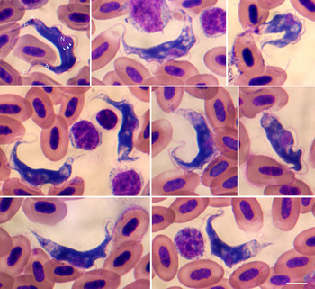
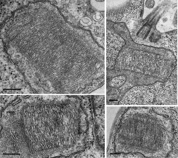
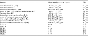
Diagnosis: Trypomastigotes scarce in thin blood films (Fig. 1). No more than 10 trypomastigotes recorded per blood film. Moderate size trypomastigotes, length 38·3 (27·6–43·4) μm, and moderately broad through the centre of their nuclei 4·6 (3·5–5·7) μm (n = 20) (Table 1). Moderately even breadth of the middle third of the trypomastigote (Fig. 1). Oval nucleus positioned centrally, the anterior part, 15·0 (13·3–17·7) μm, and posterior 13·6 (10·4–16·6) μm parts measured from the centre of the nucleus approximately even size (Table 1). The kinetoplast small and oval, not clearly stained using Diff-Quick stain. The kinetoplast 5·0 (2·9–7·1) μm from the posterior end (n = 20). The posterior end around the kinetoplast stained poorly compared to the deeply stained remainder of the trypomastigote (n = 20). The anterior end more markedly drawn out and pointed than the posterior end. Longitudinal striations observed in some trypomastigotes, particularly over the centrally positioned nucleus. In areas of deeply stained cytoplasm located outside the nucleus, striations were indistinct. The undulating membrane, pale with distinct outline, formed by the stained flagellum throughout the trypomastigote. Free flagellum originates just anterior to the kinetoplast. No dividing forms seen. No information regarding pathogenicity in birds, trypanosomes detected in clinically healthy birds. Ultrastructural investigation of the epimastigotes of T. thomasbancrofti sp. n. originated from a culture showed a kinetoplast thickness 549·4 (490·5–685·1) μm (Fig. 2).
Trypanosoma thomasbancrofti sp. n. (Kinetoplastida) on a blood smear from the Regent Honeyeater. Moderately broad trypomastigotes possess an oval nucleus (N) positioned centrally. The kinetoplast (K) is small and oval. Free flagellum (F) originates just anterior to the kinetoplast. The anterior end (A) more markedly drawn out and pointed than the posterior end (P). Longitudinal striations (arrows) observed in some trypomastigotes, particularly over the centrally positioned nucleus, in areas of deeply stained cytoplasm outside the nucleus striation are indistinct. The undulating membrane (U) is visible throughout the trypomastigote. Composite image, all at the same magnification. Scale bar, 5 µm. Diff-Quick stain. Type material G466177 at the Queensland Museum, Australia (from a type host ID: B20248 at Taronga Zoo).

Transmission electron microscopy of kinetoplasts from cultured Trypanosoma thomasbancrofti sp. n. (Kinetoplastida). Characteristic cylindrical-shaped kinetoplasts are represented by isolate RHE/B20358/AUS/2013/TB1 (genotype g1). All micrographs are to the same scale. Scale bar, 5 µm.

Morphology of trypomastigotes of Trypanosoma thomasbancrofti sp. n

s.d., standard deviation.
Discovery of T. thomasbancrofti sp. n. in the Regent Honeyeater
Initial blood smear observation of two birds (B20248, B20358) demonstrated scarce presence of trypomastigotes belonging to the genus Trypanosoma (B20248, <30 and B20358, <5 per blood smear, March 2013) (Fig. 1). Subsequently, inoculation of the blood onto a biphasic media revealed presence of epimastigotes in culture B20358 (RHE/B20358/AUS/2013/TB1) from 5 days onward (Regent Honeyeaters, Cohort 1; Supplementary Table S1). Amplification and direct PCR amplicon sequencing of SSU rDNA with Trypanosoma-specific primers demonstrated presence of a 2105 nt sequence with top blastn hits belonging to passerine bird Trypanosoma spp. sequences. Culture B20248 did not reveal the presence of epimastigotes (28 days). Ultrastructural investigation of the epimastigotes of T. thomasbancrofti sp. n. (B20358) based on TEM showed a distinct kinetoplast thickness that measured 549·4 µm (490·5–685·1, s.d. 44·7, n = 24) (Fig. 2).
We then tested six birds for the presence of T. thomasbancrofti sp. n. using repeated blood sampling tested by culture (April/May 2013; Regent Honeyeaters, Cohort 2; Supplementary Table S1). Blood was inoculated onto a biphasic medium three times, following a 7-day interval. Epimastigotes were detected as early as 4 days after inoculation. Blood from B20358 was culture positive on all three attempts. Blood from B20331 was culture positive on a single occasion, 6 days post inoculation, with low numbers of clumping epimastigotes not observed on subsequent examinations. The remaining blood cultures were negative. We then used the repeated blood Trypanosome culture approach on six birds as the gold standard test for a 2-way contingency table. Sensitivity (Se) and specificity (Sp) of single Trypanosome culture was 50% (95% CI, 3–50%) and 100% (76–100%), respectively. Positive predictive value (PPV) was 100% (5·7–100%) and negative predictive value (NPV) was 80% (61–80%) for a single blood culture.
Overall, the initial Trypanosoma-prevalence was 29·4% (5/17) in captive birds based on culture tests during 2013/2014 (B20213, B20217, B20331, B20358, B30214; Supplementary Table S1). In 2013, seven birds were tested using single culture attempt, where two birds revealed the presence of Trypanosoma sp. (July, 2013; Regent Honeyeaters, Cohort 3; Supplementary Table S1). In 2014, six birds were tested using single culture attempt and one bird (B30214) revealed presence of trypanosomes, 8 days post inoculation (October/November, 2014; Regent Honeyeaters, Cohort 4; Supplementary Table S1), subsequent sequencing confirmed T. thomasbancrofti sp. n. Comparison of the SSU rDNA of these two T. thomasbancrofti sp. n. genotypes revealed 6 nucleotide/in-del differences between the genotype g1 (culture RHE/B20358/AUS/2013/TB1; GenBank: KT728373) and the genotype g2 (culture RHE/B30214/AUS/2014/TB2; GenBank: KT728395).
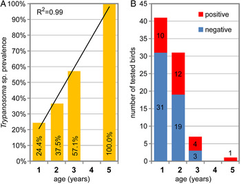
In 2015, Regent Honeyeaters considered for release (n = 81) were screened for the presence of Trypanosoma spp. using culture followed by SSU rDNA PCR and direct sequencing. From 81 birds screened, 27 birds (33·3%, 27/81) were found to be culture positive for trypanosomes (Supplementary Table S2). Only a single blood smear revealed scarce presence of (<2) trypomastigotes (B40305). Dividing the birds into age groups based on date of birth, we demonstrated increased prevalence with age (Fig. 3). Birds born in 2014 (age 1 year) had prevalence of 24·4% (10/41), birds born in 2013 (age 2 years) had prevalence of 37·5% (12/32) and birds born 2012 (age 3 years) had prevalence of 57·1% (4/7). A single 5-year-old bird born in 2010 (B30152) was Trypanosoma positive. DNA was isolated from all 27 Trypanosoma-positive cultures and SSU rDNA was amplified from 25 samples. SSU rDNA sequences matching T. thomasbancrofti sp. n. were amplified in 20 samples (20/25), and found to be identical to SSU rDNA sequence from B20358 (genotype g1) collected in 2013 (RHE/B20358/AUS/2013/TB1; GenBank: KT728373). In fact, one of the 20 samples was from the same B20358 bird, confirming that T. thomasbancrofti sp. n. remains genetically stable (genotype g1) over a 2-year period. For the sample from B20358, we isolated DNA from the culture, 3 and 6 days post inoculation, and confirmed that only unambiguous SSU rDNA of T. thomasbancrofti sp. n. was present. Five SSU rDNA sequences (from B20348, B30187, B30238, B40102, B40384) were distinct from T. thomasbancrofti sp. n., nevertheless the top blastn hits belonged to passerine bird Trypanosoma cf. avium SSU rDNA sequences. Four SSU rDNA sequences were identical to each other (B20348, B30187, B30238, B40102) and constitute genotype g3 with 18 nucleotide/in-del differences from T. thomasbancrofti sp. n. SSU rDNA sequences. Sequence from B40384 represents Trypanosoma cf. avium genotype g4 and differ in 12 nucleotide/in-del from genotype g3 (Supplementary Table S2).
Bird age correlation with prevalence of trypanosomes in the Regent Honeyeater at the Taronga Zoo. Bird blood samples were collected and cultured to detect presence of trypanosomes. (A) Birds (n = 81) were divided into age groups and their prevalence for evaluation of correlation (coefficient of determination, R 2). (B) The graph shows total number of birds tested born in an individual year, each bar is divided according to the trypanosome diagnostic results.

In total, we have tested 92 individual captive birds (Supplementary Tables S1 and S2). Five birds were tested in 2013/2014 and subsequently in 2015, testing T. thomasbancrofti sp. n. positive with SSU rDNA sequence matching RHE/B20358/AUS/2013/TB1. Only birds which maintained or gained trypanosomes were recorded, no birds that cleared trypanosomes were recorded. Bird B20358 remained positive from 2013 to 2015. Birds B20332 and B20390 were negative on three occasions in 2013, but positive in the 2015 screening. Moreover, B20390 tested negative in 2014. Birds B30403 and B30404 were negative in 2014, but positive in 2015.
Following the release in 2015, we collected blood samples from 18 Regent Honeyeaters captured at the release site and screened for the presence of Trypanosoma spp. using culture, followed by SSU rDNA PCR and direct sequencing. From the 18 birds screened, four birds (22·2%, 4/18) were culture positive for Trypanosoma (Supplementary Table S3). Two blood smears revealed scarce presence of (<2) trypomastigotes (B20305, B20239) and only one (B20239) was later positive in culture. Amplification and sequencing of SSU rDNA demonstrated presence of the T. thomasbancrofti sp. n. genotype g1 in two birds (B40304, B40442) and T. thomasbancrofti sp. n. genotype g2 in a single bird (B40584), the T. cf. avium genotype g3 in a single bird (B20239). Three of these birds, (B40584, B40304 and B40442), tested negative for Trypanosoma spp. in 2015 prior to release, suggesting that these birds acquired the infection in the wild. Seven tested negative in the wild, but tested positive while captive at Taronga Zoo. A single wild bird tested negative for trypanosomes.
Trypanosoma cf. avium in the Noisy Miner (M. melanocephala)
To investigate the possibility that T. thomasbancrofti sp. n. from Regent Honeyeaters is also present in other bird species, we collected blood from Noisy Miners, captured in close proximity to Regent Honeyeater aviaries at Taronga Zoo. Blood smears from nine birds revealed no hemozoic trypomastigote, a single bird had intraerythrocytic Haemoproteus sp. (#6) and three birds, (#2, #5, #7), had microfilaria present. Blood collected from 11 birds (May 2013) was inoculated onto the biphasic medium as pooled samples from two birds per culture flask. Trypanosomes were demonstrated in three pools on day 6 post inoculation (Supplementary Table S1). Subculturing of the trypanosomes onto a fresh media was unsuccessful and the trypanosomes were not observed in the cultures by day 28 post inoculation.
DNA was isolated from the culture from Noisy Miner pool #9/#10 (NM/#9#10/AUS/2013/TA1), followed by PCR amplification with Trypanosoma-specific primers confirmed presence of Trypanosoma sequence (SSU r|DNA, 2099 nt). Querying the new sequence against publically available trypanosome sequences confirmed high similarity with Trypanosoma from passerine birds and 100% identity with the Trypanosoma cf. avium SSU rDNA sequences (genotype g3) from the Regent Honeyeaters (B20348, B30187, B30238, B40102).
Phylogeny of bird Trypanosoma species
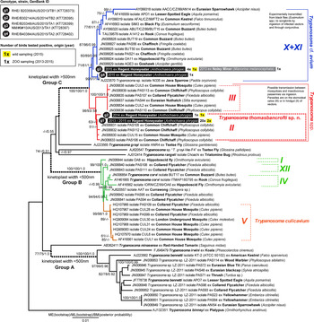
For the purpose of the phylogeny we define four distinct and newly defined Trypanosoma SSU rDNA genotypes (g1–g4) (Fig. 4). Phylogenetic inference of SSU rDNA demonstrates that g1–g4 clustered within Group C sensu Zídková et al. (Reference Zídková, Cepicka, Szabová and Svobodová2012) and the overall phylogeny was comparable with previously published trees (Zídková et al. Reference Zídková, Cepicka, Szabová and Svobodová2012) (Fig. 4). Both closely related SSU rDNA sequences (g1, g2) of T. thomasbancrofti sp. n. obtained from 24 Regent Honeyeaters clustered with closely related sequences belonging to an unnamed Trypanosoma sp. in the Group C-II sensu Zídková et al. (Reference Zídková, Cepicka, Szabová and Svobodová2012) (Fig. 4). The sequences of Trypanosoma cf. avium (g3) from one Noisy Miner and four Regent Honeyeaters were a sister group to the T. avium sequences in the Group C-X + XI sensu Zídková et al. (Reference Zídková, Cepicka, Szabová and Svobodová2012) (Fig. 4). The single SSU rDNA sequence (g4) from one Regent Honeyeater (B40384; RHE/B40384/AUS/2015/TA2) was almost identical (one mismatch over 2102 nt, 99·95% identity) to a published Trypanosoma sp. isolate N335 from the Java Sparrow (Padda oryzivora) (AJ223570) (Haag et al. Reference Haag, O'hUigin and Overath1998). For birds in Australia, Hamilton et al. (Reference Hamilton, Stevens, Gaunt, Gidley and Gibson2004) published a Trypanosoma sp. SSU rDNA sequence (AJ620557) from the Australian Currawong (Strepera sp.), however, it is clearly distinct from all our new Trypanosoma spp. sequences and belongs to the Group B sensu Zídková et al. (Reference Zídková, Cepicka, Szabová and Svobodová2012) (Fig. 4). Currently, no other sequences from Australian bird trypanosomes are publically available for comparison.
Phylogenetic tree of SSU rDNA sequences of avian Trypanosoma species. The tree was inferred using the Minimum Evolution [ME, Kimura 2-parameter (K2P) distances] method. The percentage of replicate trees in which the associated taxa clustered together in the bootstrap test using ME (1000 replicates) and Maximum Likelihood (ML, 100 replicates), and Bayesian Method (BM, posterior probability) are shown next to the branches. The ML model (K2P) included a discrete gamma distribution and allowed for invariable sites. The BM was based on K2P model with estimated discrete gamma distribution and proportion of invariable sites. The tree is drawn to scale, with branch lengths in the same units as those of the evolutionary distances used to infer the phylogenetic tree. Branch annotation (groups A–C) and clusters within ‘clade C’ are according to Zídková et al. (Reference Zídková, Cepicka, Szabová and Svobodová2012). Trypanosoma binneyi served as an outgroup.

The alignment of the Group C-II, including isolates PAS105, PAS112, PAS106 from the Common Chiffchaff (P. collybita), CUL15 from the common house mosquito (C. pipiens complex) and both T. thomasbancrofti sp. n. genotypes (2108 residues) from Regent Honeyeaters revealed only nine singletons with overall distance of 0·001. Such high overall sequence identity (99·57%) of the group suggests that all belong to the same species, despite the fact that the Common Chiffchaff and the common house mosquito were collected from Central Europe (Czech Republic). A possible transmission between mosquitoes and insectivorous passerines by ingestion was proposed for the Group C-II/III with parasites located on mosquito stomodeal valves (Group C-III) or in the hindgut (Group C-II) of vectors (Votýpka and Svobodová, Reference Votýpka and Svobodová2004; Zídková et al. Reference Zídková, Cepicka, Szabová and Svobodová2012).
The phylogenetic clades of avian Trypanosoma species based on gGAPDH gene trees share common origin except T. bennetti (Hamilton et al. Reference Hamilton, Stevens, Gaunt, Gidley and Gibson2004; McInnes et al. Reference McInnes, Gillett, Ryan, Austen, Campbell, Hanger and Reid2009). Five Trypanosmoma isolates were further characterized at gGAPDH locus (Supplementary Fig. 1). Three isolates with SSU rDNA sequences (g1) of T. thomasbancrofti sp. n. from Regent Honeyeaters were identical to each other or differed by a single nucleotide difference (B20358, B30188, 11006). The isolate with SSU rDNA sequences (g2) of T. thomasbancrofti sp. n. (11008) was 96% identical at gGAPDH from those isolates represented by SSU rDNA sequences (g1). An isolate belonging to T. cf. avium from a Regent Honeyeater (B30187, SSU rDNA sequence – g3) was 92% identical to T. thomasbancrofti sp. n. at gGAPDH.
DISCUSSION
The present study describes a new trypanosome species, T. thomasbancrofti sp. n., isolated from the endangered Regent Honeyeater. The newly described trypanosome falls within the Group C-II sensu Zídková et al. (Reference Zídková, Cepicka, Szabová and Svobodová2012) of avian trypanosomes sharing high (>99%) SSU rDNA sequence identity. One of the most conspicuous features of trypanosomes is its compact mitochondrial DNA forming a characteristic kinetoplast (Yurchenko et al. Reference Yurchenko, Hobza, Benada and Lukes1999; Votýpka et al. Reference Votýpka, Oborník, Volf, Svobodová and Lukeš2002). The thickness of the kinetoplast in avian trypanosomes was suggested as a characteristic feature of distinct species and lineages (Votýpka et al. Reference Votýpka, Szabová, Radrová, Zídková and Svobodová2012; Zídková et al. Reference Zídková, Cepicka, Szabová and Svobodová2012). The kinetoplast thickness 549±45 (491–685) μm of T. thomasbancrofti sp. n. is near identical to Group C-II isolates CUL15 and PAS112 (546; 458–668 µm) and PAS112 (527; 446–676 µm), respectively (Zídková et al. Reference Zídková, Cepicka, Szabová and Svobodová2012). The high sequence identity (>99·5%) and the kinetoplast ultrastructure of T. thomasbancrofti sp. n. are the key differentiating characteristics from other Trypanosoma spp. Twenty four newly obtained sequences forming two genotypes (g1 and g2) clustered along with the four previously described isolates (PAS105, PAS106, PAS112 and CUL15) of the Group C-II sensu Zídková et al. (Reference Zídková, Cepicka, Szabová and Svobodová2012) formed a monophyletic group representing the new species, T. thomasbancrofti sp. n. None of the named species of Trypanosoma spp. could be directly associated with T. thomasbancrofti sp. n.
Our findings support the opinion of Valkiūnas et al. (Reference Valkiūnas, Iezhova, Carlson and Sehgal2011), stating the authors’ early descriptions that avian trypanosomes are specialists and therefore highly bird host specific is incorrect and such labelled species need careful re-evaluation. In the case of the endangered Regent Honeyeater, the ultrastructure and DNA sequence strongly suggests that T. thomasbancrofti sp. n. is not geographically restricted and host specific, but likely, an intercontinentally distributed generalist trypanosome species. As such, we propose to adopt the name for the entire Group C-II that includes trypanosomes isolated from the Common Chiffchaff (P. collybita) distributed throughout Europe, Asia and Africa. Generally, there is little evidence of host–trypanosome specificity, because identical or close to identical SSU rDNA sequences of bird trypanosomes are amplified from diverse birds over large geographical distances (Sehgal et al. Reference Sehgal, Jones and Smith2001; Votýpka and Svobodová, Reference Votýpka and Svobodová2004; Zídková et al. Reference Zídková, Cepicka, Szabová and Svobodová2012). The mechanisms of trypanosome ecological plasticity warrants the attention of parasitologists and evolutionary biologists, because our example of closely related trypanosome SSU rDNA sequences from Australia's Regent Honeyeater and Europe's Common Chiffchaff suggests an intercontinental distribution.
One of the more challenging aspects encountered when working with trypanosomes is their diagnosis in avian hosts. Stained blood smears are notoriously ineffective; therefore more sensitive culture and PCR are used in studies examining trypanosome diversity (Sehgal et al. Reference Sehgal, Jones and Smith2001; Votýpka et al. Reference Votýpka, Szabová, Radrová, Zídková and Svobodová2012; Zídková et al. Reference Zídková, Cepicka, Szabová and Svobodová2012). To evaluate reliability of a single blood sampling to detect trypanosomes via culture, a technique adopted throughout this study, we tested birds weekly over a 3-week period. Comparison of a diagnostic culture against PCR would be an alternative approach, but again the analysis would be incomplete due to the absence of true positive and negative birds. The low sensitivity with high specificity of using culture as diagnostics presented here requires careful interpretation because only six birds were used in the repeated culturing experiment due to ethical and logistical considerations. Culture technique can theoretically return positive result even if a single trypanosome is present in the inoculum (50 µL of fresh avian blood). The higher NPV of 80% demonstrates the sensitivity of returning a negative result in the absence of trypanosomes in the inoculum. On the other hand, the wide 95% CI of the positive predictive value (PPV) may reflect either extremely low parasitaemia of a persistent infection or only a transient infection that did not establish. At this time we have no means to distinguish between the two.
This study revealed presupposed association of bird age with increased prevalence of Trypanosoma spp. The longest living captive Regent Honeyeaters have reportedly lived for 17 years, nevertheless the realistic life expectancy is 12–14 years (unpublished data, Larry Vogelnest). Trypanosoma species are common parasites of birds, yet the parasitaemia is often extremely low to be detected on blood smear compared with blood culture examination shown to be 20 times more sensitive in the case of the Regent Honeyeaters. The knowledge on the persistence of Trypanosoma spp. in birds is incomplete. On one hand, we demonstrated that a single bird was infected with the same genotype of T. thomasbancrofti sp. n. over a 2-year period, thereby arguing for persistence of infection. On the other hand, in our data from recaptured birds following release, we noted a number of birds that possibly eliminated parasitaemia based on results from culture testing. Birds that potentially eliminated pre-release parasitaemia possessed both T. thomasbancrofti sp. n. (genotypes 1, 2) as well as T. cf. avium (genotype 3) suggesting that the bird immune system could clear the infection. Alternatively, the infection was below the detectable level of the culturing technique. Repeated testing was not possible to confirm either elimination or persistence. Sampling of the released birds demonstrated that birds which tested negative prior to release acquired Trypanosoma spp. in the wild. The data confirm that T. thomasbancrofti sp. n. is present at the site of release. The phenomenon of elimination vs persistent Trypanosoma infection will require further experimental scrutiny. It remains to be elucidated whether parasitaemia is a good indicator of the presence of Trypanosoma spp. in the bone marrow.
In Australia, birds are known hosts of Trypanosoma spp., yet their diversity is unknown as studies applying molecular tools are absent (O'Donoghue and Adlard, Reference O'Donoghue and Adlard2000). Two named species have been described to date, T. anellobiae and T. myzanthase, based on trypomastigotes from bird blood smears (Cleland and Johnston, Reference Cleland and Johnston1910; Mackerras and Mackerras, Reference Mackerras and Mackerras1960; Bennett et al. Reference Bennett, Squires-Parsons and Poldmaa1994c). The original description of T. anellobiae is from the Little Wattlebird (Anthochaera chrysoptera), but it was later re-described from the Noisy Miner (Bennett et al. Reference Bennett, Squires-Parsons and Poldmaa1994c). Besides these named species, several trypomastigote forms are described, but their further taxonomic status is not defined (Cleland and Johnston, Reference Cleland and Johnston1911; Breinl, Reference Breinl1913; Mackerras and Mackerras, Reference Mackerras and Mackerras1960). Mackerras and Mackerras (Reference Mackerras and Mackerras1960) reviewed all previously illustrated forms of trypomastigotes recorded from Australian birds, but could not ascertain how to classify Trypanosoma spp. from different bird species or families, because of unknown host specificity. In general, Trypanosoma species identification, merely through trypomastigote morphology, is in most cases regarded as insufficient. Trypanosoma species in birds in Australia are no different to those from other continents where the majority of original descriptions based on trypomastigote morphology are difficult or even impossible to use for species identification and comparisons. Nevertheless, T. anellobiae and T. myzanthas, differ primarily in the position of their kinetoplast and were distinguished as follows ‘T. myzanthae differs from T. anellobia in shape, being usually broader, in the position of undulating membrane, usually wound around the body, in the central position of the nucleus, and in the longer, thicker, free flagellum’ (Mackerras and Mackerras, Reference Mackerras and Mackerras1960). Trypomastigotes of T. anellobia are narrow and approximately 35 µm long, displaying kinetoplasts 3 µm from the posterior end (Cleland and Johnston, Reference Cleland and Johnston1910). The broader trypomastigotes of T. myzanthae (19–25 µm) have kinetoplasts <1·5 µm from the posterior end (Mackerras and Mackerras, Reference Mackerras and Mackerras1960). Based on the original morphological description of Trypanosoma spp. alone, these were later considered ambiguous. The deteriorated status of the type slide held at the Australian Museum, deposited by Cleland and Johnston (Reference Cleland and Johnston1910), led Bennett et al. (Reference Bennett, Squires-Parsons and Poldmaa1994c) to designate a neotype. The neotype of T. anellobia was described from the Noisy Miner and is considered conspecific with the original type material from the Little Wattlebird despite the trypomastigote size averaging 25·6 (s.d. 2·3) μm (Bennett et al. Reference Bennett, Squires-Parsons and Poldmaa1994c). The above seemingly complicates the situation of trypanosomes in Australian birds, because the Noisy Miner is the type host of the T. myzanthae described by Mackerras and Mackerras (Reference Mackerras and Mackerras1960). It is likely that Noisy Miners are hosts to more than one species of trypanosome and isolation of these trypanosomes is considered necessary to resolve the relationship to trypanosomes from Regent Honeyeaters and advance the intercontinentally growing database of bird Trypanosoma SSU rDNA sequences.
We cultured and sequenced SSU rDNA of T. cf. avium from the Noisy Miners and Regent Honeyeaters. Based on the above, there are at least three reasons why the situation in Noisy Miners as the type hosts for both T. anellobia and T. myzanthae presents a confusing case. (i) Trypanosoma myzanthae is not considered in a differential diagnosis of the neotype T. anellobia by Bennett et al. (Reference Bennett, Squires-Parsons and Poldmaa1994c). (ii) We were not able to detect any trypomastigotes in our set of blood smears from Noisy Miners to perform morphological comparison to T. anellobia and T. myzanthae. (iii) The SSU rDNA sequence obtained from Noisy Miners suggests a close relationship with T. avium group. Historically, the T. avium group contained trypanosomes of owls and raptors, nevertheless, songbirds were later confirmed as the hosts of T. avium (Baker, Reference Baker, Lumsden and .1976; Podlipaev, Reference Podlipaev1990; Dirie et al. Reference Dirie, Wallbanks, Molyneux and McNamara1991; Votýpka et al. Reference Votýpka, Oborník, Volf, Svobodová and Lukeš2002; Zídková et al. Reference Zídková, Cepicka, Szabová and Svobodová2012). A member of the T. avium group was experimentally transmitted from black flies (Simulium/Eusimulium spp.) to songbirds by ingestion of infected vectors and through conjunctiva (Votýpka and Svobodová, Reference Votýpka and Svobodová2004). In Australia, old records of suspect T. avium group trypanosomes exist for the Brahminy Kite (Haliastur indus girrenera), the Grey Falcon (Falco hypoleucos) and the Boobook Owl (Ninox boobook) (Breinl, Reference Breinl1913; O'Donoghue and Adlard, Reference O'Donoghue and Adlard2000). Currently, none of these owl or raptor trypanosomes are available for comparison with either our new sequences or the intercontinentally growing database of SSU rDNA (Votýpka et al. Reference Votýpka, Oborník, Volf, Svobodová and Lukeš2002; Votýpka and Svobodová, Reference Votýpka and Svobodová2004; Zídková et al. Reference Zídková, Cepicka, Szabová and Svobodová2012). It is likely, that T. avium complex species exist in Australia, because it infects a broad range of avian hosts and has been suggested to be intercontinentally distributed (Bennett, Reference Bennett1961; Votýpka et al. Reference Votýpka, Oborník, Volf, Svobodová and Lukeš2002; Zídková et al. Reference Zídková, Cepicka, Szabová and Svobodová2012). It was noteworthy that we did not detect dual infection in any of the studied cultures. Whether absence of dual infection was an artefact of the culture is not known. It remains to be elucidated if birds infected with one trypanosome remain resistant to a super-infection with a different trypanosome.
The current results demonstrate that the screening approach applied to the captive bird population is suitable for screening of wild birds. Single culture was sufficient to demonstrate parasitaemia during the repeated culture experiment and is therefore recommended for wild bird screening. Additional genotyping of cultured trypanosomes from Regent Honeyeaters will be required to demonstrate conspecificity with T. thomasbancrofti sp. n. and absence of other Trypanosoma spp. belonging to other bird clades in either wild or captive birds.
Currently, no information exists on the effect of T. thomasbancrofti sp. n. on its hosts; however, all five trypanosome positive captive birds remained clinically healthy for over 12 months (Vogelnest, personal communication). Nevertheless, an argument that infected birds should not be released because the trypanosome species present in captive birds might be alien has been considered. If it is alien, the parasite might give rise to disease in free-living birds. However, given the limited evidence of pathogenicity and the worldwide distribution of Clade C-II and therefore T. thomasbancrofti sp. n. there is insufficient evidence to justify not releasing positive birds. The finding that at least some birds acquired T. thomasbancrofti sp. n. infection after their release demonstrates natural trypanosome circulation in their wild environment. The revelation that both the free living Noisy Miners and captive as well as wild Regent Honeyeaters serve as hosts of an identical T. cf. avium confirms previous suggestions that T. avium complex are capable of infecting wide range of birds including passerines (Bennett and Fallis, Reference Bennett and Fallis1960; Bennett, Reference Bennett1961; Bennett et al. Reference Bennett, Earle and Squires-Parsons1994a, Reference Bennett, Siikamaki, Ratti, Allander, Gustafsson and Squires-Parsonsb; Votýpka et al. Reference Votýpka, Oborník, Volf, Svobodová and Lukeš2002; Votýpka and Svobodová, Reference Votýpka and Svobodová2004; Votýpka et al. Reference Votýpka, Szabová, Radrová, Zídková and Svobodová2012; Zídková et al. Reference Zídková, Cepicka, Szabová and Svobodová2012).
We suggest that T. thomasbancrofti sp. n. has an intercontinental distribution. Current data are unable to identify whether the trypanosome is native or introduced into Australia. However, if the parasite is a generalist infecting migratory songbirds, then most likely, the parasite should be considered native to Australia. Alternatively, the parasite may have been introduced with invasive birds, such as the Common Myna (Acridotheres tristis), a known reservoir for avian malaria in Australia (Clark et al. Reference Clark, Olsson-Pons, Ishtiaq and Clegg2015). In Australia, the Common Myna has not been extensively surveyed for trypanosomes, but a record of an unknown Trypanosoma sp. exists from its native Sri Lanka (Crusz, Reference Crusz and Fernando1984). A small survey of Common Mynas in South East Queensland, Australia, showed no presence of trypanosomes in blood smears (N. Clark, personal communication). To evaluate if T. thomasbancrofti sp. n. was present in Australia prior the introduction of the Common Myna in the 19th century, we would need to consider testing preserved material from before such introduction. Museum materials of frogs have been successfully sampled to show that some myxozoan parasites in Australia were not present in frogs prior to the introduction of the Cane Toad (Rhinella marina) to Australia in 1935 (Hartigan et al. Reference Hartigan, Phalen and Šlapeta2010). Trypomastigotes, however, are rare in birds’ blood and do not concentrate in specific body cavities, thereby preventing its detection, unlike the myxozoa that accumulate in gallbladders of frogs. It is unlikely we can obtain conclusive evidence for the introduction of T. thomasbancrofti sp. n., retrospectively. Furthermore, museum material is unlikely to serve as a good proxy to evaluate absence of the parasite prior to introduction of exotic birds.
With almost one hundred individual birds tested for trypanosomes, we have tested the equivalent of one quarter of the remaining population of less than 400 wild Regent Honeyeaters. In conclusion, the trypanosomes in the Regent Honeyeaters represent the most intensive assessment of avian trypanosomes in Australia. However, the lack of baseline data on parasite diversity in Australia, such as diversity of Trypanosoma spp., limits our ability to ascertain the risk of whether parasites differ between source and destination sites and to what extent they may cause disease. It remains only an assumption that other Australian passerine birds are hosts for T. thomasbancrofti sp. n. either in the captive population or the remaining localities of the currently fragmented distribution of wild Regent Honeyeaters. The distribution of mosquitoes, in particular of the genus Culex (for example, C. pipiens group, C. annulirostris), as suspect vectors of T. thomasbancrofti sp. n. overlaps both captive and wild locations (Hemmerter et al. Reference Hemmerter, Šlapeta and Beebe2009; Russell, Reference Russell2012). Ingestion of infected mosquitoes as the likely route of transmission makes any preventative measures complex, if not impractical, when considering captive breeding programmes.
Taxonomic Summary
Trypanosoma thomasbancrofti sp. n. Šlapeta
Trypanosomatida: Trypanosomatidae
Type host: Regent Honeyeater, Anthochaera phrygia (Shaw, 1794) (Aves: Passeriformes: Meliphagidae); syn. Xanthomyza phrygia (Shaw, 1794).
Type locality: Zoo breeding population at Taronga Zoo, Mosman, Sydney, New South Wales, Australia. Animals are living at Taronga Zoo or were released.
Site of infection: Blood plasma.
Type material/hapantotype: Blood smear from Regent Honeyeater, Anthochaera phrygia (ID: B20248, collected on 1 February 2013) deposited under accession number G466177 at the Queensland Museum, South Brisbane QLD 4101, Australia. Parahapantotypes: blood smears G466180 (ID: B40305, collected on 17 February, 2015) and G466181 (ID: B20358, collected on March 14, 2013).
The ZooBank LSID: urn:lsid:zoobank.org:act:D8949216-F714-4CCB-850D-CAE6884CC8EC
Prevalence: Using birds considered for release in 2015, 33·3% (27/81) bird bloods tested culture positive for Trypanosoma spp., and 24·4% (20/81) were successfully genotyped as T. thomasbancrofti sp. n. in the captive population at Taronga Zoo in 2015. Only a single blood smear, 1·2% (1/81), demonstrated presence of trypomastigotes.
Nucleotide sequence data: The sequence of the small subunit ribosomal RNA gene (SSU rDNA) and glycosomal glyceraldehyde phosphate dehydrogenase gene (gGAPDH) from the Regent Honeyeater (ID: B20358, currently at Taronga Zoo) is available in GenBank™ under Accession No. KT728373, KU323392 and designated as RHE/B20358/AUS/2013/TB1). Identical SSU rDNA of T. thomasbancrofti sp. n. was identified in an additional 19 and 2 Regent Honeyeaters, A. phrygia, held at Taronga Zoo and released into the wild, respectively (Supplementary Tables S2 and S3; GenBank: KT728374-KT728392, KT728393-KT728394). Near identical SSU rDNA (>99·5%) with T. thomasbancrofti sp. n. was identified in a single Regent Honeyeater, A. phrygia originated from Taronga Zoo and in one released wild bird (GenBank: KT728395).
Additional hosts: T. thomasbancrofti sp. n. was identified, based on the high sequence identity (>99·5%) with SSU rDNA of T. thomasbancrofti sp. n., in the Common Chiffchaff, Phylloscopus collybita (isolate PAS105, PAS112, PAS106, GenBank: JN006831, JN006832, JN006833) and from the Common House Mosquito, Culex pipiens complex (CUL15, GenBank: JN006830) in Central Europe.
Etymology: The specific epithet “thomasbancrofti” is given in honour from the surname of Thomas Lane Bancroft (1860–1933) who undertook many parasitological investigations in Australia and whom was the first to note trypanosomes in Australian endemic bird blood smears.
Remarks: This is the first Trypanosoma species described from the Regent Honeyeater (A. phrygia). In Australia, Trypanosoma myzanthase Mackerras and Mackerras, Reference Mackerras and Mackerras1960 and Trypanosoma anellobiae Cleland and Johnston, Reference Cleland and Johnston1910 are described from passerine (Passeriformes) birds and honeyeaters (Melliphagidae). The original description of T. anellobiae is from the Little Wattlebird (A. chrysoptera), a honeyeater from the same genus Anthochaera. Trypomastigote of T. thomasbancrofti sp. n., however, is distinct from both described species in shape and position of kinetoplast. T. anellobiae is very narrow and approximately 35 µm in length with kinetoplast 3 µm from the posterior end. T. myzanthase is broad, 19–24 µm in length, with rather large and distinct kinetoplast situated <1·5 µm from the posterior end. Our species shape is amid the two described species and the kinetoplast is further from the posterior end compared to T. anellobiae and T. myzanthase. Intercontinentally, there are several avian Trypanosoma spp. described from various bird groups including passerine (Passeriformes) birds. Absence of type material, insufficient illustration of type material and cryptic morphology make it difficult to compare and identify avian Trypanosoma species at an intercontinental scale. Molecular characterization enabled us to show that Regent Honeyeaters, A. phrygia, held at Taronga Zoo are hosts for four Trypanosoma spp. SSU rDNA genotypes. Two SSU rDNA genotypes (g1, g2) are considered T. thomasbancrofti sp. n., while the remaining two represent T. cf. avium. Therefore applying techniques such as culture, TEM, genetic characterization and comparison with isolates from sympatric bird species, aids in species identification.
SUPPLEMENTARY MATERIAL
To view supplementary material for this article, please visit http://dx.doi.org/S0031182016000329.
ACKNOWLEDGEMENTS
We thank Taronga Zoo's bird keepers for their care of the birds. Taronga Wildlife Hospital veterinary nurses and veterinarians Drs Hulst, Vinette Herrin and Tobias for assistance with sample collection and Tammy McDonogh for ongoing laboratory support. Dean Ingwersen Woodland Bird and WA Programme Manager and Regent Honeyeater recovery coordinator Birdlife Australia, Glen Johnson Senior Biodiversity Officer and Elizabeth Wemyss Project Biodiversity Officer Hume Region Regional Services Department of Environment, Land, Water & Planning (DELWP) are acknowledged for their assistance in recapturing wild and pre-released captive bred birds for blood sampling. We thank Cameron Webb (ICPMR Westmead, Medical Entomology, University of Sydney) for discussions on mosquitoes at the Zoo, Nick Clark (Griffith University) for discussion around Common Mynas and Stefan Saverimuttu for assistance with Noisy Miner sample collection at Taronga Zoo during his B.Sc. (Vet) candidature. We acknowledge the support from the facilities at the Australian Microscopy & Microanalysis Research Facility node (Sydney Microscopy and Microanalysis) at the University of Sydney and we thank Ms. Naveena Gokoolparsadh for her expert consultations.
FINANCIAL SUPPORT
This study was in part funded through the Faculty of Veterinary Science, University of Sydney diagnostic laboratory (JS, DM, KG). Captive bred pre-release health screening and blood sampling at Taronga Zoo for the 2015 release was funded in part by the NSW Office of Environment and Heritage (LV, MS, PT). VMA is supported by the International Postgraduate Research Scholarship (IPRS) and an Australian Postgraduate Award (APA) tenable at The University of Sydney.