Introduction
Enlarging the external auditory meatus is a vital step in any exploratory procedure that leaves an open mastoid cavity. Such enlargement ensures good aeration, minimising moisture accumulation and consequent microbial colonisation. It also affords good visibility, aiding monitoring for recurrent squamous or mucosal disease. Finally, it ensures easy passage of microsuction instruments for aural toilet, which is often regularly required in these patients. Indeed, the latter may obviate the need for repeated general anaesthesia in small children.
Despite widespread recognition of the importance of external auditory meatus enlargement, it is often the most neglected part of the mastoid operation, and as such is one of the prime causes of a continually discharging mastoid cavity.
The endaural approach initially popularised by LempertReference Lempert1 has rapidly become the favourite of many otologists. It is certainly in keeping with the philosophy of modern mastoid surgery, involving as it does a minimalist approach, a small incision through which is removed only as much bone as necessary to evacuate all the diseased epithelium, and a primary or staged reconstruction of the ossicular chain. Early squamous disease is usually confined to the attic or posterior mesotympanum. Access to these areas, and indeed to the majority of the mastoid bone, is eminently possible via an endaural approach.
We propose a novel, simple method of creating a meatoplasty via an extension from the endaural incision.
Technique
The patient's head is appropriately positioned in a head ring, and the ear is dressed and prepped with aqueous antiseptic solution. The proposed site of the endaural skin incision is accurately marked along the skin crease identified by pushing the crus of helix anteriorly with an index finger. Following aural toilet, a solution of local anaesthetic plus vasoconstrictor (2 per cent lidocaine with 1:80 000 adrenaline) is then infiltrated into this site and also very slowly (so as not to raise a skin blister) into the anterosuperior aspect of the external auditory canal, at the junction of the bare and hair-bearing skin. If performed correctly, the superoposterior canal wall skin should blanch without the need for a second canal injection.
The endaural incision is then fashioned in the usual way. If necessary, a temporalis fascia graft may be harvested.
The site of the next incision, along the anterior border of the conchal cartilage, is marked. It is made more prominent by gripping the antihelix and pushing the concha anteriorly (Figure 1). As demonstrated in Figure 1, it meets the endaural incision and extends the full width of the anterior conchal margin. Again, this, as well as the lateral skin of the conchal bowl, is infiltrated with local anaesthetic plus vasoconstrictor.
The conchal margin is made more prominent by gripping the antihelix and pushing the concha anteriorly.

The incision is deepened onto the anterior border of the conchal cartilage. Skin and soft tissue are elevated off both sides of the cartilage and a bean-shaped segment is excised with dissecting scissors (Figure 2). Care should be taken to avoid button-holing the skin on the lateral aspect of the conchal cartilage.
Skin and soft tissue are elevated off both sides of the conchal cartilage, and a bean-shaped segment is excised with dissecting scissors.

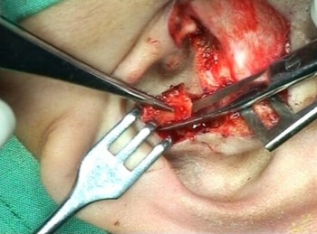
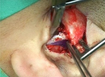
A triangle is shaded in with a marker pen on the skin of the canal side of the second incision. Its apex is at the inferior point of the second incision, which makes up one of its sides, and its base is the endaural incision, being approximately 5 mm in length (Figure 3). This triangle of skin and underlying soft tissue is resected (Figure 4). The excised conchal cartilage may be used later for canal wall reconstruction if necessary. The mastoid exploration can now proceed, after raising the conchomeatal flap and placing self-retaining retractors.
A triangle of skin is shaded in on the canal side of the conchal incision. Its base, 5 mm in length, is at the endaural incision.

The triangle of skin is resected together with the underlying soft tissue.

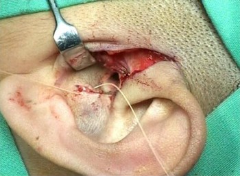
After completion of the mastoidectomy and tympanoplasty, the edges of the second and third incision are sutured with interrupted fine absorbable sutures (4/0 Vicryl Rapide). We prefer to close this incision at the end of the procedure, to avoid breaking the sutures when placing the self-retaining retractors (Figures 5 and 6).
After completion of (attico)mastoid exploration, the incision is closed with interrupted 4/O Vicryl Rapide sutures.

The appearance at the end of the procedure.

An online video demonstration of the procedure is available on The Journal of Laryngology & Otology website, as detailed in Appendix 1.
Results
To date, we have used this technique successfully in 64 patients. In all patients, we successfully achieved our goal of an adequately sized meatus with good access for microsuction, as well as obtaining an acceptable cosmetic result for the patient.
Discussion
A narrow lateral external auditory canal is usually caused by an abnormality of the skin and underlying soft tissue (e.g. congenital narrowing or cicatrisation following inflammation) or cartilage (e.g. excessive anterior projection of the conchal cartilage). Medially, the bony tympanic ring may be a significant contributor. Frequently, several factors are present simultaneously. Treating the bony component of canal narrowing should be part of the (attico)mastoid procedure; however, skin, soft tissue and cartilage components are often neglected. Indeed, an inadequate meatoplasty is one element of an important triad of causes for a continually discharging mastoid. The other two elements are a relatively high facial ridge and incomplete epithelialisation (with formation of granulation tissue, mucosal or squamous webbing).Reference Wormald and Nilssen2–Reference Sade, Weinberg, Berco, Brown and Halevy4
Meatoplasty was originally described by Stacke in the late nineteenth century, and rapidly became popularised by the forefathers of mastoid surgery (e.g. Korner and Siebenmann) in the ensuing years.Reference Phillips and Phillips5 Numerous variations of these original techniques have since been described. Nevertheless, an almost universal feature of a modern meatoplasty, and of our technique, is the excision of a piece of anterior conchal cartilage along with adjacent soft tissue.
Various techniques are used to widen the diameter of the ring of lateral skin. Essentially, this may be dealt with in two ways.
The first technique involves the use of radial incisions to create flaps based on the outer circumference of the external canal. These are elevated, thinned and pressed onto the bony canal after removing underlying soft tissue. This is the principle behind the classic Portmann three-flap technique.Reference Tos6
The second technique involves dividing the ring and bridging the resulting gap with a rotated skin flap. Unlike the first technique, the second is in principle less likely to re-stenose, and has been the focus of recent papers. Osborne and MartinReference Osborne and Martin7 have described rotating a superiorly based pre-tragal flap into the endaural wound after partial excision of the concha. Likewise, Martin-Hirsch and SmeltReference Martin-Hirsch and Smelt8 reported the use of a superior flap originating more medially in the redundant conchal skin. Banerjee et al. Reference Banerjee, Moir, Jervis and Narula9 described the use of an inferiorly based transposition flap, although inferoposterior widening occasionally results in inferior cosmesis.Reference Osborne, Terry and Gandhi10
An alternative technique of enlarging the ring is via local skin transposition, providing width at the expense of depth. This is epitomised by a Z-plasty used posteriorly on the conchal skin.Reference Murray, Jassar, Lee and Veitch11
These techniques, and many others, have produced good outcomes in their author's hands. However, at first glance they appear to be relatively complex procedures which demand the creation of viable flaps. In contrast, our technique does not rely on the creation of flaps but, rather, merely on simple excision of a small triangle of redundant skin and soft tissue overlying the excised conchal cartilage. It is thus a relatively straightforward technique which is vascularly robust. The transverse suture line heals with a barely perceptible scar. Our technique widens the canal in a posterior direction, and the resulting post-operative appearance is aesthetically pleasing. Trainees who pass through our unit have consistently commented on the ease of learning this meatoplasty technique.
Although described above for an endaural incision, our technique may be easily modified to enable its performance from a post-aural approach. In this case, the endaural incision is abbreviated so that it does not transcend lateral to the junction of the root of the helix and the superior tragus. The remainder of the procedure is as described above.
We perform the meatoplasty at the beginning of the procedure in order to minimise surgical errors that may arise from fatigue after a potentially lengthy tympanomastoid exploration.
A final note should be made regarding the surgical treatment of a narrow canal wall secondary to chronic otitis externa. Good ventilation prevents moisture accumulation and consequent microbial overgrowth and inflammation. An adequate meatoplasty facilitates this and, indeed, if performed relatively early in cases refractory to medical management, may halt chronic disease and obviate the need for a more medial canalplasty.Reference Martin-Hirsch and Smelt8, Reference Kumar and Smelt12, Reference Hunsaker13 The latter often involves bony widening with extra skin coverage obtained by either free skin grafts or complex, multistage, distant (usually post-aural) rotation flaps. Our technique would be ideal in this situation also.
Conclusion
We describe a novel technique for meatoplasty, which is an essential prerequisite for a healthy mastoid cavity and may halt disease progression in cicatricial chronic otitis externa. Our technique may be performed via either endaural or postaural approaches. It is relatively easy to perform and achieves a harmonious balance between patency and cosmesis.
Appendix 1. Supplementary video material
A video clip of the procedure is available on The Journal of Laryngology & Otology website, at http://journals.cambridge.org/sup_S0022215110001842sup001.