I. INTRODUCTION
The surface structure of scaffold plays a critical role in osteoconduction, osteoinduction, and osteogenesis for bone tissue engineering (BTE). Reference Yang, Xu, Kotaki and Wang1,Reference Cao, Zhou, Xue, Li, Yang, Long and Ji2 A suitable surface structure could enhance the bonding strength at implant-bone interface and accelerate cell adhesion and proliferation, thereby shortening the healing time. Reference Cheng and Teoh3,Reference Gittens, McLachlan, Olivares-Navarrete and Cai4 It has been demonstrated that a multilayer stereo porous structure on the surface of the scaffold could stimulate the production of osteoblasts and reduce the chances of rejection. Reference Bagherzadeh, Latifi, Najar, Tehran and Kong5–Reference Choi and Sofranko7 The multilayer porous structure is believed to provide more contact area for cell adhesion and implant-bone interface in comparison with smooth surface, since the vacant volume of porosity can be colonized by osteoblasts. Reference Khan and Newaz8–Reference Bignon, Chouteau, Chevalier, Fantozzi, Carret, Chavassieux, Boivin, Melin and Hartmann10 To improve the biocompatibility of scaffolds for BTE applications, different approaches have been adopted in an effort to obtain the optimal surface structure, including plasma-ion beam treatment, Reference Ahmed, Rho, Lee, Kim, Kim, Jang, Moon and Lee11,Reference Bax, McKenzie, Weiss and Bilek12 electric discharge, Reference Stráský, Havlíková, Bačáková, Harcuba, Mhaede and Janeček13 surface grafting, Reference Venugopal, Prabhakaran, Zhang, Low, Choon and Ramakrishna14,Reference Chen and Su15 vapor deposition, Reference Yamazaki, Maeda, Obata, Inukai, Kato and Kasuga16 and flame treatment. Reference He, Chen, Farson, Lannutti and Rokhlin17
There is a general consensus that the surface structure of the scaffold is associated with the surface chemical composition and crystal characteristics. Reference Rey, Combes, Drouet and Glimcher18,Reference Dohan Ehrenfest, Coelho, Kang, Sul and Albrektsson19 Calcium phosphate ceramics including hydroxyapatite (HAP) and β-tricalcium phosphate (β-TCP) are now considered as one of the most promising bone replacement materials because of the excellent bioactive and osseoconductive properties. Reference Veljović, Zalite, Palcevskis, Smiciklas, Petrović and Janaćković20–Reference Cao and Kuboyama24 Moreover, HAP/β-TCP biphasic calcium phosphate (BCP) has received increasing interest as scaffold material due to a more controllable degradability than pure HAP or β-TCP. Reference Elayaraja, Chandra, Joshy, Suganthi, Asokan and Narayana Kalkura25–Reference Emadi, Roohani Esfahani and Tavangarian27 In view of the higher dissolution rate than HAP in phosphoric acid (H3PO4), Reference Shiota, Shibata, Yasuda and Matsuo28,Reference Abe, Okazaki, Hiasa and Yasuda29 β-TCP in the outer layer of BCP scaffold could be selectively etched leaving micro/nanometer-sized pores on the scaffold surfaces. The porous structure could be controlled by adjusting the HAP/β-TCP weight ratio in the scaffolds.
The present study aimed to obtain various micro/nanometer-sized porous surface structures of BCP scaffolds and investigate the influence on biocompatibility. Multilayer stereo micro/nanometer-sized porous surface structures were built by selective etching of the BCP scaffolds with different HAP/β-TCP weight ratios using phosphoric acid. The surface structures and compositions were characterized by scanning electron microscopy/energy dispersive x-ray (SEM/EDX), x-ray diffraction (XRD) and an optical profiler system. Human osteosarcoma MG-63 cells were selected to study the influence of micro/nanometer-sized porous surface structure on the biocompatibility of scaffold. The results were exhibited and discussed in terms of distribution and number of MG-63 cells on the scaffold surfaces.
II. MATERIALS AND METHODS
A. Samples preparation
Commercially pure β-TCP 99.8% (Kunshan Chinese Technology New Materials Co., Ltd., Kunshan, China) and HAP 99.5% (Specification MHAP04, Nanjing Emperor Nano Material Co., Ltd., Nanjing, China) powder were utilized in this study. The HAP powder was fabricated by the supplier using sol–gel method through the reaction of (CH3O)3PO and Ca(NO3)2•4H2O. Reference Kaygili, Tatar and Yakuphanoglu30 Its essential parameters were as follows: Long needle-like particles with a width of about 20 nm and length of about 150 nm, as well as a Ca/P ratio of 1.67 ± 0.02. The β-TCP powder was synthesized by the supplier from aqueous precipitation method through the reaction of (NH4)2HPO4 and Ca(NO3)2 using ammonium hydroxide solution for pH control. Reference Kannan, Pina and Ferreira31 It had an average size of 200 nm and a Ca/P ratio of 1.50 ± 0.03. The HAP/β-TCP composite powders were obtained by mechanically mixing HAP and β-TCP powder with weight ratios of 90/10, 80/20, 70/30, 60/40, and 50/50.
BCP scaffolds were fabricated using a previously developed selective laser sintering (SLS) system Reference Shuai, Gao, Nie, Hu, Zhou and Peng32–Reference Shuai, Nie, Gao, Feng, Zhuang, Zhou and Peng34 with laser power of 18 W, scan speed of 200 mm/min, and laser beam of 0.5 mm. The composite powders were firstly placed on the substrate with a layer thickness of 0.15 mm. The laser beam then selectively scanned and sintered the powders under computer control resulting in a layer thickness of 0.11 mm due to densification during the sintering process. When finished, a new layer of powders was placed on the former layer. The above process was repeated many times until the sintering of scaffolds was completed. BCP scaffolds were obtained after the unsintered powders were brushed off.
Then the scaffolds were immersed in 2% H3PO4 solution for 7 min at 30 °C without agitation, by which β-TCP in the outer layer of BCP scaffolds could be selectively etched leaving micro/nanometer-sized pores on the scaffold surfaces. Subsequently, the treated BCP scaffolds were repeatedly washed with deionized water and dried with a stream of compressed air at 120 °C for 1 min. Multilayer stereo micro/nanometer-sized porous structures were finally constructed on the surfaces of BCP scaffolds.
B. Characterizations
The evolution of micro/nanometer-sized porous surface structures was characterized using a Veeco Wyko® NT9100 optical profiler system (Plainview, NY) equipped with Vision® software (Plainview, NY). The surface morphologies and element compositions were analyzed using a scanning electron microscope (JSM-6490LV, JEOL Ltd., Japan) coupled with EDX. For SEM observation, all samples were coated with gold in advance. The phase compositions on the scaffold surfaces were studied using x-ray diffractometer (XRD; model No. D/MAX 2550V, Rigaku, Tokyo, Japan) with wave length of 0.15418 nm, 2θ range of 20–60° and scan speed of 8°/min.
The specific surface area and pore size distribution on the scaffold surface were determined from nitrogen adsorption/desorption isotherms at −196 °C using a Quadrasorb SI-3MP system (Quantachrome Inc., Boynton Beach, FL). Prior to the measurements, the scaffolds were degassed at 150 °C overnight under vacuum. The multipoints Brunauer–Emmett–Teller method (BET) was utilized to calculate the specific surface area. Pore size distribution on the scaffold surface was derived from the desorption isotherm based on the Barrett–Joyner–Halenda (BJH) model.
C. Cell culture
The human osteosarcoma MG-63 cells derived from American Type Culture Collection (ATCC) were used to assess the cellular responses to the prepared surface structures of BCP scaffolds. Incubation was performed in 10-cm dishes of Dulbecco's modified Eagle's medium (DMEM, Gibco) supplementing with 10% fetal bovine serum (FBS, Gibco, Grand Island, NY) and 1% antibiotic/antimycotic (penicillin G sodium 100 U/mL, streptomycin 100 U/mL, amphotericin B 0.25 μg/mL). The MG-63 cells were trypsinized using 0.1% trypsin and 0.1% ethylene diamine tetraacetic acid (EDTA) for 5 min, centrifuged at 400g for 5 min, and resuspended in the medium.
For determination of the cell adhesion and proliferation, the prepared scaffold samples in duplicate were placed in 12-welled tissue culture polystyrene plates. All samples were sterilized in 70% ethanol overnight and rinsed extensively with phosphate-buffered saline (PBS), followed by treatment under ultraviolet light overnight and drying under sterile conditions at 37 °C for 2 h. Subsequently, 1 mL medium of cell suspension at a concentration of 2 × 10⁴ cells/mL was added to each well and maintained at 37 °C in a humidified atmosphere containing 5% CO2. The culture medium was renewed every 3 days to ensure a constant nutrition supply throughout the experimental period. After 7 days of culture, the culture medium was removed and nonadherent MG-63 cells were washed with PBS (pH 7.4). Subsequently, the samples were fixed with 2.5% glutaraldehyde and dehydrated in graded series of ethanol (70, 80, 90, 95, and 100%), then dried and sputter-coated with gold for observation of surface morphologies using SEM.
The duplicate samples were used to determine cell numbers using cell-counting assay. The MG-63 cells remaining attached to the surface of the scaffolds were washed with PBS and detached with EDTA solution containing 0.25% trypsin at 37 °C for 5 min. A flow cytometry was used to count the number of adherent cells.
D. Statistical analysis
Experiments were carried out using the same batches of MG-63 cells cultures and reproducibility of the data are confirmed by repeating the experiments at least five times. Data were analyzed by analysis of variance (ANOVA) and P-values were calculated using Student's t-test for multiple comparisons. Differences were considered significant at P < 0.05.
III. RESULTS AND DISCUSSION
A. Surface structure
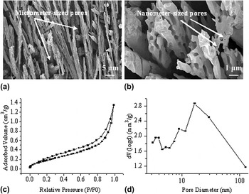
The porous structure on the surface of BCP scaffold (HAP/β-TCP 60/40) after etching is shown in Fig. 1. Many micrometer-sized needles were presented on the scaffold surface as the β-TCP in the outer layer dissolved in phosphoric acid [Fig. 1(a)]. The original locations of β-TCP were replaced by micro/nanometer-sized pores which were uniformly distributed over the scaffold surface [Fig. 1(b)]. The nitrogen adsorption/desorption isotherms in Fig. 1(c) exhibited a type II isotherm pattern with a type H3 hysteresis loop. The continuous accretion of nitrogen adsorption at the P/P 0 from 0.8 to 1.0 indicated the appearance of macroporosity. As can be seen from Fig. 1(d), there were well-defined nanometer-sized pores on the scaffold surface with a specific surface area of 1.38 m2/g. The nitrogen adsorption data indicated the hierarchical pore size distribution at 3–5 and 10–100 nm, as well as pore size above 100 nm. These findings suggested that there were both nanometer-sized pores and micrometer-sized pores on the scaffold surface after etching.
Characterization of porous surface structure after etching: (a) low magnification and (b) high magnification SEM micrographs; (c) nitrogen adsorption/desorption isotherms; (d) pore size distribution curve.

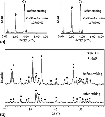
The surface compositions obtained by EDX and XRD are shown in Fig. 2. It can be seen in Fig. 2(a) that the surface of the scaffold was mainly composed of Ca and P. The Ca/P ratio before etching was calculated as 1.59 ± 0.03 while the Ca/P ratio after etching was 1.67 ± 0.02, indicating the micrometer-sized needles as HAP (stoichiometric value of 1.67). Reference Akazawa, Murata, Hino, Nagano, Shigyo, Nomura, Inano, Itabashi, Yamagishi, Nakamura, Takahashi, Iida and Kashiwazaki35 XRD diffractograms in Fig. 2(b) show a combination of the peaks for β-TCP [Joint Committee on Powder Diffraction Standards (JCPDS) 09-0169] and HAP (JCPDS 09-0432) on the scaffold surface before etching. Reference Figueiredo, Coimbra, Cabrita, Guerra and Figueiredo36 In contrast, the diffractogram after etching was dominated by the peaks of HAP with the absence of β-TCP. This further confirmed the complete dissolution of β-TCP from the surface of BCP scaffold.
(a) EDX and (b) XRD diffractograms on the surface of BCP scaffold before and after etching.

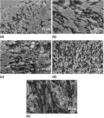
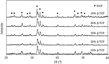
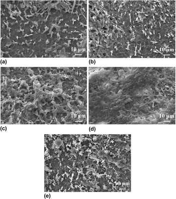
The evolution of surface structures of BCP scaffolds with different β-TCP content in the range of 10–50% after selective etching is shown in Fig. 3. The prepared surface structures changed regularly with the variation of β-TCP content. A few micrometer-sized pores between the needles formed by etching were found on the surface of the scaffold containing 10% β-TCP in Fig. 3(a). The number of surface pores increased with the β-TCP content in scaffold (20 or 30%) in Figs. 3(b) and 3(c). The micrometer-sized needles were uniformly distributed on the scaffold surface when the β-TCP content reached 40% in Fig. 3(d). The micrometer-sized needles were embedded in the outer layer of scaffold with an average size of 0.9 μm estimated by linear intercept method. On the surface of the scaffold containing 50% β-TCP, parts of needles were dissolved in phosphoric acid resulting in the decrease of micro/nanometer-sized pores in Fig. 3(e) when compared with the scaffold containing 40% β-TCP. XRD analysis was carried out on the etched surfaces of BCP scaffolds with different β-TCP content, as shown in Fig. 4. It can be seen that β-TCP were completely dissolved from the scaffold surfaces in all cases since no peaks of β-TCP were observed in all the diffractograms.
SEM micrographs of surface structures of BCP scaffolds with different β-TCP content after etching: (a) 10; (b) 20; (c) 30; (d) 40, and (e) 50%.

XRD diffractograms on the surface of BCP scaffold with different β-TCP content after etching.

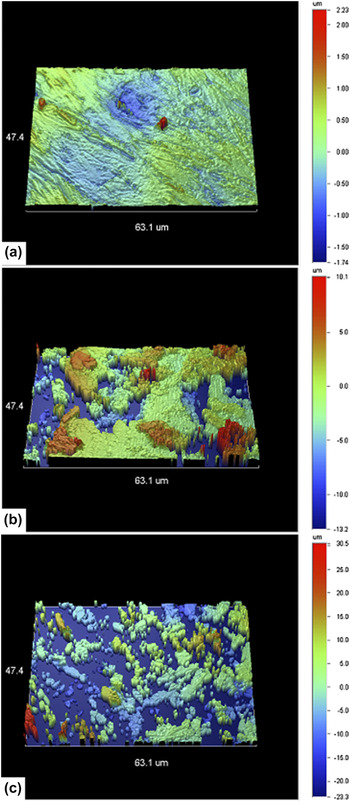
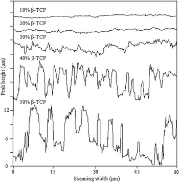
The three-dimensional (3D) surface profiles of scaffolds with different β-TCP content after etching are shown in Fig. 5. The micrometer-sized needles corresponding to HAP were randomly distributed on the surfaces of scaffolds. There was a significant difference between the surface profiles of scaffolds with different β-TCP content. The height distributions on surface structures were extracted and summarized for comparing using Vision® software, as shown in Fig. 6. The surface structures revealed periodic fluctuations in the needle height, which had an average value increasing with β-TCP content. The scaffold with 40% β-TCP had the largest number of micro/nanometer-sized needles on the surface with an average height of 7.53 ± 0.02 μm, compared with 0.59 ± 0.02 μm of 10% β-TCP, 1.31 ± 0.01 μm of 20% β-TCP, 3.76 ± 0.01 μm of 30% β-TCP, and 12.09 ± 0.03 μm of 50% β-TCP.
3D surface profiles of BCP scaffolds with different β-TCP content after etching: (a) 10; (b) 30, and (c) 50%.

Height distributions on various micro/nanometer-sized porous surface structures.

B. Cell morphology
The SEM observations of MG-63 cells on various surface structures after 7 days of culturing were presented in Fig. 7. The appearance of MG-63 cells on the surfaces varied with surface structure. A few MG-63 cells grew on the surface of the scaffold containing 10% β-TCP with underdeveloped filopodia extensions in Fig. 7(a). Besides, the cells exhibited a mixed morphology between spherical and spread at the initial adhesion stage, which was indicative of very thin extensions of cell spreading. Reference Bass, Belamie, Grosso, Boissiere, Coradin and Sanchez37,Reference Janshoff, Kunze, Michaelis, Heitmann, Reiss and Wegener38 The morphology of adherent cells on the surface of the scaffold containing 20% β-TCP was similar to that on the scaffold containing 10% β-TCP, while exhibited greater ruffling on the surface in Fig. 7(b). Cell fusion was also observed and cells were found to bridge the pores, leading to the cell invasion of multilayer micro/nanometer-sized porous surface structure. The grown cells were well spread and appeared to form a continuous monolayer on the surface of the scaffold containing 30% β-TCP in Fig. 7(c). This indicated that the cells had migrated into the pores and proliferated promptly. The highest cellular density was observed on the surface of the scaffold containing 40% β-TCP in Fig. 7(d). It appeared to form multiple layers on the whole scaffold surface. Numerous cells had extensions that covered distances in excess of 10 μm, implying vigorous cell metabolism. Abundant extracellular matrix formed and the cells displayed typical slabstone-like appearance. Reference Hu, Ding, Chen, Liu, Zhang, Ding and Wang39,Reference Leng, Ding, Zhang and Wang40 These phenomenon suggested excellent cell-adherence and proliferation ability of the surface structure. The scaffold containing 50% β-TCP showed a relatively slower cellular growth rate since cells were found to be randomly distributed on the edge of pores, as shown in Fig. 7(e). Thin and long extensions were elongated on the scaffold surface which was similar to the surface morphology of scaffold containing 20% β-TCP. On the basis of the above analysis, the surface structure of BCP scaffold containing 40% β-TCP was more favorable for the growth of MG-63 cells than the other four types of surface structure.
SEM micrographs of MG-63 cells cultured on the BCP scaffolds with different β-TCP content after etching: (a) 10; (b) 20; (c) 30; (d) 40, and (e) 50%.

C. Cell number
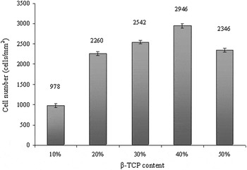
The number of adherent cells on each etched surface of BCP scaffolds after 7 days culturing is displayed in Fig. 8. The number was calculated as the sum of adherent cells based on analyses per unit area (1 mm2) of scaffold surface. The results indicated a close relationship between cell number and surface structure. The surface structure of scaffold containing 40% β-TCP showed high phenotypic expression of MG-63 cells with the largest number of cells on the scaffold surface, which were approximately 2.5 times that of the scaffold containing 10% β-TCP. The cell number of scaffold containing 50% β-TCP was close to that of scaffold containing 20% β-TCP, but less than that of scaffold containing 40% β-TCP. The decrease of cell attachment and proliferation might be attributed to the damage of micro/nanometer-sized porous surface structure caused by the dissolution of excessive β-TCP.
MG-63 cell number on the surface structures of the BCP scaffolds with different β-TCP content after etching.

The results demonstrated that the surface structure of scaffold was a key contributing factor for cell adhesion and proliferation. The MG-63 cells grew along the periphery of pores on the multilayer micro/nanometer-sized porous surface and tended to elongate in the direction toward the center of scaffold. The micro/nanometer-sized pores provided location for cell adhesion and migration and resulted in multilayer growth. However, excessive β-TCP in BCP scaffold led to a decrease of micro/nanometer-sized pores on the scaffold surface. The etched surface of the scaffold containing 40% β-TCP showed the optimal structure for MG-63 cell seeding and growth in virtue of the largest cell number and highest cellular density.
Recently, Houmard et al. Reference Houmard, Fu, Genet, Saiz and Tomsia41 fabricated HAP/β-TCP composite scaffolds by robocasting. Immersion tests in static simulated body fluid (SBF) and water showed no significant weight loss of the scaffolds after 5 months, while the dissolution of Ca and P for the 60HAP/40β-TCP scaffolds was ∼20 times higher than HAP. Daculsi et al. Reference Daculsi, Miramond, Borget and Baroth42 developed a smart calcium phosphate bioceramic scaffold for BTE. In vivo experiment indicated higher colonization by osteogenic cells on the scaffold with HAP/β-TCP ratio of 20/80. Arinzeh et al. Reference Arinzeh, Peter, Archambault, van den Bos, Gordon, Kraus, Smith and Kadiyala43 used porous HAP/β-TCP (65/35) ceramic as the carrier for mesenchymal stem cells (MSCs) to heal a critical-sized bone defect. The results indicated enhanced repair of bone defect and no adverse immune response was detected. Afterward, Arinzeh et al. Reference Arinzeha, Tranb, Mcalary and Daculsi44 investigated various compositions of HAP/β-TCP ceramic for bone regeneration using MSCs. And 20/80 HAP/β-TCP was found to have the greatest effect on bone formation. Kadiyala et al. Reference Kadiyala, Jaiswal and Bruder45 evaluated the ability of MSCs to heal a critical-sized bone defect after loading onto a porous HAP/β-TCP ceramic. The defects showed significant bone formation at 4 weeks and were completely healed by 8 weeks. Wang et al. Reference Wang, Zhao, Lin and Huang46 studied the osteoconductivity of bone marrow stromal cells (BMSCs) in polycaprolactone/calcium phosphate cement (PCL/CPC) composites. Results showed that a PCL/CPC mass ratio of 70/30 could allow BMSCs for active attachment, proliferation, and osteogenesis.
These studies mainly focused on the evaluation of various HAP/β-TCP ratios of BCP scaffolds for bone repair using different type of cells, which provided guidance for optimal selection of HAP/β-TCP ratio. Our study aimed to obtain various micro/nanometer-sized porous surface structures of BCP scaffolds and investigate the influence on biocompatibility using MG-63 cells, instead of evaluating various HAP/β-TCP ratios on the biocompatibility. BCP scaffolds were first fabricated by a previously developed SLS system. After immersion in phosphoric acid solution, β-TCP in the outer layer of BCP scaffolds were selectively etched off the scaffold surfaces, leaving micro/nanometer-sized pores and HAP on the surfaces. As confirmed by XRD and EDX results, the phase composition on the scaffold surface after etching was independent of the HAP/β-TCP ratios before etching due to the complete dissolution of β-TCP. As a result, different HAP/β-TCP ratios (90/10, 80/20, 70/30, 60/40, and 50/50) before etching resulted in the same phase composition on the scaffold surfaces after etching but different micro/nanometer-sized porous surface structures of BCP scaffolds, which showed significant impact for cell adhesion and proliferation.
IV. CONCLUSIONS
In this study, selective etching treatment was used to construct the multilayer stereo micro/nanometer-sized porous surface structure of bone scaffold. The original locations of β-TCP in the outer layer of scaffold were replaced by micro/nanometer-sized pores after selective etching. SEM analysis revealed various porous surface structures by adjusting the β-TCP content in the BCP scaffolds. The needle height on the surface obtained by optical profiler showed periodic fluctuations and increased from 0.59 ± 0.02 μm to 12.09 ± 0.03 μm when β-TCP content rose from 10 to 50%. The cell number and density gradually increased as more micro/nanometer-sized pores formed on the scaffold surface after etching. However, excessive β-TCP led to the damage of micro/nanometer-sized porous surface structure and impeded the cell interaction. The BCP scaffold containing 40% β-TCP was found to have an optimal porous surface structure for cell adherence and proliferation. These findings were expected to provide guidelines for the preparation of micro/nanometer-sized porous surface structure and further improve the biocompatibility of bone scaffold in vivo.
ACKNOWLEDGMENTS
This work was supported by the following funds: (1) The Natural Science Foundation of China (51222506, 81372366); (2) Hunan Provincial Natural Science Foundation of China(14JJ1006); (3) Program for New Century Excellent Talents in University (NCET-12-0544); (4) The Fundamental Research Funds for the Central Universities (2011JQ005, 2012QNZT015); (5) Project supported by the Fok Ying-Tong Education Foundation, China (131050); (6) Shenzhen Strategic Emerging Industrial Development Funds (JCYJ20130401160614372); (7) The Open-End Fund for the Valuable and Precision Instruments of Central South University; (8) The faculty research grant of Central South University (2013JSJJ011, 2013JSJJ046); (9) Hunan Provincial Innovation Foundation for Postgraduate.