Introduction
Identifying patients with increased risk for post-operative complications or lingering fluid overload at discharge following congenital heart surgery is a vexing challenge for providers. Reference Trivedi, Diaz-Castrillon and Morell1 While the most common cause for readmission following congenital heart surgery is unrelated to the procedure (e.g., infection, gastrointestinal complications), management of fluid status is seemingly one of the most modifiable factors to reduce risk of post-operative surgical complications. Reference Kogon, Jain, Oster, Woodall, Kanter and Kirshbom2 Atrial septal defect closure has long been shown to have an increased risk for post-pericardiotomy syndrome and pericardial effusions for unclear reasons. Reference Elias, Glatz and O’Connor3–Reference Timmis, Gordon and Ramos5 Children following Fontan completion have very high medical care utilisation rates in the post-operative period following discharge, including frequent emergency room evaluations and hospital readmission. Reference Cashen, Petersen and Rood6,Reference Kodnad, Tabaie, Rosenblum and Kamaleswaran7 Conditions tightly linked with fluid overload, that is, congestive heart failure and pleural effusion, are the most common reasons for re-admissions to critical care units following Fontan surgery. Reference Cashen, Petersen and Rood6
Lung ultrasound can identify and semi-quantify pulmonary oedema, as a marker of fluid overload or pulmonary inflammation, by the presence, number, and appearance of acoustic artefacts called “B-lines” within targeted lung fields. Reference Demi, Wolfram and Klersy8 Lung ultrasound is thus emerging, particularly in adult patients, as a non-invasive tool for providers during the evaluation of suspected fluid overload, decompensated heart failure, or increased risk for re-admission. Reference Demi, Wolfram and Klersy8–Reference Platz, Merz, Jhund, Vazir, Campbell and McMurray14 The Association for European Paediatric and Congenital Cardiology Imaging Working Group recently recommended that there is sufficient evidence in the published literature to support the systematic use of lung ultrasound for the diagnosis and evaluation of neonates and children with cardiac disease. Reference Cantinotti, di Salvo and Voges15–Reference Wu, Hou and Lu18 Lung ultrasound around congenital heart surgery seems to have the potential to contribute additive prognostic value beyond traditional risk factors to predict adverse outcomes with current paediatric literature mostly focused on pre-operative or immediate perioperative risk prognostication. Reference Cantinotti, Giordano and Scalese19–Reference Song, Kim, Lee, Kang, Kim and Kim22 The prognostic utility of lung ultrasound during discharge testing in adults with heart failure has been consistently demonstrated, Reference Rivas-Lasarte, Maestro and Fernandez-Martinez13,Reference Platz, Merz, Jhund, Vazir, Campbell and McMurray14 but there are scant publications about lung ultrasound and discharge risk stratification in paediatrics or CHD. Additionally, there is inconsistency in paediatric literature regarding who performs and interprets lung ultrasound, for example, radiology versus cardiology and clinician- versus technician-performed. Reference Cantinotti, di Salvo and Voges15
This paper presents a prospective study evaluating the prognostic power of lung ultrasound performed in a manner applicable to the workflow of a paediatric echocardiography laboratory to predict important post-discharge adverse clinical events in children following congenital heart surgery.
Materials and method
A prospective, single-blinded observational trial was performed at a high-volume paediatric heart centre from January 2023 to December 2023. Subjects aged 2 months to 17 years were consecutively identified by investigators from the daily cardiovascular surgery schedule. Young infants were not enrolled since researchers believed a substantively different enrolment and scanning process would be needed based on recent specific recommendations for lung ultrasound in neonates. Reference Demi, Wolfram and Klersy8,23,Reference Wolfram, Miller and Demi24 Enrolment inclusion criteria for type of surgery were confined to Fontan completion of single-ventricle palliation (including additional major cardiac surgery, e.g., branch pulmonary arterioplasty or atrial septectomy) and primary repair of septal defects, that is, atrial septal defect, ventricular septal defect, or atrioventricular septal defect repair (including surgery typically associated with septal defect repairs, such as atrioventricular valve repair or outflow tract resection). Subjects undergoing major cardiac surgery beyond primary septal defect repair were excluded, for example, repair of tetralogy of Fallot, valve replacement, semilunar valve repair, or aortic coarctation repair. Inclusion diagnoses were selected to provide a clinically relevant profile of post-operative risks following congenital heart surgery, Reference Kogon, Jain, Oster, Woodall, Kanter and Kirshbom2 but they were limited to ensure feasibility of consecutive enrolment within limits of available research staff. Non-surgical exclusion criteria were primary respiratory disorder, active lower tract respiratory infection, tracheostomy in place, chest tubes in place, prolonged post-operative hospitalisation >30 days, and hospital discharge >3 days after lung ultrasound.
Approval for the study design was obtained from the Institutional Review Board at Baylor College of Medicine, in accordance with ethical guidelines set forth for human subjects research. Subjects’ legal guardians provided consent for enrolment, and subjects older than 7 years of age provided assent. Consented subjects underwent lung ultrasound as they neared expected discharge following chest tube removals. Lung ultrasounds were performed by registered cardiac sonographers after undergoing a training curriculum conducted by the primary investigator (AFR). The curriculum consisted of a synchronous didactic followed by proctored scanning sessions until competency was achieved. To decrease heterogeneity of imaging technique and due to limited lung ultrasound safety guidance of lower frequency probes in children, lung ultrasounds were strictly performed with 12-MHz sector probes. Imaging was performed on standard echocardiography systems (GE E-95 and Philips Epic) within the 12 MHz probe frequency agility range (9 MHz to 12 MHz), and harmonics were turned off to promote visualisation of reverberation artefacts. For subjects enrolled after 3/2023, the mechanical index goal was set initially to ∼ 0.4–0.7 to be in line with new consensus recommendations. Reference Demi, Wolfram and Klersy8,23,Reference Wolfram, Miller and Demi24 Sonographers scanned bilateral superior anterior lung fields with a probe placed in the second or third rib space in the mid-clavicular line with the probe indicator towards the subject’s head. The focal point (as applicable) was set at the pleural line. Depth was adjusted to optimise the appearance of pleural line, A-lines, and B-lines. With the image centred on the rib space bracketed by rib shadows, two 4-second clips of each lung were stored. Posterolateral lung ultrasound or chest ultrasound evaluations for pleural effusion were not performed. De-identified images were coded with study numbers and were stored on the research ultrasound image database (GE Viewpoint). The primary investigator (AFR) reviewed de-identified lung ultrasounds and scored each lung in accordance with a previously published paediatric 4-point scale based on the number of B-lines (Table 1). Reference Cantinotti, Marchese and Giordano25 Subject lung scores were summed to create a total lung ultrasound score for the subject. Subjects’ summed scores were used for comparison to clinical outcomes. For inter-rater reliability analysis, the second investigator (TD) reviewed the first 24 subjects’ lung ultrasounds using the same 4-point scale for each lung after undergoing training consisting of asynchronous didactic on the scoring system.
Lung ultrasound scoring system

Lung ultrasounds were performed on standard echocardiography systems with harmonics off to promote visualisation of reverberation artefacts. 12-MHz sector probe (frequency agility range: 9 MHz to 12 MHz) was placed in the second or third rib space in the mid-clavicular line with probe indicator towards the subject’s head. With the image centred on the rib space bracketed by rib shadows, two 4-second clips of each lung were stored. Each lung scored based on the number of appearances of B-lines (adapted from Cantinotti et al, Diagnostics 2022). Reference Girona-Alarcon, Cuaresma-Gonzalez and Rodriguez-Fanjul20
Clinicians on discharging inpatient and post-discharge outpatient primary teams were blinded to lung ultrasound results. Enrolled subjects underwent standard outpatient follow-up and medical care per institutional standards and clinicians’ discretion. Typically, post-operative patients have an electrocardiogram, echocardiogram, and two-view chest radiography just prior to hospital discharge. Following discharge, post-operative patients generally see the congenital heart surgery team within 5–7 days after discharge for clinic visit and chest radiography and then follow-up with primary cardiologist within 1 month for clinic visit and an echocardiogram.
Subjects’ electronic medical records were reviewed to identify the primary outcome and other important perioperative clinical outcomes. The primary outcome was the composite subsequent development of new pericardial (small or greater) or pleural (moderate or greater) effusion following lung ultrasound. Primary outcomes were identified in formal reads of echocardiography or chest X-ray performed after hospital discharge and interpreted by hospital cardiology and radiology attending staff, respectively. Due to inconsistency of reporting and/or size quantification, echocardiography reports were not used to identify pleural effusions in this study. Subject demographics, surgical details, and clinical outcomes identified for comparison included gestational age at birth, pre-operative pulmonary artery pressure (pre-Fontan cardiac catheterisation), post-surgical chest tube duration, ventricular systolic function at hospital discharge echocardiography, atrioventricular valve regurgitation at hospital discharge echocardiography, discharge medications, and post-operative hospital length of stay. A diuretic medication score was devised with one point given for each mg/kg/day of furosemide and one-point for each 10 mg/kg/day of chlorothiazide.
Statistical analysis
Continuous variables were summarised as median (IQR) or mean (range) and compared using Wilcoxon rank-sum tests; categorical variables were compared using Fisher’s exact or chi-square tests. Associations between lung ultrasound scores and perioperative variables were assessed with non-parametric (Spearman’s) correlation and group comparisons. Odds ratios with 95% confidence intervals were calculated for the relationship between lung ultrasound score and the primary outcome. Receiver operating characteristic analysis was used to evaluate discriminatory ability, and a post hoc cut-off of ≥3 was selected as optimal. A p-value < 0.05 was considered significant. Analyses were performed in Stata (StataCorp, College Station, TX).
Results
From the daily cardiovascular surgery schedule, investigators consecutively identified 150 potential subjects between January 2023 and December 2023 (Figure 1). Consent was obtained, and lung ultrasound was attempted for 102 subjects. Movement and non-compliance prevented lung ultrasound imaging in two subjects. Lung ultrasound images were interpreted by the investigator for 96/100 studies; the image quality was too poor for interpretation in four subjects (ventricular septal defect – 1, atrial septal defect – 1, and Fontan – 2). These subjects were mostly older patients (ages: 14 years, 13 years, 11 years and 6 months). After lung ultrasound, an additional 10 subjects were excluded (updated excluded diagnosis – 1 and prolonged length of stay – 9) yielding 86 subjects who met complete criteria and had adequate imaging for enrolment and comparison to outcomes.
Subject recruitment consort diagram.

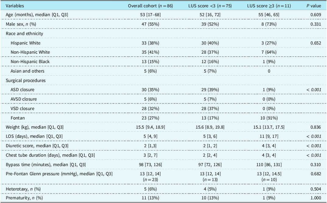
The median age for enrolled subjects was 53 months [IQR: 17–68] and median weight 15.5 kg [IQR: 9.4–18.9]. Primary surgical procedures included atrial septal defect repair (n = 30), ventricular septal defect repair (n = 28), atrioventricular septal defect repair (n = 5), and Fontan completion (n = 23) (Table 2). The majority of Fontan surgeries were non-fenestrated (19/23, 83%), and 14/23 (61%) had major additional cardiac surgery beyond Fontan completion. Subject median length of stay was 5 days [IQR: 4–9] with a median chest tube duration of 3 days [IQR: 2–7]. Discharge diuretic score median was 2 [IQR: 1–3] (Table 2). Clinical follow-up ranged from 6 weeks to 53 weeks.
Enrolled subject demographics and clinical outcomes

AVSD = atrioventricular septal defect; ASD = atrial septal defect; VSD = ventricular septal defect; LUS = lung ultrasound; LOS = length of stay.
Median time from enrolled subjects’ lung ultrasound to day of discharge was 1 day [IQR: 0–1]. Median mechanical index was 0.6 [IQR: 0.5–0.8]. Mean score was 0.9 (range 0–5), and inter-rater agreement on a subset of 24 subjects was excellent (k = 0.92, p < 0.001). Lung ultrasound scores were significantly higher in post-Fontan subjects versus septal defect closure cohorts (mean 1.9 versus 0.5, respectively, p < 0.001). Lung ultrasound score was correlated with hospital length of stay (ρ = 0.29, p = 0.0066), discharge diuretic score (ρ = 0.38, p < 0.001), and chest tube duration (ρ = 0.25, p = 0.021). There were no significant associations between lung ultrasound score and age (ρ = –0.17, p = 0.11), weight (ρ = –0.20, p = 0.07), bypass time (ρ = 0.15, p = 0.17), cross-clamp time (ρ = –0.02, p = 0.88), or pre-Fontan pulmonary artery mean pressure (ρ = 0.11, p = 0.61).
Primary outcome occurred in 12 subjects (atrial septal defect – 4 and Fontan – 8), including 7 readmissions to the hospital (atrial septal defect – 1 and Fontan – 6). Of the Fontan patients with primary outcome (all pleural effusions), 6/8 had lung ultrasound score ≥3. Two Fontan patients were readmitted for pleural effusion after prolonged admission with low discharge lung ultrasound score (lengths of stay 15 days, 28 days and lung ultrasound score 1,0, respectively). Four subjects (atrial septal defect – 4) developed post-discharge pericardial effusions. One required hospital readmission and had a discharge lung ultrasound score of 3; the others had a discharge lung ultrasound score of 0 and were managed as outpatient. Three subjects (ventricular septal defect – 3, all lung ultrasound score <3) had pericardial effusions at the time of lung ultrasound which did not prevent discharge. These were considered non-events since all had resolution or improvement at follow-up evaluation and did not develop subsequent additional effusions.
Post hoc analysis identified a lung ultrasound score cut-off of 3 that provided the best prognostic performance. Eleven subjects had a lung ultrasound score ≥3 (Fontan – 10, atrial septal defect – 1), and 7/11 had a primary outcome (64%). The odds ratio for the primary outcome for score ≥3 was 24.5 (5.3–113, p < 0.0001). Score <3 had a negative predictive value of 93%. For subjects with score <3, 5/75 had primary outcomes with 2 pleural effusions (Fontan – 2) and 3 small (outpatient) pericardial effusions (atrial septal defect – 3). In the post-Fontan cohort, the odds ratio for the primary outcome with score ≥3 was 8.3 (1.2–59.0, p < 0.036). Secondary analysis revealed lung ultrasound score was the best predictor of pericardial or pleural effusion large enough to require hospital readmission. For readmission, each point of lung ultrasound score higher is associated with an odds ratio of 2.23 (95% CI 1.32–3.76, p = 0.003) with an area under the receiver operator curve = 0.8192 (Figure 2).
Receiver operator curves for lung ultrasound score and clinical outcomes.

Discussion
Our study supports previous research in children and adults that lung ultrasound findings are associated with important clinical outcomes in post-operative cardiac patients, particularly within the high-risk, post-Fontan subject cohort. A novel finding from this research is that a reliably interpretable lung ultrasound can be obtained using a high-frequency phased-array cardiac transducer, ideally during the performance of a paediatric echocardiogram. Similar to previous paediatric research, children with worse lung ultrasound findings in our prospective study had longer hospital lengths of stay, prolonged chest tube output, and increased diuretic needs, suggesting validity in the ability of lung ultrasound to identify sicker patients and likely increased risk for worse outcomes. Reference Cantinotti, di Salvo and Voges15–Reference Ozdemir, Tepe and Ozlu17,Reference Cantinotti, Giordano and Scalese19–Reference Kaya, Dilli and Sarikaya21 These findings were particularly driven by post-Fontan patients having worse lung ultrasound scores. Of note, lung ultrasound scores ≥3 were exceedingly rare in subjects following simple defect closure corresponding with a low rate of post-operative complications and readmissions.
Importantly, our study results demonstrated lung ultrasound to have promising prognostic capability to predict post-operative effusions when performed on patients who were otherwise deemed suitable for discharge after congenital heart surgery. Although it was not the primary outcome, lung ultrasound was the best predictor of pericardial or pleural effusions which were large enough to lead to readmission. Although a post hoc finding, the lung ultrasound score cut-off of 3 is clinically intuitive, since in a bilateral process there needs to be at least one lung ultrasound score ≥2, which corresponds to “moderate B-lines, moderate congestion” per the Cantinotti scoring. Reference Cantinotti, Marchese and Giordano25 Unilateral lung ultrasound scores of 2 or 3 with ipsilateral score of 0 would more likely present focal pathology inconsistent with post-operative fluid overload. The predictive capability of lung ultrasound for post-operative effusions persisted within the high-risk, post-Fontan subject cohort, although with more imperfect characteristics. While it is important to remember that B-lines on lung ultrasound are inherently non-specific findings from a broad spectrum of parenchymal pathologies, Reference Martelius, Heldt and Lauerma26 scoring pulmonary oedema based on the number of B-lines in lung fields is less qualitative than assessments of pulmonary oedema derived from chest X-ray or physical examination. Thus, lung ultrasound scoring presents a more objective option to assess otherwise unsuspected risk for adverse events in the post-operative period after congenital heart surgery.
Lung ultrasound performed with cardiac probes and a focused anterior scanning protocol has practical applicability for modern paediatric echocardiography laboratories. Nearly, all subjects scanned had bilaterally interpretable images (96%, 96/100), and there were only two toddler-aged subjects whose non-compliance and movement prevented image acquisition. The near-perfect interrater reliability supports the reproducibility of the imaging strategy and the ability to include lung ultrasound in additional clinical investigations. Of note, the subjects with unacceptable imaging were some of the oldest subjects in the cohort. We suspect limiting ourselves to the 12-MHz probe may have prevented adequate imaging in some of these circumstances. Whether lower-frequency sector probes could have improved image quality for these larger subjects with deeper scanning depths could be a topic for additional research.
Our study may show the manner in which lung ultrasound can be performed prior to discharge and predict complications, but how lung ultrasound results can be incorporated into decision-making to reduce post-operative effusions after congenital heart surgery remains unclear. Even assuming lung ultrasound findings are due to pulmonary oedema and not another aetiologies, the decisions to increase diuretics, delay discharge, or arrange earlier outpatient clinic follow-up may not correct the underlying pathophysiology for pulmonary oedema and fluid overload. For example, the BLUSHED-AHF trial successfully targeted and decreased B-lines via goal-directed medical therapy in the first 48 hours of hospitalisation in adult acute heart failure patients, but it did not improve length of stay or 30-day mortality. Reference Pang, Russell and Ehrman27 At this stage of understanding, lung ultrasound findings with moderate congestion in a lung segment (more than approximately 5 and/or multiple coalescing B-lines per rib space) at discharge following congenital heart surgery can be considered a quantifiable method to prompt comprehensive clinical re-evaluation of discharge suitability and outpatient follow-up plans. But, more specific goal-directed medical therapies based on lung ultrasound findings in children could be a topic of future research. Our study suggests low lung ultrasound scores may confer lower complications, but it should not provide false reassurances resulting in the relaxing of post-discharge follow-up diligence. Particularly, our study does not contribute clarification to the mystery of increased risks of pericardial effusions or post-pericardiotomy syndrome following atrial septal defect surgery, which could be due to other post-operative factors aside from fluid overload. Reference Indrebo, Berg and Holmstrom28,Reference Lehto and Kiviniemi29 Lung ultrasound score in our research did not predict small outpatient pericardial effusions, but it did seem to predict the one readmission in the series following atrial septal defect repair for post-pericardiotomy syndrome.
In our series, there was no correlation between lung ultrasound score and subject weight or age. This contrasts with prior research which, importantly, enrolled infant children as well. Reference Kaskinen, Martelius, Kirjavainen, Rautiainen, Andersson and Pitkanen16,Reference Cantinotti, Giordano and Scalese19 Our study did not enrol young infants <2 months of age, in part due to our surgical criteria and, in part, due to potential safety or diagnostic accuracy implications of lung ultrasound in neonates. While the clinical implications are debated, a unique susceptibility of lung tissue to develop pulmonary capillary haemorrhage after exposure to certain types of ultrasound waves has long been described in neonatal animal models during controlled laboratory experiments dating back to the 1990s. Reference Child, Hartman, Schery and Carstensen30,Reference Miller31 Guidelines statements discuss that this may result in confounding, falsely positive B-lines in smaller patients. Reference Demi, Wolfram and Klersy8 To date, there are no reported instances of clinically significant pulmonary capillary haemorrhage occurring in humans exposed to medical ultrasound in clinical settings. Reference Demi, Wolfram and Klersy8,Reference Jagla, Krzeczek, Buczynska, Zakrzewska and Kwinta32,Reference Meltzer, Adsumelli and Risher33 To minimise the risk of this confounder or adverse effect, the American Institute for Ultrasound in Medicine, as well as two recent guideline statements from 2023, Reference Demi, Wolfram and Klersy8,Reference Wolfram, Miller and Demi24 recommend adjusting the mechanical index to 0.4–0.7 in adults/children and to <0.4 in neonatal lung ultrasound.
Cantinotti et al. seemed to have set the current standard for paediatric cardiology regarding how to grade individual ribs spaces, Reference Cantinotti, di Salvo and Voges15,Reference Cantinotti, Marchese and Giordano25 but there is a lack of consensus in the literature related to cumulative scoring and for how many lung segments should be scanned. For practicality purposes, our protocol included two superior–anterior scanning regions to reduce scan time and to generally ease coordination with typical paediatric echocardiography imaging protocols. Our lung ultrasound protocol diagnostic target was residual fluid overload or post-operative inflammation, which are more generalised, systemic clinical findings. Diagnostic targets with specifically localised lung pathologies such as pneumonia, pneumothorax, or atelectasis would require more extensive protocols, including more anterior, lateral, and posterior lung fields. Comparison of the test characteristics and practicalities of more lung regions or anterior versus posterolateral lung ultrasound scanning areas in the post-operative period is potentially interesting future research.
Limitations
Our observational study necessitated broad heterogeneity of diagnoses to complete enrolment within a reasonable time frame at a single, congenital heart centre. An inherent weakness to our observational study design is that we did not standardise patient management decisions or perform uniform, active detection of post-operative complications beyond chart review. The increased variability of diuretic management and prolonged hospitalisations of the post-Fontan patients likely increased the confounding impacts of medical decisions and impacted the lung ultrasound predictability. We did not include comparative chest radiography in our study to avoid potential biases, particularly without a gold standard comparison, for example, chest CT. We did not have the resources to perform pre-operative lung ultrasound or include an independent marker of fluid status at the time of discharge, for example, brain natriuretic peptide. Therefore, it is unclear if lung ultrasound findings correlated with pre-existing lung pathology (e.g., pulmonary oedema and pulmonary fibrosis), post-operative fluid overload, inflammation, or other causes of increased B-lines. Our study was underpowered for comparison of lung ultrasound scoring before and after the change in limitations of mechanical index, but this alteration in protocol may have influenced the overall results of the study. Sonographer blinding was incomplete since they were able to ascertain cardiac diagnoses during standard echocardiography, usually obtained prior to lung ultrasound; physician interpretation was blinded from the clinical data.
Conclusion
This single-centre, prospective observational trial provides additional evidence that use of lung ultrasound in paediatric heart disease is reliable and associated with important post-operative complications after congenital heart surgery. This study potentially supports a role for lung ultrasound amongst other discharge testings to identify unsuspected risk for post-operative effusions and re-admission in patients otherwise deemed suitable for discharge after congenital heart surgery. Further, better-controlled research will be needed to determine how lung ultrasound results can inform diuretic, follow-up visit, or discharge management decisions.
Acknowledgements
None.
Financial support
This research received no specific grant from any funding agency, commercial, or not-for-profit sectors.
Competing interests
None.
Ethical standards
The authors assert that all procedures contributing to this work comply with the ethical standards of the relevant national guidelines on human experimentation and have been approved by the institutional committees of Baylor College of Medicine.