Introduction
Congenital cholesteatoma classically presents as a pearly, whitish mass behind a normal, intact tympanic membrane, generally in the anterosuperior mesotympanum.Reference Bennett, Warren, Jackson and Kaylie1 In contrast to the far more prevalent acquired cholesteatoma, patients with congenital cholesteatoma by definition lack a history of previous otological disease. Perforation, trauma, surgery, otorrhoea or repeated infections have to be excluded through careful history taking. This can pose a challenge, as the disease may eventually cause rupture of the tympanic membrane with otorrhoea and apparent infection. Although the acquired form is far more predominant, several authors have claimed an increase in the incidence of congenital cholesteatoma in recent years.Reference Potsic, Korman, Samadi and Wetmore2–Reference Kazahaya and Potsic4 The existing theories of congenital cholesteatoma do not presuppose a heritable mechanism. We describe a case of male siblings with congenital cholesteatoma, both of whom were diagnosed prior to three years of age.
Case report
A 34-month-old boy was referred to paediatric otolaryngology following several months of bloody, left-sided otorrhoea refractory to oral and ototopical antibiotics. At the time of referral, treatment had been ongoing for approximately six months without significant or sustained improvement. Prior to the onset of otorrhoea, the patient had a single episode of otitis media, on the contralateral side, at age five months. This was described as a ‘TM [tympanic membrane] pink/red with fluid/poor landmarks’ and had resolved on interval examination. All subsequent ear examination were described as normal until the onset of left-sided otorrhoea at age 28 months.
An initial course of topical and oral systemic antibiotics prescribed by the paediatrician failed to control the symptoms and the patient was referred to an otolaryngologist. After several additional courses of ototopical antibiotic drops, a computed tomography (CT) scan was obtained, which was consistent with an extensive left-sided cholesteatoma. Referral to a tertiary care centre was then made.
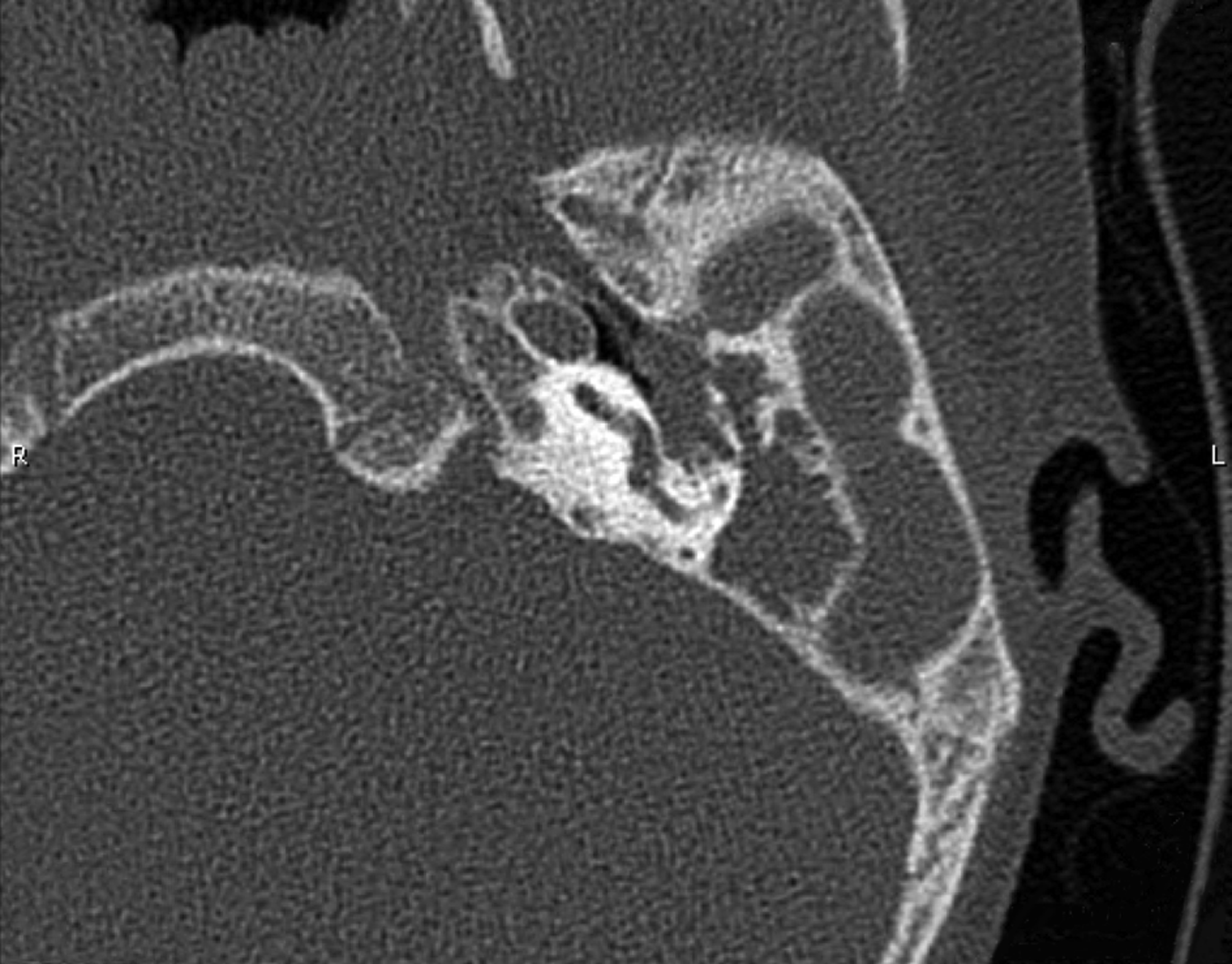
Physical examination demonstrated a small area of granulation tissue in the left external auditory canal, originating from the anterior tympanic membrane. The tympanic membrane was largely intact with the exception of the small area of granulation; the entire middle ear was filled with a white mass. Examination of the right tympanic membrane and middle ear was within normal limits. A moderate, left-sided conductive hearing loss was present. The CT scan was reviewed and revealed extensive cholesteatoma with ossicular erosion and destruction of the mastoid air cell system (Figure 1).
Non-contrast axial CT scan of the left temporal bone. CT = computed tomography

The extent of the disease at a young age, the limited history of otitis media and the absence of a retraction pocket with an essentially intact tympanic membrane suggested congenital cholesteatoma.
The patient was brought to the operating theatre for surgical management. Cholesteatoma was encountered just deep to the mastoid cortex, and was found to be filling the entire middle ear and mastoid process, with extensive destruction of the mastoid air cell system. The fallopian canal and otic capsule bone remained intact.
The family history was notable for an older brother who had been diagnosed with congenital cholesteatoma at age two years and who had been treated uneventfully with canal wall up mastoidectomy. The surgery report described a well-encapsulated cholesteatoma cyst within the anterosuperior mesotympanum with an intact tympanic membrane. No other family members, including a younger male sibling, have any history or current evidence of ear disease.
Discussion
This case joins a single, previous report by Lipkin et al.Reference Lipkin, Coker and Jenkins5 which described congenital cholesteatoma in multiple family members, suggesting that, in rare cases, genetic predisposition may play a role in the formation of congenital cholesteatoma.
A key problem in the study of this condition is the identification of a representative patient population. The condition is rare and diagnosis is challenging; with progression of the disease, perforation of the eardrum may occur from medial to lateral, making the distinction between acquired and congenital lesions difficult. Conversely, early diagnosis requires astute observation by the otoscopist, as a white lesion behind an intact tympanic membrane can easily be overlooked.
• We describe congenital cholesteatoma in two siblings
• Although several different theories exist, the exact aetiology of congenital cholesteatoma remains unclear
• The present case report suggests that, in rare cases, genetic predisposition may play a role in the formation of the disease
What all theories concerning the aetiology of this condition have in common is that, since the tympanic membrane remains intact, an explanation other than retraction and encapsulation of the tympanic membrane epithelium is needed. The most widely accepted theories at this time are the epithelial rest theory described by Teed, and the epidermoid formation theory described by Michaels, who suggested that fetal epidermoid formations persist and eventually expand to produce the pathogenic lesion.Reference Persaud, Hajioff, Trinidade, Khemani, Bhattacharyya and Papadimitriou6–Reference Michaels8 Other interpretations include the epithelial migration theory by Aimi,Reference Aimi9 with its focus on the absence of the tympanic annulus (normally forming a barrier between the external and middle ear during fetal development) as a pathway for the invasion of the epithelium. Northrop et al. advocated the amniotic fluid contamination theory, which suggested that epidermal cells in the amniotic fluid are introduced through the eustachian tube into the middle ear, where they lodge and proliferate.Reference Northrop, Piza and Eavey10 The ‘acquired’ inclusion theory by Tos doubted the congenital pathogenesis and suggested that, through a retraction, several cells of the keratinised squamous epithelium of the tympanic membrane may be left behind on the ossicles and are the source of disease.Reference Tos11
While certainly a rare occurrence, this case of siblings with likely congenital cholesteatoma does suggest that heredity may play a role. Fastidious documentation of ear examinations, including onset, duration and laterality of findings, can help to distinguish cases of acquired cholesteatoma from congenital cholesteatoma that has caused perforation. With more accurate identification of true congenital cases, future studies to elucidate the pathogenesis of this disease may be enhanced.