Implications
Increased intestinal permeability (IP) can lead to health disorders and reduced feed efficiency in chickens. We aimed to develop a model of increased IP by administering gram-negative bacterial toxin lipopolysaccharide (LPS) to chickens. This model could then be applied in an attempt to develop a new lactulose, mannitol and rhamnose (LMR) sugar permeability method to determine IP. An established model of increased IP and novel biomarkers to monitor IP changes could be applied to the study of nutritional interventions aimed at improving the health and welfare of chickens. However, further research is required to determine the optimal LPS dose.
Introduction
Increased IP results when enterocytes linked by tight junctions (TJs) fail to prevent microbes or toxins entering the systemic circulation (Li et al., Reference Li, Zhang, Chen, Yang, Zhang, Lu, Zhou and Wang2015). Human studies of increased IP have been associated with enteric infections, inflammatory bowel disease, obesity, type-1 diabetes, liver diseases, irritable bowel syndrome and celiac disease (Skouroliakou et al., Reference Skouroliakou, Ntountaniotis, Kastanidou and Massara2016). Limited in vivo research has shown that increased IP can lead to increased bacterial translocation (Chen et al., Reference Chen, Tellez, Richards and Escobar2015), coccidiosis (Chapman, Reference Chapman2014) and lameness (Wideman et al., Reference Wideman, Hamal, Stark, Blankenship, Lester, Mitchell, Lorenzoni and Pevzner2012) in chickens. These diseases can impact health and welfare leading to reduced efficiency and economic loss (Zuidhof et al., Reference Zuidhof, Schneider, Carney, Korver and Robinson2014).
Increased IP models are well established in humans (Ni et al., Reference Ni, Chen, Zhu, Shi, Shen, Gong, Gao, Yu, Li and Li2015; Piton et al., Reference Piton, Cypriani, Regnard, Patry, Puyraveau and Capellier2015), pigs (Zhu et al., Reference Zhu, Wu, Jiang, Zheng, Wang, Yang, Ma, Gao and Hu2015), rats and mice (Williams et al., Reference Williams, Duckworth, Watson, Frey, Miguel, Burkitt, Sutton, Hughes, Hall, Caamano, Campbell and Pritchard2013; Ruan et al., Reference Ruan, Liu, Zhou, Mi, Liu, Wu, Yao, Assaad, Deng, Hou, Wu and Yin2014). Dextran sodium sulphate (DSS) ingestion, fasting (Kuttappan et al., Reference Kuttappan, Berghman, Vicuna, Latorre, Menconi, Wolchok, Wolfenden, Faulkner, Tellez, Hargis and Bielke2015; Vicuna et al., Reference Vicuna, Kuttappan, Tellez, Hernandez-Velasco, Seeber-Galarza, Latorre, Faulkner, Wolfenden, Hargis and Bielke2015b), dexamethasone (Vicuna et al., Reference Vicuna, Kuttappan, Galarza-Seeber, Latorre, Faulkner, Hargis, Tellez and Bielke2015a) and wire floor stress (Wideman et al., Reference Wideman, Hamal, Stark, Blankenship, Lester, Mitchell, Lorenzoni and Pevzner2012) have recently been employed as models of increased IP in chickens. These studies have been conducted in one strain of chickens (Cobb). However, it has been determined that the IP of different strains is affected differently by DSS ingestion (Perše and Cerar, Reference Perše and Cerar2012). Lipopolysaccharide, found in the outer coat of gram-negative bacteria (Tran, Reference Tran and Whitfield2009), has been well established as a model to increase IP in mice (Williams et al., Reference Williams, Duckworth, Watson, Frey, Miguel, Burkitt, Sutton, Hughes, Hall, Caamano, Campbell and Pritchard2013), rats (Ruan et al., Reference Ruan, Liu, Zhou, Mi, Liu, Wu, Yao, Assaad, Deng, Hou, Wu and Yin2014) and pigs (Zhu et al., Reference Zhu, Wu, Jiang, Zheng, Wang, Yang, Ma, Gao and Hu2015). Although LPS has been used to induce systemic inflammation in chickens (Hu et al., Reference Hu, Guo, Li, Yan, Bun and Huang2011; Tan et al., Reference Tan, Liu, Guo, Applegate and Eicher2014), limited research has been conducted regarding its effect on IP as reviewed by Gilani et al. (Reference Gilani, Howarth, Kitessa, Forder, Tran and Hughes2016). Lipopolysaccharide, at doses of 0.25 to 1 mg/kg BW, has been used to increase IP in chickens (Wu et al., Reference Wu, Zhou, Wu, Zhang and Wang2013; Li et al., Reference Li, Zhang, Chen, Yang, Zhang, Lu, Zhou and Wang2015).
In addition, there is limited literature of in vivo biomarkers that could potentially be utilized to evaluate IP changes in chickens (Gilani et al., Reference Gilani, Howarth, Kitessa, Forder, Tran and Hughes2016). Lactulose, rhamnose and mannitol sugars have been validated in rats (Ruan et al., Reference Ruan, Liu, Zhou, Mi, Liu, Wu, Yao, Assaad, Deng, Hou, Wu and Yin2014), pigs (Wijtten et al., Reference Wijtten, Verstijnen, van Kempen, Perdok, Gort and Verstegen2011) and humans (Cox et al., Reference Cox, Lewis and Cooper1999; van Wijck et al., Reference van Wijck, Verlinden, van Eijk, Dekker, Buurman, Dejong and Lenaerts2013; Tran et al., Reference Tran, Hawkes, Graham, Kitchen, Symonds, Davidson and Butler2015), and could be applied to chickens. Lactulose, a high molecular weight (342 Da) sugar, traverses the epithelium paracellular, through the TJs of enterocytes in small quantities. Rhamnose and mannitol are lower molecular weight (182 Da) sugars which are transcellular absorbed directly into the blood. Increased lactulose to rhamnose (van Wijck et al., Reference van Wijck, Verlinden, van Eijk, Dekker, Buurman, Dejong and Lenaerts2013; Tran et al., Reference Tran, Hawkes, Graham, Kitchen, Symonds, Davidson and Butler2015) or L/M ratio (Araujo et al., Reference Araujo, Yunta, Terre, Mereu, Ipharraguerre and Bach2015; Shaikh et al., Reference Shaikh, Rajan, Forsyth, Voigt and Keshavarzian2015) due to increased permeability of lactulose and reduced absorbance of rhamnose or mannitol (due to damaged surface area for absorption) represents a biomarker of increased IP.
Recently, diamine oxidase (DAO), d-lactate (Wu et al., Reference Wu, Zhou, Wu, Zhang and Wang2013; Li et al., Reference Li, Zhang, Chen, Yang, Zhang, Lu, Zhou and Wang2015) and fluorescein isothiocyanate dextran (FITC-d) (Vicuna et al., Reference Vicuna, Kuttappan, Tellez, Hernandez-Velasco, Seeber-Galarza, Latorre, Faulkner, Wolfenden, Hargis and Bielke2015b) concentrations in blood have been utilized as in vivo biomarkers of increased IP in chickens. In addition, increased concentrations of zonula occludens (ZO-1) have been observed in celiac disease, diabetic and insulin-resistant human patients (Klaus et al., Reference Klaus, Motal, Burger-Klepp, Marschalek, Schmidt, Lebherz-Eichinger, Krenn and Roth2013). The aims of the current study were to evaluate whether the LMR sugar test could be used to evaluate changes in permeability following LPS administration at doses of 0.5 mg/kg BW (with fasting) and 1 mg/kg BW (with and without fasting) in chickens. The second aim was to compare the LMR sugar test with blood concentrations of FITC-d, DAO, d-lactate and ZO-1 as potential biomarkers of increased IP.
Material and methods
Animal management
Day-old male Ross 308 broiler chicks were obtained from a local hatchery (Baiada, Gawler, Australia) and raised on indoor floor pens on wood shavings for 14 days. Birds were fed a commercially available broiler feed (Ridley Agri Products Pty., Murray Bridge, SA, Australia). All birds were provided with ad libitum feed and water with 16 h day and 8 h night cycle. The birds were randomly allocated to two groups, control and LPS treatment. On day 14, birds were allocated in pairs to metabolic cages for 2 days. After 2 days acclimatization, birds were weighed and administered either LPS (Escherichia coli 055:B5; Sigma-Aldrich, Sydney, Australia) or sterile saline intraperitoneally at 16, 18 and 20 days of age.
Experimental protocol
All protocols were approved by the Animal Ethics Committees of the University of Adelaide and the Primary Industry Regions South Australia. All animal studies were performed in compliance with the Australian Code of Practice for the Care and Use of Animals for Scientific Purposes.
Oral solutions and gavages
Birds were orally gavaged via a feeding tube (round tip at the end) fitted to a disposable syringe. The tube was carefully inserted ~6 to 8 cm into the oesophagus. Lactulose, mannitol and rhamnose sugar solution was prepared with 25 g lactulose, 5 g mannitol and 5 g rhamnose (Sigma-Aldrich) dissolved in 100 ml milli-Q water (MilliPore, Molsheim, France; 18.2 MΩ cm at 25°C). Fluorescein isothiocyanate dextran (4000 Da molecular weight; Sigma-Aldrich) was dissolved in milli-Q water (2.2 mg FITC-d in 1 ml).
Experiment 1
In the first experiment, 36 birds were distributed into control and LPS groups and were intraperitoneally administered either LPS or saline at 0.5 mg/kg BW on days 16, 18 and 20. Birds in each group were distributed further into sub-groups according to their blood collection time points of 0, 30, 60, 90, 120, 180 min (n=3 per time point). Following their final injection, birds in both groups were fasted for 19.5 h (overnight). The next day, (day 21) all groups except time point 0 were orally gavaged with LMR sugar solution (2 ml/bird) and blood collected from the brachial vein.
Experiment 2
In the second experiment (n=36), similar procedures to the first experiment were followed except for the LPS dose and time points. The dose of LPS in this experiment was increased to 1 mg/kg BW and birds in the control and LPS groups were distributed only into three time points of 60, 90 and 120 min (n=6 per time point). Oral gavage and blood collection procedures were as for the first experiment.
Experiment 3
In the final experiment, 32 birds were randomly allocated into control and LPS groups and were intraperitoneally administered 1 mg/kg BW LPS or sterile saline. Only one time point (90 min) was used (n=16). Contrary to the first two experiments, birds were not fasted. In addition, birds were gavaged twice, first with FITC-d at time 0 min (2.2 mg/ml per bird) and second with LMR sugar at 60 min (2 ml/bird). Blood was collected from the brachial vein at 90 min after LMR gavage, and then allocated to serum and plasma vacutainers (BD, Sydney, NSW, Australia).
Processing blood samples
After collection, blood samples for plasma and serum were placed on ice and at room temperature, respectively, and centrifuged at 3000 r.p.m. for 10 min. The supernatant was collected and plasma and serum were stored in 0.5 aliquots at −20°C until further analysis.
High-performance ionic chromatography analysis
Lactulose, mannitol and rhamnose standards were prepared using fasted chicken plasma (sourced from a local slaughter house), and spiked to the following concentrations 125, 62.5 and 31.25 µM and stored at −20°C until further analysis. After thawing plasma and samples, 15% tri-chloroacetic acid (TCA; Sigma-Aldrich) was added, centrifuged at 13 000 r.p.m. for 10 min and the supernatant mixed with an equal volume of mixed-bed ion exchange matrix amberlite MB-1 resin (Sigma-Aldrich) for 30 min. The supernatant was transferred to Thomson single stepTM 0.2 µm nylon filter vials (Adelab Pty Ltd, Adelaide, SA, Australia) and analysed using the Dionex ICS-4000 (Thermo Fisher Pty Ltd, Scoresby, Victoria, Australia) high-performance ionic chromatography (HPIC) system as described by Tran et al. (Reference Tran, Hawkes, Graham, Kitchen, Symonds, Davidson and Butler2015).
Fluorescein isothiocyanate dextran, diamine oxidase, d-lactate and zonula occludens tight junction proteins in blood
Fluorescein isothiocyanate dextran analysis was conducted as previously described (Vicuna et al., Reference Vicuna, Kuttappan, Tellez, Hernandez-Velasco, Seeber-Galarza, Latorre, Faulkner, Wolfenden, Hargis and Bielke2015b). In brief, standards (0, 0.0001, 0.001, 0.01, 0.1, 1.0 and 10 µg/ml) were prepared using the same FITC-d sugars as for oral gavage. Standards and serum samples were run in triplicate and concentration of FITC-d in serum was calculated by a standard curve and a Synergy MX plate reader (BioTek Instruments, Bedfordshire, UK) at excitation and emission wavelengths of 485 and 530 nm, respectively. ELISA kits for DAO, d-lactate and TJ proteins ZO-1 were obtained from MyBioSource, Inc. (San Diego, California, USA). All standards and plasma samples were measured in duplicate in a microplate reader at 450 nm wavelength (Bio-Rad Laboratories Inc., California, USA) following the manufacturer’s instructions.
Statistical analysis
All statistical analyses were conducted using SPSS 22 (IBM SPSS; IBM Corp., Armonk, New York, USA). BW, concentrations of individual sugars (LMR) (µmol/l) and ratios (L/R and L/M), FITC-d, DAO, d-lactate and TJs were compared using one-way ANOVA. Interaction between LPS dose and time points were measured by two-way ANOVA. Data from all experiments were normally distributed and significance (P) was measured at 0.05.
Results
BW
The BW increment of the LPS-treated group were significantly lower than control during days 16 to 18 for all three experiments and during 18 to 20 days in the first experiment only (Table 1). In addition, at day 16, BW of control and LPS-treated birds were not different significant in all three experiments (data not shown).
Average BW increment±SEM of control and lipopolysaccharide (LPS)-treated chickens during 16 to 18 and 18 to 20 days of age

a,bDifferent superscripts show the significance level P<0.05.
Lactulose, rhamnose and mannitol concentrations and their ratios
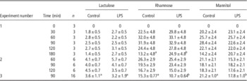
Lactulose, rhamnose and mannitol sugar concentrations in blood (µmol/l) for control and LPS treatment groups were not significantly different (Table 2). However, rhamnose concentration was significantly higher at 180 min for the LPS-treated group in the first experiment compared with the control group. Similarly, rhamnose and mannitol values were significantly lower in the third experiment in LPS-treated group compared with the control group.
Chicken plasma values of lactulose, rhamnose and mannitol (µmol/l) (mean±SEM) in three experiments

LPS, lipopolysaccharide.
a,bAll values are P>0.05 except where stated as different superscripts.
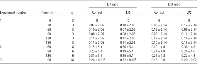
The ratios of L/R and L/M were not statistically different between control and LPS treatment in experiments 1 and 2 (Table 3). However, the L/R ratio in the third experiment was significantly higher in the LPS-treated group compared with control due to significantly lower concentration of rhamnose in the LPS-treated group. Although mannitol concentration was also significantly lower in the third experiment, it did not change the L/M ratio.
Chicken plasma lactulose to rhamnose (L/R) and mannitol (L/M) ratios (mean±SEM)

LPS, lipopolysaccharide.
a,bAll values are P>0.05 except where stated with different superscripts.
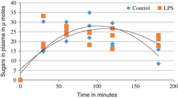
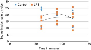
Lactulose, rhamnose and mannitol levels peaked at 90 to 120 min in the blood post oral gavage in the first experiment (0, 30, 60, 90, 120 and 180 min) as shown in Figure 1. This was repeated in the second experiment, with only three time points of 60, 90 and 120 min, as shown in Figure 2, confirming the trend that the optimal time point for collecting sugars was 90 min post oral gavage.
Lactulose, rhamnose and mannitol sugars in chicken plasma at 0, 30, 60, 90, 120 and 180 min (n=3 per time point) post oral gavage. LPS, lipopolysaccharide.

Lactulose, rhamnose and mannitol sugars in chicken plasma at 60, 90 and 120 min (n=6 per time point) post oral gavage. LPS, lipopolysaccharide.

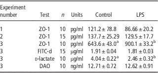
Zonula occludens concentration for LPS-treated chickens was not significantly different than control in the first two experiments. However, ZO-1 concentration in the third experiment was significantly higher for LPS-treated chickens (Table 4). Diamine oxidase, d-lactate and FITC-d concentrations were measured in the third experiment only. d-lactate values decreased significantly due to the LPS treatment. Fluorescein isothiocyanate dextran and DAO were not significantly different for LPS treatment compared with control (Table 4).
Zonula occludens, diamine oxidase (DAO), d-lactate and fluorescein isothiocyanate dextran (FITC-d) (mean±SEM) of control and lipopolysaccharide (LPS) treated chickens

a,bAll values are P>0.05 except where stated as different superscripts.
Discussion
This is the first study to utilize the lactulose, rhamnose and mannitol sugar test to evaluate IP changes following LPS challenge in chickens. The concentrations of LMR (and their ratios) in the first two experiments did not indicate any IP changes following LPS treatment. The interaction between the LPS dose and time points for individual sugars was not statistically significant. In addition, birds in these experiments were fasted for 19.5 h and recently it has been shown that 24 h fasting increased IP in chickens (Kuttappan et al., Reference Kuttappan, Berghman, Vicuna, Latorre, Menconi, Wolchok, Wolfenden, Faulkner, Tellez, Hargis and Bielke2015; Vicuna et al., Reference Vicuna, Kuttappan, Tellez, Hernandez-Velasco, Seeber-Galarza, Latorre, Faulkner, Wolfenden, Hargis and Bielke2015b). To exclude the possible interference of fasting with LPS, a third experiment at 1 mg/kg BW without fasting was conducted. The L/R ratio for the LPS treatment was significantly increased compared with control, unlike the L/M ratio. Araujo et al. (Reference Araujo, Yunta, Terre, Mereu, Ipharraguerre and Bach2015) and Shaikh et al. (Reference Shaikh, Rajan, Forsyth, Voigt and Keshavarzian2015) argued that L/M and L/R ratios should be considered along with the plasma concentrations of the individual sugars to better understand the sugar permeability test. The increased L/R ratio in the current study was due to the decreased rhamnose concentration, rather than increased lactulose permeation. In addition, rhamnose and mannitol are of similar molecular size, suggesting that both sugars and their ratios should behave similarly with respect to permeation (Wang et al., Reference Wang, Llorente, Hartmann, Yang, Chen and Schnabl2015). However, in no other studies have both sugars been studied together. In summary, the unaffected lactulose concentrations in the current study suggested that fasting did not obscure LPS effects; and LPS at doses of 0.5 and 1 mg/kg BW did not provide a reliable method to increase IP in chickens. Before gavage with sugars, blood plasma did not contain lactulose, rhamnose or mannitol (Figure 1), indicating no endogenous sugars in the blood. The plasma concentrations showed peaks for mannitol and rhamnose at 90 min and a peak concentration for lactulose at 120 min. In other time-course experiments these sugars peaked at 120 min in dogs (Sørensen et al., Reference Sørensen, Proud, Rutgers, Markwell, Adam and Batt1997) and in humans (Cox et al., Reference Cox, Lewis and Cooper1999), comparable with the current lactulose results, but not for rhamnose and mannitol.
In the current study, BW decreased after the first LPS injection in all three trials consistent with previous poultry studies (Hu et al., Reference Hu, Guo, Li, Yan, Bun and Huang2011; Tan et al., Reference Tan, Liu, Guo, Applegate and Eicher2014). Although feed intake was not measured in these trials, others have shown that LPS induced a systemic inflammation and reduced feed intake (Wu et al., Reference Wu, Zhou, Wu, Zhang and Wang2013). This may have been the reason for the reduced BW observed in the current study. However, BW did not reduce after the second LPS injection, consistent with other studies (Shini et al., Reference Shini, Kaiser, Shini and Bryden2008; Wu et al., Reference Wu, Zhou, Wu, Zhang and Wang2013). It is possible that chickens in the LPS treatment group may have overcome their initial shock resulting in similar growth between both groups. This may also explain why the sugar permeability test did not show statistical difference between control and LPS treatment.
Diamine oxidase is an endo-cellular enzyme of 250 kDa (Sun et al., Reference Sun, Gao, Li, Li, Li, Wu, Yeo and Jin2015) and has been utilized in rats for increased IP (Ruan et al., Reference Ruan, Liu, Zhou, Mi, Liu, Wu, Yao, Assaad, Deng, Hou, Wu and Yin2014), pigs (Xun et al., Reference Xun, Shi, Zhou, Hou, Cao and Zhao2015) and chickens (Lei et al., Reference Lei, Li, Yu, Rajput and Li2013; Wu et al., Reference Wu, Zhou, Wu, Zhang and Wang2013; Li et al., Reference Li, Zhang, Chen, Yang, Zhang, Lu, Zhou and Wang2015). Diamine oxidase concentrations in the third experiment for control and LPS treatment were inconsistent with previous studies. This may have been due to mild intestinal inflammation induced by LPS as previously reported (Wang et al., Reference Wang, Zhang, Wen, Jiang, Wang and Zhou2012). In addition, different strains of chickens used (Arbor Acres v. Ross 308 in the current study) may have also had an effect. d-lactate is produced by bacterial fermentation and have been employed in humans (Ni et al., Reference Ni, Chen, Zhu, Shi, Shen, Gong, Gao, Yu, Li and Li2015; Shi et al., Reference Shi, Wu, Wan, Liu and Su2015) and in chickens (Wu et al., Reference Wu, Zhou, Wu, Zhang and Wang2013). d-lactate concentration in the current study was not consistent with the previous study and could have been due to different strains of chickens, or different ELISA kits (porcine origin compared with chicken-specific kit in the current study).
Zonula occludens plays an important role in IP (Fink, Reference Fink2003). Zonula occludens values, in the first two experiments were not significantly different compared with control, whereas increased significantly in the third experiment. These variations and limited published data may need further investigations. Fluorescein isothiocyanate dextran has a high molecular weight of 40 kDa and increased serum concentrations indicate increased IP in chickens (Kuttappan et al., Reference Kuttappan, Berghman, Vicuna, Latorre, Menconi, Wolchok, Wolfenden, Faulkner, Tellez, Hargis and Bielke2015; Vicuna et al., Reference Vicuna, Kuttappan, Galarza-Seeber, Latorre, Faulkner, Hargis, Tellez and Bielke2015a and Reference Vicuna, Kuttappan, Tellez, Hernandez-Velasco, Seeber-Galarza, Latorre, Faulkner, Wolfenden, Hargis and Bielke2015b). However, DSS ingestion and fasting were utilized as models of increased IP, compared with the LPS model in the current studies. Fluorescein isothiocyanate dextran concentrations in the third experiment revealed that LPS at 1 mg/kg BW did not increase IP.
Conclusions
The lactulose, rhamnose and mannitol sugar concentrations in plasma did not indicate increased IP due to the LPS treatment. As LPS and the sugar test have not been previously utilized in poultry, DAO, d-lactate and FITC-d biomarkers were used to confirm that LPS at a dose of 1 mg/kg BW did not increase IP in the current study. In addition, it was noted that fasting of 19.5 h did not change the outcome of LPS administration on IP. Further studies are required to validate the lactulose, rhamnose and mannitol sugar permeability tests and to determine effective dose and LPS administration procedure to increase IP.
Acknowledgements
The authors would like to thank the Australian Poultry CRC for sponsoring this PhD project and Nutreco N.V. for their support.