Milk is a rich source of dietary proteins, being composed of caseins and whey proteins. Beside their nutritional values, milk proteins play an important role in promotion of health and prevention of diseases (Meisel, Reference Meisel1998, Reference Meisel2005). Milk protein-derived bioactive peptides are frequent components of food additives used for the formulation of functional foods (Huth et al. Reference Huth, Layman and Brown2004). They are inactive within the sequence of milk proteins but they can be released in vivo by digestive proteases or in vitro by enzymatic hydrolysis either by digestive, microbial, plant proteases or by fermentation using different Lactic Acid Bacteria (LAB) starter cultures with proteolytic properties (Pescuma et al. Reference Pescuma, Hébert, Rabesona, Drouet, Choiset, Haertlé, Mozzi, Font de Valdez and Chobert2011). The bioactive peptides derived from milk proteins display various biofunctionalities such as antioxidant activities, anticancer activities, reduction of blood pressure (ACE), opioid activities, mineral binding, growth stimulation and antimicrobial activities (Fiat et al. Reference Fiat, Migliore-Samour, Jollès, Drouet, Sollier and Caen1993; Tirelli et al. Reference Tirelli, de Noni and Resmini1997; Clare & Swaisgood, Reference Clare and Swaisgood2000; Meisel, Reference Meisel2004). Hence caseins may play different biological functions after being hydrolyzed with different proteases. According to recent publications casein-derived bioactive peptides may decrease the risk of heart disease, diabetes and cancer (McLachlan, Reference McLachlan2001; Rival et al. Reference Rival, Fornaroli, Boeriu and Wichers2001; Aimutis, Reference Aimutis2004).
Such reports stimulate interest in the functional foods, which have health promoting properties thanks to the preventive and therapeutic activities of casein peptides. Bioactive peptides moderating the cardiovascular diseases are of special interest since these diseases affect about one third of adult human population. Angiotensin Converting Enzyme (ACE, EC 3.4.15.1) is a peptic dipeptide hydrolase playing an important role in regulation of blood pressure. Both whey- and casein-derived peptides have shown ACE-inhibitory activities (López-Fandiño et al. Reference López-Fandiño, Otte and van Camp2006). The most effective ACE inhibitors have been identified to be produced from whole-, β- and αS-caseins. Studies have shown that bioactive peptides derived from bovine whole casein hydrolyzed with pepsin and trypsin have high ACE-inhibitory activity (Pihlanto-Leppälä et al. Reference Pihlanto-Leppälä, Koskinen, Phlola, Tupasela and Korhonen2000). Maruyama (Reference Maruyama1982) has also reported that hydrolysis of casein inhibited the ACE activity in vitro.
Lipid oxidation is one of the main reasons for sensory deterioration of foods during food processing and storage leading to the production of off-flavors as well as toxic reaction products (Halliwell, Reference Halliwell2001). A number of diseases such as Alzheimer, diabetes, atherosclerosis, rheumatoid arthritis and cancer result from uncontrolled oxidative stresses by excess of free radicals and other reactive oxygen species present in organism (Abuja & Albertini, Reference Abuja and Albertini2001; Liu et al. Reference Liu, Raina, Smith, Sayre and Perry2003; Collins, Reference Collins2005). The majority of antioxidants used in food industry are artificial (BHA, BHT, and N-propyl gallate) and their use may present risk and cause side-effects in vivo. Therefore, it is recommended to use natural antioxidants isolated from natural sources including foods, like polyphenols extracted from plants (Okada & Okada, Reference Okada and Okada1998). It has been shown that some amino acids, peptides and proteins also display antioxidant activities. Among them, casein-derived peptides have been studied for their antioxidant activity (Jung et al. Reference Jung, Kim and Kim1995). Therefore, their use as antioxidants could have additional advantages over other natural mostly polyphenolic antioxidants (Abd El-Salam et al. Reference Abd El-Salam, El-Shibiny and Buchheim1996; Holt, Reference Holt and Fox1997; Clare & Swaisgood, Reference Clare and Swaisgood2000; Wong & Kitts, Reference Wong and Kitts2003).
Camel milk differs from bovine milk in composition and structure of its protein components, which influences its functional and biological properties (El-Hatmi et al. Reference El-Hatmi, Girardet, Gaillard, Yahyaoui and Attia2007). Caseins are the major proteins also in camel milk and β-CN constitutes about 65% of total camel caseins (Kappeler et al. Reference Kappeler, Farah and Puhan2003). The extent of hydrolysis of camel caseins was shown to be greater than that of bovine caseins when treated in parallel with pancreatic enzymes (Salami et al. Reference Salami, Yousefi, Ehsani, Dalgalarrondo, Chobert, Haertlé, Razavi, Saboury, Niasari-Naslaji and Moosavi-Movahedi2008). Despite that recent studies suggest that camel milk could have significant therapeutic attributes such as anti-cancer and anti-diabetic properties (Agrawal et al. Reference Agrawal, Swami, Beniwal, Kochar, Sahani, Tuteja and Ghouri2003; Magjeed, Reference Magjeed2005). Search for dairy bioactive peptides has focused until now mainly on bovine and to smaller extent on ovine and caprine milk proteins. The bioactivity of peptides obtained from camel milk casein has not been studied so far. Until recently, most of the camel milk was used as food for shepherd families and for their animals. Nowadays the commercial production of camel milk is more developed. Therefore, this study was undertaken to investigate the antioxidant and ACE-inhibitory activities of camel caseins after their proteolysis by digestive proteases to evaluate the potential benefits for human health from consumption of camel milk.
Materials and Methods
Camel milk was provided by the Department of Clinical Sciences, Faculty of Veterinary Medicine, University of Tehran (Iran). Chymotrypsin (EC 3.4.21.1, activity 45 units/mg protein), trypsin (EC 3.4.21.4, activity 13 500 units/mg protein), pepsin (EC.3.4.21, activity 32 620 unit/mg protein) from porcine gastric mucosa, 6-hydroxy-2,5,7,8-tetramethylchroman-2-carboxylic acid (Trolox), 2,20-azinobis(3-ethylbenzthiazoline-6-sulfonic acid) (ABTS), rabbit lung acetone powder, N-(3-[2-furyl]acryloyl)-l-phenylalanylglycylglycine (FAPGG) and Captopril were obtained from Sigma-Aldrich Chemie Gmbh (Munich, Germany). Other chemicals were of analytical grade (Sigma-Aldrich) and used without further purification. All solutions, prepared with double-distilled water, were kept at 4°C before further use.
Camel casein preparation
Camel milk was warmed to 37°C and skimmed immediately by centrifugation (5000 g, 15 min). pH of skim milk was adjusted to 4·6 with 1 N HCl. The solution was mixed at 37 °C for 30 min and caseins (CNs) were precipitated and separated from whey proteins by centrifugation (5860 g, 60 min, 4°C), then washed three times with distilled water, lyophilized, and stored at −20°C until use.
Purification of camel β-CN
Camel β-CN was purified following a modified purification procedure (Rasmussen et al. Reference Rasmussen, Due and Petersen1995; Kauf & Kensinger, Reference Kauf and Kensinger2002; Barzegar et al. Reference Barzegar, Yousefi, Sharifzadeh, Dalgalarrondo, Chobert, Ganjali, Norouzi, Ehsani, Niasari-Naslaji, Saboury, Haertlé and Moosavi-Movahedi2008). One gram of lyophilized powder of whole casein fraction was dissolved in 50 ml of 20 mm sodium acetate buffer, pH 6·6, containing 4 m urea, 35 mm EDTA, 10 mm β-mercaptoethanol and 1 mm PMSF (phenylmethanesulfonyl fluoride). DEAE-cellulose (10 g) was pre-equilibrated in 60 ml of the above buffer. The casein mixture was centrifuged at 45 000 g for 20 min at 4°C. The supernatant was collected and mixed with DEAE-cellulose at 4°C for 30 min. Batch purification procedure was performed for camel β-CN purification. The mixture of DEAE-cellulose and whole casein was packed into a column. The flow through was collected in three fractions. The column was washed with 100 ml of the same buffer and the elution was collected in 2 fractions. The column was washed again with the same buffer containing 1 m NaCl and the elution was collected in one fraction. The collected fractions were loaded onto a 15% sodium dodecyl sulfate polyacrylamide gel electrophoresis (SDS-PAGE). The first four fractions that contained mostly β-CN were mixed together and were dialyzed twice against 2 l of 20 mm imidazole buffer, pH 7·3, containing 10 mm β-mercaptoethanol and 0·01% sodium azide. The fractions were further dialyzed against 2 l of the above buffer containing 4 m urea and then were loaded onto DEAE-cellulose column. The stepwise gradient used was 20 mm imidazole buffer, pH 7·3, containing 10 mm β-mercaptoethanol and 4 m urea, and different concentrations of NaCl (0–0·25 m). Flow-rate was 0·5 ml/min and each fraction contained approximately 3 ml. The protein content of each fraction was determined using Bradford method. The protein content was plotted against fraction numbers and the fractions with high protein concentration were loaded onto a 15% SDS-PAGE. The fractions with purity higher than 98% were collected, dialyzed against double distilled water and lyophilized until further use.
Preparation of casein hydrolysate samples
Camel whole casein and β-CN were dissolved in 20 mm phosphate buffer at pH 7·8 in the case of trypsin and chymotrypsin, and 20 mm sodium acetate buffer, pH 2·0, for pepsin. The incubation was carried out at 37°C, at an enzyme/substrate ratio of 1/100 (w/w), up to 2 h for trypsin and chymotrypsin and 30 min for pepsin. For the mixture of enzymes, first camel whole casein and β-CN were treated with pepsin for 30 min after which the pH was adjusted to 7·8 and trypsin and chymotrypsin were added and the incubation was carried out up to 2 h. After each hydrolysis the mixtures of protein and enzymes were heated for 15 min at 85°C to inactivate the proteases. After each experiment, CNs and their hydrolysates were separated by centrifugation for 15 min at 1680 g, at 4°C using ultrafiltration (UF) membranes (Amicon Ultra-15, Millipore, cutoff of 10, 5, and 3 kDa). Permeate and retentate of each stage of filtration were collected, lyophilized, and stored at −20°C until further use.
Determination of the ACE-inhibitory activity
The modified method of Vermeirssen et al. (Reference Vermeirssen, Van Camp and Verstraete2002) was used for the determination of ACE-inhibitory activity. The rabbit lung acetone extract was prepared by dissolving 1 g of rabbit lung acetone powder in 10 ml 50 mm Tris-HCl buffer, pH 8·3 containing 5% (v/v) glycerol. The mixture was held overnight at 4°C and ultra-centrifuged for 40 min at 40 000 g. The clear brown supernatant, which has high ACE activity was collected and stored at 4°C until further use. The assay was carried out in sterile honeycomb micro plates. In each well, 150 μl furanacrylolyl tripeptide (1 mm), dissolved in 50 mm Tris-HCl buffer (pH 8·3) containing 400 mm NaCl, 50 μl of each protein, peptide fraction, or water were mixed and pre-incubated for 2 min at 37°C. ACE extract (10 μl) was added to the mixture and the absorbance at 340 nm was measured using an ELISA reader Expert 96 (ASYS Hi-tech, Eugendorf, Austria) over a period of 25 min at 37°C. For determination of the IC50 value for ACE-inhibitory activity of each peptide fraction, the activity of ACE in units/l was calculated according to the following formula (Harjanne Reference Harjanne1984):

where V t is the final assay volume; V s is the volume of ACE present in the assay; d is the light path (cm); Δε is the absorbance change at 340 nm obtained by the complete hydrolysis of 1 mm of FAPGG to FAP and GG and ΔA/min is the decrease in absorbance at 340 nm per minute. The IC50 value was determined from a plot of percentage of ACE inhibitory activity against the peptide concentration. The results are mean values of triplicates.
Measurement of antioxidant activity
The antioxidant activities of camel whole casein and β-CN, and of their hydrolysates, were determined by Trolox equivalent antioxidant capacity (TEAC) method according to the method described by Re et al. (Reference Re, Pellegrini, Proteggente, Pannala, Yang and Rice-Evans1999) and Salami et al. (Reference Salami, Yousefi, Ehsani, Razavi, Chobert, Haertlé, Saboury, Atri, Niasari-Naslaji, Ahmad and Moosavi-Movahedi2009). To prepare the ABTS•+ radical, 7 mm ABTS solution was oxidized in water by treatment with 2·45 mm potassium persulfate (molar ratio of 1:0·5) for 12–16 h in the dark. The ABTS•+ solution was diluted in 5 mm phosphate buffer, pH 7·4, to give an absorbance of 0·70±0·2 at 734 nm. Sample (10 μl) was added to 1 ml of reagent and incubated at 25°C. Scavenging of the ABTS•+ radical was monitored by an absorbance decrease at 734 nm using a spectrophotometer (Shimadzu, Model UV-3100, Kyoto, Japan). A solvent blank was run in each assay. The water-soluble vitamin E analogue Trolox was used as standard. The results are mean values of experiments carried out in triplicates.
Protein and peptide assay
The protein concentration was determined by the Bradford method using bovine serum albumin (BSA) as the protein standard (Marshall & Williams, Reference Marshall and Williams1993). Each measurement was carried out three times. The peptide concentration in whole casein and β-CN hydrolysates and their UF fractions was determined by the OPA (o-phthaldialdehyde) method using tryptone as standard (Minervini et al. Reference Minervini, Algaron, Rizzello, Fox, Monnet and Gobbetti2003).
Statistical analysis
The results of this study are presented as mean value with standard deviations. Statistical analysis were performed using t test and P value <0·05, with the assumption of two-sample unequal variance.
Results and Discussion
Purification of camel β-CN
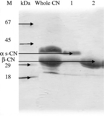
Camel β-CN was purified in a two-step purification procedure using anion-exchange resin (DEAE-cellulose). Purified β-CN was collected. Fig. 1 shows the SDS-PAGE (Gel 15%) profile of the purified camel β-CN.
SDS-PAGE profiles for purification of camel β-CN. After different steps of purification, β-CN was purified from other caseins. Lane 1 corresponds to the fraction having high content of αS-CN and lane 2 corresponds to the purified β-CN obtained from DEAE-cellulose column. M: molecular mass markers.

Determination of the ACE-inhibitory activity
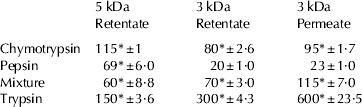
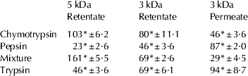
Camel whole casein and β-CN were hydrolyzed with each digestive enzyme (pepsin, chymotrypsin, trypsin) or with the 3 proteases as described in Materials and Methods section. The hydrolysates were fractionated according to their size with UF membrane filters with molecular mass cutoff of 10, 5 and 3 kDa. Peptide fractions obtained from hydrolysis of camel whole casein and β-CN showed a high inhibition of ACE activity (Table 1) compared with non-hydrolyzed whole casein and β-CN (IC50>700 μg protein/ml). The lowest IC50 value (20 and 23 μg protein/ml in the case of 3 kDa retentate and permeate, respectively) was obtained for whole casein hydrolysate after pepsinolysis. Pihlanto-Leppälä et al. (Reference Pihlanto-Leppälä, Koskinen, Phlola, Tupasela and Korhonen2000) reported IC50 value of more than 1000 μg/ml for bovine milk protein hydrolysate using pepsin. When whole casein was hydrolyzed with chymotrypsin, obvious ACE inhibitory activity was observed (Table 1a). The low value of IC50 obtained in this study showed a notable ACE inhibitory activity compared with other milk proteins hydrolysates. Cheung et al. (Reference Cheung, Wang, Ondetti, Sabo and Cushman1980) have reported that peptides with hydrophobic C-terminal amino acids had high ACE-inhibitory activity. In the case of β-CN, the lowest IC50 values were observed in the 5-kDa retentate obtained after action of pepsin and in the 3 kDa permeate obtained after proteolysis with the 3 proteases (Table 1b). López-Expósito et al. (Reference López-Expósito, Quirós, Amigo and Recio2007) reported high ACE-inhibitory activity of ovine casein peptides produced with pepsin. Pepsin cleaves peptide bonds involving hydrophobic and aromatic amino acids residues. Camel β-CN has 46 possible potential cleavage sites for pepsin, which is more numerous than tryptic and chymotryptic sites (http://www.expasy.org). There is no specific and no exact relationship between structure and activity of ACE-inhibitory peptides. Number of different peptides adopting a variety of structures and showing ACE-inhibitory activities have been identified in the recent years. In general, the two main common structural features of peptides showing ACE-inhibitory activity are the presence of hydrophobic amino acid in the C-terminal region and the presence of numerous prolines in their sequence (Otte et al. Reference Otte, Shalaby, Zakora, Pripp and El-Shabrawy2007). Some correlation between the ACE efficiency of these peptides and their structural homology with bradykinin should be mentioned. It has been shown that when proline is the antepenultimate amino acid, it enhances the binding of peptide to ACE (Gómez-Ruiz et al. Reference Gómez-Ruiz, Ramos and Recio2004). The most abundant amino acid in camel β-CN is proline accounting for 17% of the total amino acids in the sequence. When camel β-CN is subjected to pepsin some of the peptides that are produced have prolines as the antepenultimate amino acid. This may explain the significant ACE-inhibitory activities of peptic peptides of camel β-CN i.e. IC50 value <50. A high ACE-inhibitory activity was also observed for tryptic peptides of camel β-CN, these observations may be explained by the presence of high amount of prolines in its primary structure. Tauzin et al. (Reference Tauzin, Miclo and Gaillard2002) reported high ACE-inhibitory value for tryptic peptides of bovine αS2-casein. The presence of positively charged amino acid lysine or arginine at the C-terminal part of a peptide sequence after trypsinolysis also makes the resulting peptides able to inhibit ACE activity. The most efficient inhibitors of ACE from milk proteins hydrolysates have been reported to have values of IC50<50 μg/ml, which are in the range of those found in this study (López-Expósito et al. Reference López-Expósito, Quirós, Amigo and Recio2007).
ACE-inhibitory activities of crude peptide fractions from camel whole casein (IC50* (μg/ml))
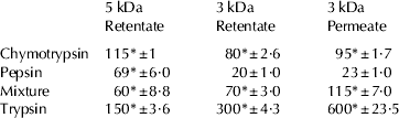
Mixture: hydrolysate obtained after the action of pepsin, trypsin and chymotrypsin
* Data marked with an asterisk are significantly different (P<0·05)
ACE-inhibitory activities of crude peptide fractions from camel β-casein (IC50* (μg/ml))
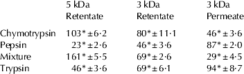
* IC50 is the concentration of an ACE-inhibitor needed to inhibit 50% of ACE activity
*Data marked with an asterisk are significantly different (P<0·05)
Mixture: hydrolysate obtained after the action of pepsin, trypsin and chymotrypsin
Antioxidant activities of camel whole and β-casein before and after hydrolysis
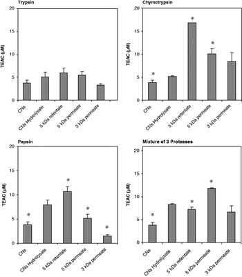
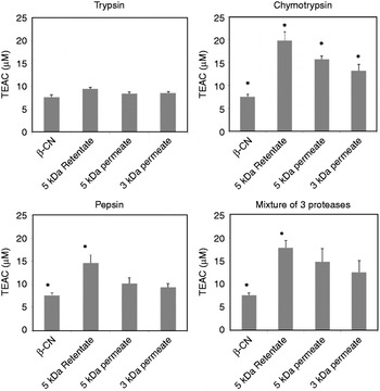
In this study, the antioxidant activity of camel whole casein and β-CN before and after hydrolysis was calculated measuring TEAC values, based on the consumption of coloured ABTS radicals. Treatment of camel whole casein and camel β-CN with gastrointestinal enzymes increased the antioxidant activity of the digests significantly in most cases. The peptide fractions obtained from camel whole casein when treated with trypsin did not show a high antioxidant activity and in case of 3 kDa permeate; it did not show a significant difference. This result shows the importance of the C-terminal amino acids in determining the antioxidant properties of a peptide fraction. When the totally hydrolyzed solutions of these caseins were fractionated, the antioxidant activity of each fraction increased compared with the whole hydrolysates. Both chymotrypsin-treated camel whole casein and β-CN showed the highest antioxidant activity. Chymotrypsin cleaves proteins after the hydrophobic and aromatic amino acids. The highest antioxidant activity was observed for peptides with molecular weight of 5–10 kDa (Fig. 2). This finding suggests that the antioxidant activities of peptides depend not only on their amino acid composition but also on the size and sequence of their amino acids. Studies have shown that free amino acids have smaller antioxidant activity than casein hydrolysates, which means that the primary structure of casein plays an important role (Silk et al. Reference Silk, Marrs and Addison1973; Imondi & Stradley, Reference Imondi and Stradley1974). The main mechanism by which peptides act in the food system is by free radical scavenging. The most effective amino acids with antioxidant activity and the best free radical scavengers are Cys, Trp, Tyr, Met, Phe, His, Ile, Leu and Pro. Not only the amino acid composition of a protein is important but also its accessibility and the positioning of the amino acids (Elias et al. Reference Elias, McClements and Decker2005, Reference Elias, Kellerby and Decker2008; Arcan & Yemenicioğlu, Reference Arcan and Yemenicioğlu2007). Camel milk caseins consist of αS-, β- and κ-caseins. Camel β-CN hydropathicity index is −0·339, which is the highest hydrophobicity among camel caseins. The most effective amino acids with highest antioxidant activities are hydrophobic and aromatic. Therefore, camel β-CN has considerable antioxidant activity even before it is hydrolysed. Additionally, the high content of these amino acids in camel β-CN make β-CN prone to hydrolysis by chymotrypsin and pepsin. After its hydrolysis, the peptides generated by chymotrypsinolysis and pepsinolysis (Fig. 3), showing mostly hydrophobic and aromatic amino acids in C-terminal position had the highest antioxidant activities.
Measurement of size-dependent antioxidant activities of camel whole casein and its peptides. Whole casein (CNs) and peptide fractions obtained after enzymatic hydrolysis followed by size fractionation were used for measuring antioxidant activities. The data are expressed as Trolox equivalent antioxidant capacity (μm). Chymotrypsin, trypsin, pepsin and their mixture were used for the proteolysis of caseins. The results are mean values of experiments carried out in triplicates. *Data marked with an asterisk are significantly different (P<0·05).

Measurement of size-dependent antioxidant activities of camel β-casein and its peptides. β-CN and peptide fractions obtained after enzymatic hydrolysis followed by size fractionation were used for measuring antioxidant activities. The data are expressed as Trolox equivalent antioxidant capacity (μm). Chymotrypsin, trypsin, pepsin and their mixture were used for the proteolysis of caseins. The results are mean values of experiments carried out in triplicates. *Data marked with an asterisk are significantly different (P<0·05).

Conclusion
In conclusion, both camel whole casein and β-CN showed high ACE-inhibitory activity following hydrolysis with pepsin and with the mixture of 3 proteases. The results obtained in this study suggest that β-CN may play an important role in camel milk casein as a natural source of anti hypertensive agents. Both camel whole casein and β-CN showed a significant antioxidant activity when hydrolyzed with chymotrypsin. The results of this study may encourage the use of camel milk caseins and derived peptides as anti hypertensive and antioxidant agents.
The support of the Research Council of the University of Tehran, Iran National Science Foundation (INSF), Center for International Research and Collaboration (ISMO), French Embassy in Tehran and the French Ministry of Foreign Affairs, by funding of Gundishapur project #18647RM are gratefully acknowledged.