Introduction
Mammals that undergo heat stress (HS) in summer have disturbed their reproductive functions such as spermatogenesis, oocyte maturity and early embryonic cell death (Hansen, Reference Hansen2009). For the high milk-producing dairy cows, HS prior to artificial insemination affects the conception rate after artificial insemination (Chebel et al., Reference Chebel, Santos, Reynolds, Cerri, Juchem and Overton2004).
Many researchers have indicated that the stable temperature for HS ranges from 40.0 °C to 42.0 °C with full, half or other periods of bovine in vitro maturation (IVM) (Edwards & Hansen, Reference Edwards and Hansen1996, Reference Edwards and Hansen1997; Roth & Hansen, Reference Roth and Hansen2004a, Reference Roth and Hansenb; Reference Roth and Hansen2005; Edwards et al., Reference Edwards, Saxton, Lawrence, Payton and Dunlap2005; Ju et al., Reference Ju, Jiang, Tseng, Parks and Yang2005; Soto & Smith, Reference Soto and Smith2009; Zhandi et al., Reference Zhandi, Towhidi, Nasr-Esfahani, Eftekhari-Yazdi and Zare-Shahneh2009). It has been proposed that HS values more like the circadian variation of the core body temperature of the cows are more realistic, because the temperatures for HS examined experimentally in vitro seemed to be higher than that experienced by cows actually exposed to HS (Rivera & Hansen, Reference Rivera and Hansen2001).
Oxidative stress is mediated by reactive oxygen species (ROS), which are generated as by-products of normal cellular metabolism and serve as key signal molecules in various physiological and pathological processes (Al-Gubory et al., Reference Al-Gubory, Fowler and Garrel2010). This situation results in an imbalance of the intracellular redox potential (Deleuze & Goudet, Reference Deleuze and Goudet2010). Heat-stressed bovine embryos in vitro involve increased intracellular ROS (Sakatani et al., Reference Sakatani, Kobayashi and Takahashi2004). This increase also seems to occur in bovine oocytes (Lawrence et al., Reference Lawrence, Payton, Godkin, Saxton, Schrick and Edwards2004). Elevated ROS of bovine oocytes due to excessive glucose during IVM have a negative effect on the embryos up to the blastocyst stage (Hashimoto et al., Reference Hashimoto, Minami, Yamada and Imai2000). However, the detrimental effect(s) of the ROS can be mitigated with antioxidants for heat-stressed bovine oocytes and embryos in vitro (Ealy et al., Reference Ealy, Drost, Barros and Hansen1992; Lawrence et al., Reference Lawrence, Payton, Godkin, Saxton, Schrick and Edwards2004; Sakatani et al., Reference Sakatani, Suda, Oki, Kobayashi, Kobayashi and Takahashi2007, Reference Sakatani, Yamanaka, Kobayashi and Takahashi2008).
It is well known that cysteine is a low-molecular-weight thiol compound, a sulfuric amino acid, and that it stimulates glutathione (GSH) synthesis via the γ-glutamyl cycle in oocytes and embryos (Meister & Tate, Reference Meister and Tate1976; de Matos et al., Reference de Matos, Furnus, Moses, Martinez and Matkovic1996; Luberda, Reference Luberda2005; Viet-Linh et al., Reference Viet-Linh, Dang-Nguyen, Nguyen, Manabe and Nagai2009). GSH is the most abundant non-protein thiol in mammalian cells (Franco & Cidlowski, Reference Franco and Cidlowski2009), maintains cellular redox status, and protects the cell from ROS (Meister, Reference Meister1983). Increased GSH in matured oocytes is essential to form a male pronucleus after fertilization and to promote early embryo development in mouse and hamster (Luberda, Reference Luberda2005).
Apoptosis is a process of programmed cell death, and is a homeostatic maintenance process involved in numerous biological systems (Franco & Cidlowski, Reference Franco and Cidlowski2009). Apoptosis of cells is induced by excess ROS and/or depletion of glutathione (Franco & Cislowski, Reference Franco and Cidlowski2009; Circu & Aw, Reference Circu and Aw2010). Heat stress during IVM can induce apoptosis in bovine oocytes (Roth & Hansen, Reference Roth and Hansen2004a; Zhandi et al., Reference Zhandi, Towhidi, Nasr-Esfahani, Eftekhari-Yazdi and Zare-Shahneh2009). For bovine embryos in vitro, HS increased the ROS with a reduction in the GSH level, and this change was alleviated by the addition of β-mercaptoethanol, a thiol antioxidant compound (Sakatani et al., Reference Sakatani, Yamanaka, Kobayashi and Takahashi2008). From this finding, we infer that HS may have a connection with ROS, apoptosis and the GSH level of bovine oocytes, and that we might be able to alleviate oxidative stress by the addition of cysteine during IVM.
Therefore, in the present study, we investigated the effects of the addition of cysteine on IVM whilst exposing the cells to fluctuating temperatures. The HS mimicked the vaginal temperature of dairy cows during the summer with regard to nuclear maturation and subsequent embryonic development up to the blastocyst stage of bovine oocytes. We also evaluated the ROS and GSH levels of the oocytes, and the apoptosis reaction level of the oocytes and cumulus cells.
Materials and methods
Chemicals
All chemicals in the present study were purchased from Sigma-Aldrich Co. unless otherwise stated.
Oocyte collection
Bovine ovaries were obtained from a local abattoir and transported to the laboratory in sterilized saline (23–25 °C) containing 100 U/ml of penicillin and 100 μg/ml streptomycin (Meiji Seika Co. Ltd). Cumulus–oocyte complexes (COCs) were aspirated from superficial follicles (2–6 mm in diameter), using an 18-G needle attached to a 5 ml disposable syringe. Then, oocytes enclosed with multi-layered cumulus cells were selected and washed three times with the maturation medium mentioned below.
In vitro maturation and HS treatment
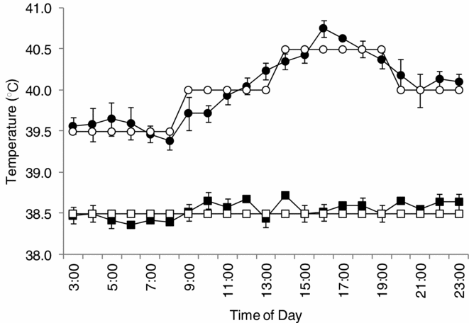
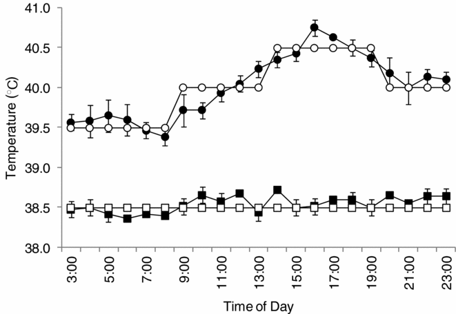
IVMD101 medium (Research Institute for the Functional Peptides, Yamagata, Japan) supplemented with 0.0, 0.6, 1.2 and 1.8 mM cysteine was used as maturation medium for the COCs. In the 0.0 mM cysteine group, the concentration of cysteine was not 0.0 mM because the IVMD101 medium already contains 0.8 μM cysteine. The COCs were transferred into a 100 μl droplet of the maturation medium (20 COCs/droplet) covered with mineral oil, and cultured under 5% CO2, 95% air with 100% humidity for 20 h. For HS treatment, the IVM temperature was adjusted to the vaginal temperature in the circadian rhythm of lactating dairy cows during hot (August–September) and cool (January–February) seasons in the Miyazaki prefecture in southwestern Japan (Fig. 1) (Nabenishi et al., Reference Nabenishi, Ohta, Nishimoto, Morita, Ashizawa and Tsuzuki2011). The incubator for the HS group was set at 39.5 °C for 5 h, 40.0 °C for 5 h, 40.5 °C for 6 h, and 40.0 °C for 4 h. Alternatively, in the control group, COCs were incubated continuously at 38.5 °C.
The heat stress (HS) temperature during in vitro maturation (IVM). The HS temperature of IVM (○) was adapted to the circadian vaginal temperature (●) of lactating dairy cows during the hot season. The (□) shows the IVM temperature for the control group, and (■) was the circadian vaginal temperature of the dairy cows during the cool season (Nabenishi et al. Reference Nabenishi, Ohta, Nishimoto, Morita, Ashizawa and Tsuzuki2011).
In vitro fertilization and embryo culture
Frozen semen in a 0.5 ml plastic straw was thawed for 30 s in a water bath at 37 °C. Next, the semen was diluted 10 times with IVF100 (Research Institute for the Functional Peptides), centrifuged (700 g) for 5 min twice at room temperature and re-diluted with IVF100 to give a final concentration of spermatozoa of 1 × 10⁷ sperm cells/ml. Next, the COCs were transferred into semen drops (20 COCs/100 μl drop), and co-incubated for 5 h at 38.5 °C, 5% CO2, 95% air, and 100% humidity. After fertilization, the putative zygotes were stripped of their cumulus cells by pipetting. The putative zygotes in each group were washed with IVD101 medium (Research Institute for the Functional Peptides) without further selection, transferred into droplets (20 putative zygotes/100 μl droplet) of IVD101 medium covered with mineral oil and cultured for 8 days under 5% O2, 5% CO2, 90% N2, and 100% humidity at 38.5 °C. The cleavage rates from the 2- to 16-cell stages on day 2 and blastocyst formation rates on day 8 after fertilization were evaluated using an inverted microscope.
Furthermore, to observe the effect of HS on oocyte fragmentation, the COCs exposed to the HS during IVM were denuded of their cumulus cells and loaded into the IVF100 medium without spermatozoa for 5 h followed by additional culture in IVD100 medium. On the eighth day after IVF100 treatment, the development rates from the 2- to 16-cell stages and up to the blastocyst stage in each group were observed.
Evaluation of metaphase II (MII) stage oocytes
After IVM, the COCs were stripped of their cumulus cells by pipetting to prepare denuded oocytes (DOs). Then, the DOs were fixed in 4% paraformaldehyde for 10 min at room temperature and stained with 5 μg/ml Hoechst 33342 in phosphate-buffered saline (PBS)(–) (pH 7.4) for 5 min at room temperature (Kim et al., Reference Kim, Jeong, Lee, Jeong, Hossein, Hyun and Hwang2010). After washing three times in 50 μl of PBS(–) supplemented with 0.1% PVA (PBS(–)-PVA) droplets, the DOs were mounted onto glass slides, and the nuclear maturation rates up to the oocyte MII stage were evaluated with a fluorescence microscope (Nikon Eclipse TS100: UV-1A Filter, 400 nm). The oocytes collected before IVM were also fixed and stained using the same method.
Evaluation of oocyte viability
The survival rates of the oocytes were evaluated using the method reported by Somfai et al. (Reference Somfai, Kashiwazaki, Ozawa, Nakai, Maedomari, Noguchi, Kaneko, Nagai and Kikuchi2008). Briefly, the DOs, derived from of each groups’ COCs before and after IVM, were stained with a 2.5 μg/ml concentration of fluorescein diacetate (FDA) for 2 min at room temperature in the dark. Then, the DOs were washed with 50 μl PBS(–)-PVA three times, and observed under a fluorescence microscope (FITC Filter, 460 nm). The oocytes that showed as bright green were considered to be living.
Measurement of ROS and GSH by fluorescence probes
The ROS and GSH contents of the DOs derived from the COCs matured with or without 1.2 mM cysteine under the presence of HS were analyzed with the method reported by Sakatani et al. (Reference Sakatani, Yamanaka, Kobayashi and Takahashi2008). Briefly, for the ROS assay, the DOs derived from each group were transferred to 50 μl droplets of 10 μM 2′7′-dichrolodihydrofluorescein diacetate (DCHFDA; Molecular Probe) in PBS(–) for 30 min at 38.5 °C. After incubation, the DOs were washed with 50 μl of PBS(–) three times, transferred to a glass slide with a small amount of PBS(–), and observed under a fluorescence microscope with a FITC filter. Images were taken by a charged-coupled device (CCD) camera and the fluorescence intensity of only the oocytes that released the first polar body were measured with the Nis Elements (Nikon). The oocytes were considered to be matured to the MII stage when they released the first polar body (Dominko & First, Reference Dominko and First1997).
For the GSH assay, the DOs were transferred to 10 μM Cell Tracker Blue (Molecular Probe) drops for 30 min at 38.5 °C. This reagent can detect only the reduced form of glutathione (Sakatani et al., Reference Sakatani, Yamanaka, Kobayashi and Takahashi2008). The DOs were then incubated for 30 min with TCM-199 plus 5% (v/v) fetal bovine serum at 38.5 °C in an incubator. After incubation, the DOs were washed with 50 μl PBS(–) supplemented with 5% polyvinylpyrrolidone (PVP) three times and observed under a fluorescence microscope with a UV-1A Filter. As for the ROS assay, only images of those DOs that released the first polar body that appeared to be matured completely were used for GSH analysis.
The DOs before IVM were also stained using a similar method and the levels of ROS and GSH were observed.
TdT-mediated dUTP nick end labelling (TUNEL) assay
A TUNEL assay was done using HS COCs matured with or without 1.2 mM cysteine, the same concentration of cysteine used for the ROS and GSH assays. In this assay, a TUNEL assay kit (In Situ Cell Death Detection Kit; Roche Applied Science) was used to assess the presence of apoptotic cells (Balboula et al., Reference Balboula, Yamanaka, Sakatani, Hegab, Zaabel and Takahashi2010). The COCs before and after IVM were fixed in 4% (w/v) paraformaldehyde solution (pH 7.4) for 10 min at room temperature, washed with 50 μl PBS(–) twice, then treated with 0.5% Triton-X in PBS(–) for 20 min at room temperature. After washing with PBS(–) supplemented with 5% PVP twice for 10 min each, the fragmented DNA ends of the cells were labelled with fluorescein-dUTP for 60 min at 37.0 °C. Then, the COCs were stained with 5 μg/ml Hoechst 33342 for 5 min at room temperature, washed three times with PBS(–) supplemented with 5% PVP for 5 min each, and transferred to slides glass with mounting solution (Vectashield, Vector Laboratories). The fluorescence of fragmented DNA ends was detected using a fluorescence microscope with a FITC filter. The obtained images were also measured with the Nis Elements. The TUNEL-positive level was assessed by the proportion of the fluorescence intensity of TUNEL-positive cells for the total area of COCs stained by 5 μg/ml Hoechst 33342. To confirm the accuracy of fluorescence intensity we used, the correlation with fluorescence intensity and the percentage of TUNEL positive cumulus cells to total cell number was assessed. Moreover, the DOs removed from the cumulus cells before and after IVM were also stained using the same method to investigate the presence or absence of any apoptotic effect on the DOs in each group.
Statistical analysis
All data were analysed using JMP software (version 8.01; SAS Institute Japan). Percentage data were subjected to arcsine transformation before analysis and data were expressed as mean ± SEM. Differences between treatments in each experiment were analysed by ANOVA followed by multiple comparisons using Tukey's test. p-values of less than 0.05 were considered statistically significant.
Results
The survival and the nuclear rates at the germinal vesicle (GV) stage in the ‘before IVM’ group were 96.7 ± 1.9%, (58/60) and 100.0 ± 0.0% (57/57), from four replicates, respectively.
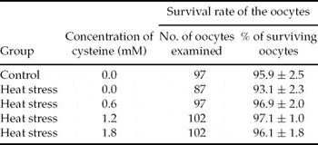
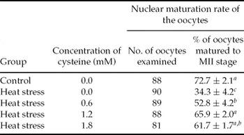
The survival and the nuclear maturation rates up to the MII stage of the oocytes are shown in Tables 1, 2 and Figure 2-1. Regarding the survival rate, no significant difference was observed among any of the groups. However, for the nuclear maturation rate up to the MII stage of mature oocytes, the rates of HS without cysteine (HS + 0.0 mM cysteine) and HS + 0.6 mM cysteine groups were significantly lower (p < 0.05) than that of the control group. However, the rates of the HS + 1.2 mM cysteine and HS + 1.8 mM cysteine groups were significantly higher (p < 0.05) than that of HS + cysteine 0.0 mM, and these rates were the same as those of the control group.
The survival rate of the heat-stressed oocytes after in vitro maturation (IVM) with various concentrations of cysteine

Data collected from five replicates represent the mean ± SE.
The nuclear maturation rate up to the metaphase II (MII) stage of heat-stressed oocytes after in vitro maturation (IVM) with various concentrations of cysteine

a –cValues with different superscripts within each column differ (p < 0.05).
Data collected from five replicates represent the mean ± SE.
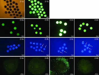
The photos of the oocytes under the heat stress (HS) for fluorescein diacetate (FDA) (2-1a, b), reactive oxygen species (ROS) (2-2a–d), glutathione (GSH) (2-3a–d) and TdT-mediated dUTP nick end labelling (TUNEL) (2-4a–d) assays. Representative images of morphology (2-1a) and FDA staining (2-1b) of matured oocytes in the control group (38.5 °C). The oocytes showing as bright green were considered living. Representative images of 2′7′-dichrolodihydrofluorescein diacetate (DCHFDA) staining the intracellular ROS levels in the before in vitro maturation (IVM) group (2-2a), control group (2-2b), HS + 0.0 mM cysteine group (2-2c) and 1.2 mM cysteine group (2-2d). Note the oocytes showing as bright green showed higher levels of intracellular ROS in the HS + 0.0 mM cysteine group (2-2c). Representative images of Cell Tracker Blue staining the intracellular GSH levels before IVM group (2-3a), control group (2-3b), HS + 0.0 mM cysteine group (2-3c) and 1.2 mM cysteine group (2-3d). Note the oocytes expressed higher levels of intracellular GSH in the HS + 1.2 mM cysteine group (2-3d). Arrows show the metaphase II (MII) stage of the oocytes with the first polar body (2-3b–d). Representative images of TUNEL labeling of cumulus–oocyte complexes (COCs) in the ‘before IVM’ group (2-4a), control group (2-4b), HS + 0.0 mM cysteine group (2-4c) and 1.2 mM cysteine group (2-4d). Higher TUNEL staining (green) in the HS + 0.0 mM cysteine group indicates a higher level of nucleus DNA fragmentation of the cumulus cells (2-4c).

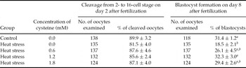
The embryonic development rates in each group are shown in Table 3. Although there was no significant difference in cleavage rates among any of the groups, the blastocyst formation rates of the HS + 0.0 mM and 0.6 mM cysteine groups were significantly lower (p < 0.05) than that of the control group. However, the rate of the HS + cysteine 1.2 mM group was significantly higher than that of HS + 0.0 mM cysteine (p < 0.05), and the rates were at the same level as the control group.
The cleavage and blastocyst formation rates of the heat-stressed oocytes treated with various concentrations of cysteine

a –c Values with different superscripts within each column differ (p < 0.05).
Data collected from seven replicates represent the mean ± SE.
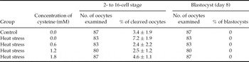
The fragmentation rate up to the 2–16-cell stage in all groups was in a range from 2.4 to 7.2%, and there were no significant differences among any of the groups. In addition, no blastocysts were observed in any of the groups (Table 4).
The fragmentation rate on day 8 after treatment with various concentrations of cysteine under heat stress

Data collected from five replicates represent the mean ± SE.
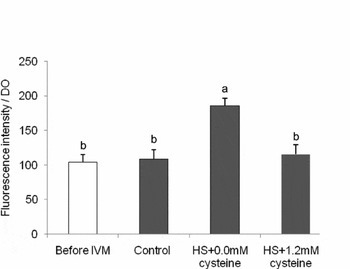
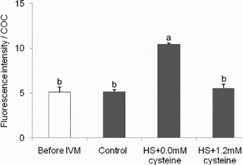
The ROS and GSH levels in the oocytes are shown in and Figs. 2-2, 2-3, 3 and 4. The ROS levels in the ‘before IVM’ and HS + 1.2 mM cysteine groups were similar to that of the control group. However, the HS + 0.0 mM cysteine group showed a significantly higher level (p < 0.05) than the other groups.
Effect of heat stress (HS) plus cysteine during maturation on intracellular reactive oxygen species (ROS) levels of the oocytes. Data represent mean ± SE. The total number of oocytes used for each treatment was: before (open bar) and after (black bars) in vitro maturation (IVM), n = 44; control, n = 22; HS + 0.0 mM cysteine, n = 15; HS + 1.2 mM cysteine, n = 12; in five replicates. Superscripts above each bar represent means that differ significantly (p < 0.05). DO, denuded oocytes.

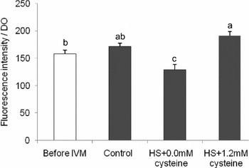
Effect of heat stress (HS) plus cysteine during maturation on intracellular glutathione (GSH) levels of the oocytes. Data represent mean ± SE. The total number of oocytes used for each treatment was: before (open bar) and after (black bars) in vitro maturation (IVM), n = 47; control, n = 22; HS + 0.0 mM cysteine, n = 20; HS + 1.2 mM cysteine, n = 13; in four replicates. Superscripts above each bar represent means that differ significantly (p < 0.05). DO, denuded oocytes.

The control group showed increased GSH levels compared with the ‘before IVM’ group. After IVM, the GSH level of the HS + 0.0 mM cysteine group was significantly decreased (p < 0.05) compared with the control group. However, the HS + 1.2 mM cysteine group showed a significantly increased GSH level compared with the HS + 0.0 mM cysteine (p < 0.05) group, and the level was the same as that of the control group.
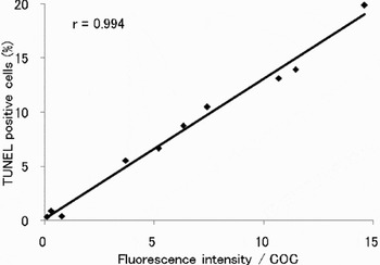
There was a significant positive correlation (r = 0.99, p < 0.01) between the fluorescence intensity and the percentage of TUNEL-positive cumulus cells to total cell number (Fig. 5). Therefore, measurement of the fluorescence intensity seemed to be feasible for the TUNEL assay.
The relationship between the fluorescence intensity and the percentage of TdT-mediated dUTP nick end labelling (TUNEL)-positive cumulus cells to total cell number. Nuclei of the cumulus cells showing green fluorescence were counted as apoptotic; the total number of nuclei per COC was determined by the Hoechst 33342 staining.

The rate of TUNEL-positive cumulus cells is shown in Figs. 2-4 and 6. The value of the ‘before IVM’ group was similar to that of the control group. Moreover, the rate of TUNEL-positive cells of the HS + 0.0 mM cysteine group was significantly higher (p < 0.05) than that of the control group. However, the rate of the HS + 1.2 mM cysteine group, which was the same as that of the control group, showed significantly reduced (p < 0.05) TUNEL-positive cells compared with the HS + 0.0 mM cysteine group. The mean ± SE of the COCs area used to measure the TUNEL level of the cumulus cells was almost the same among all groups: they were 145022.0 ± 11948.0 μm2 (n = 34), 140983.4 ± 11948.0 μm2 (n = 38), 174090.1 ± 11573.2 μm2 (n = 36) and 133961.8 ± 10073.9 μm2 (n = 39) in the before IVM, control, HS + 0.0 mM cysteine and HS + 1.2 mM cysteine groups, respectively. No significant differences were found among these groups.
Effect of heat stress (HS) plus cysteine during maturation on the apoptotic status in cumulus–oocyte complexes (COCs). Data represent mean ± SE. The total number of oocytes used for each treatment was: Before (open bar) and after (black bars) in vitro maturation (IVM), n = 34; control, n = 38; HS + 0.0 mM cysteine, n = 36; HS + 1.2 mM cysteine, n = 36; in five four replicates. Superscripts above each bar represent means that differ significantly (p < 0.05).

Conversely, TUNEL-positive nuclei in the DOs were not observed in any groups.
Moreover, the autofluorescence levels (mean ± SE) of the control groups in ROS, GSH and the TUNEL assay were 0.003 ± 0.0002, 0.003 ± 0.0002 and 0.24 ± 0.015 fluorescence density/DO or COC, respectively.
Discussion
In the present study, we adapted a fluctuating temperature to represent HS during IVM, which was increased up to 40.5 °C at maximum, mimicking the circadian vaginal temperature of dairy cows in the summer in the Miyazaki prefecture, one of the hottest regions in southwestern Japan.
Many researchers have adapted the stable temperature from 40.0 °C to 42.0 °C with full, half or other periods of bovine IVM (Edwards & Hansen, Reference Edwards and Hansen1996; Reference Edwards and Hansen1997; Roth and Hansen, Reference Roth and Hansen2004a, Reference Roth and Hansenb, Reference Roth and Hansen2005; Edwards et al., Reference Edwards, Saxton, Lawrence, Payton and Dunlap2005; Ju et al., Reference Ju, Jiang, Tseng, Parks and Yang2005; Soto & Smith, Reference Soto and Smith2009; Zhandi et al., Reference Zhandi, Towhidi, Nasr-Esfahani, Eftekhari-Yazdi and Zare-Shahneh2009). Only a few reports using the fluctuating temperature have mimicked the rectal temperature of dairy cows; effects of HS during bovine IVM, IVF or IVC with 40.5 °C or 41.0 °C as maximum temperature have been reported (Rivera & Hansen, Reference Rivera and Hansen2001; Sugiyama et al., Reference Sugiyama, McGowan, Kafi, Phillips and Young2003; Reference Sugiyama, McGowan, Phillips, Kafi and Young2007). It is thought that culture temperatures used for bovine embryos to demonstrate the effects of HS on development were higher than those of the body temperatures typically experienced by heat stressed cows (Rivera & Hansen, Reference Rivera and Hansen2001). In addition, maximal rectal temperatures in heat stressed lactating cows who experiencing infertility ranged from 40.1 to 40.5 °C (Roth et al., Reference Roth, Meidan, Braw-Tal and Wolfenson2000), which was lower than other studies using in vitro models (Roth & Hansen, Reference Roth and Hansen2004b; Reference Roth and Hansen2005; Zhandi et al., Reference Zhandi, Towhidi, Nasr-Esfahani, Eftekhari-Yazdi and Zare-Shahneh2009). In the light of these reports, it is suggested that the temperature we used may be appropriated to evaluate the influence of HS on bovine oocyte maturation in vitro.
In the present study, the survival rates of heat stressed oocytes, with or without the variable cysteine concentrations, determined by FDA staining and used genetically for membrane integrity (Arav et al., Reference Arav, Zeron, Leslie, Behboodi, Anderson and Crowe1996), were almost the same as those of the control group. Our results are consistent with the report of Edwards and Hansen (Reference Edwards and Hansen1996), who revealed that temperatures of 41.0 or 42.0 °C HS for 12 or 24 h periods during IVM did not decrease the membrane integrity of bovine oocytes.
When 1.2 mM cysteine was added to the maturation medium for heat-stressed oocytes, the blastocysts formation rate was significantly increased compared with HS-treated oocytes matured without cysteine (HS + 0.0 mM cysteine) in the present study. In addition, although the HS + 0.0 mM cysteine group showed increased ROS and TUNEL-positive levels of cumulus cells and reduced GSH levels compared with those of the control group, the HS + 1.2 mM cysteine group showed decreased levels of both ROS and TUNEL-positive levels, and increased GSH levels similar to those in the control group.
It is well known that cysteine is a low-molecular-weight thiol compound. It is a sulphuric amino acid that stimulates GSH synthesis via the γ-glutamyl cycle in oocytes and zygotes (Meister & Tate, Reference Meister and Tate1976; de Matos et al., Reference de Matos, Furnus, Moses, Martinez and Matkovic1996; Luberda, Reference Luberda2005; Viet-Linh et al., Reference Viet-Linh, Dang-Nguyen, Nguyen, Manabe and Nagai2009). High levels of GSH in matured oocytes are essential to form a male pronucleus after fertilization and promote early embryo development in the mouse and hamster (Luberda, Reference Luberda2005). Glutathione has important roles in cellular defence against oxidative aggression and redox homeostasis that are critical for proper functioning of cellular processes, including apoptosis (Circu & Aw, Reference Circu and Aw2008). Apoptosis of the cells is induced by excess ROS and/or depletion of GSH (Franco & Cislowski, Reference Franco and Cidlowski2009; Circu & Aw, Reference Circu and Aw2010). Conversely, cumulus cells play an important role in the oocyte's meiotic and developmental competence to supply energy substrates such as glucose and other factors (Tanghe et al., Reference Tanghe, Van Soom, Nauwynck, Coryn and de Kruif2002; Thompson et al., Reference Thompson, Lane and Gilchrist2007). The degree of apoptosis of cumulus cells may be predictive of low developmental potential in cattle (Yuan et al., Reference Yuan, Van Soom, Leroy, Dewulf, Van Zeveren, de Kruif and Peelman2005; Van Soom et al., Reference Van Soom, Vandaele, Goossens, de Kruif and Peelman2007). Therefore, we assumed that the addition of cysteine to the IVM medium may increase the GSH concentration of heat-stressed oocytes, with scavenging ROS of the oocytes suppressing cumulus cell apoptosis.
In the present TUNEL assay, although apoptotic reactions were detected in the cumulus cells in all groups, no apoptotic nuclei were observed within the oocytes in any group. Tatemoto et al. (Reference Tatemoto, Sakurai and Muto2000) reported that cumulus cells can protect pig oocytes from oxidative stress during IVM. In addition, cumulus cells have a thermoprotective role during bovine oocyte maturation under HS (Edwards & Hansen, Reference Edwards and Hansen1996). Furthermore, Yuan et al. (Reference Yuan, Hao, Liu, Wu, Yang, Liu, Tian, Zhu and Zeng2008) reported that pig oocytes that were attached to heat-stressed apoptotic cumulus cells during IVM inhibited the development rate of parthenogenetical embryos up to the blastocyst stage. They indicated that apoptotic cumulus cells may contribute to maturation failure of oocytes in vitro. The decrease in the nuclear maturation rates up to the MII stage in the present results may have been caused by cumulus cells being attacked by ROS and inducing their apoptosis during IVM.
In this study, although 0.6 mM cysteine did not improve the development rate up to the blastocyst stage, the 1.2 mM cysteine group showed improved development of heat-stressed oocytes. This concentration of cysteine was approximately two-fold more than that in general use (less than 0.6 mM) for bovine IVM, (Sugiyama et al., Reference Sugiyama, McGowan, Kafi, Phillips and Young2003; Ju et al., Reference Ju, Jiang, Tseng, Parks and Yang2005; Furnus et al., Reference Furnus, de Matos, Picco, García, Inda, Mattioli and Errecalde2008). Usually, standard in vitro culture conditions exert oxidative stress or an imbalance between oxidants and antioxidants. ROS are oxygen-derived molecules formed as intermediary products of cellular metabolism and they represent a major culture-induced stress (Combelles et al., Reference Combelles, Gupta and Agarwal2009; Deleuze & Goudet, Reference Deleuze and Goudet2010). In addition, Sakatani et al. (Reference Sakatani, Kobayashi and Takahashi2004) reported that heat-stressed embryos on day 0 and 2 after IVF showed increased ROS while reducing embryonic development up to the blastocyst stage compared with the control group. Maternal HS resulted in increased ROS activity in oviducts and embryos in the mouse (Ozawa et al., Reference Ozawa, Hirabayashi and Kanai2002; Matsuzuka et al., Reference Matsuzuka, Ozawa, Nakamura, Ushitani, Hirabayashi and Kanai2005a, Reference Matsuzuka, Sakamoto, Ozawa, Ushitani, Hirabayashi and Kanaib). As a result, it is postulated that the increased ROS production by HS may not be well scavenged when using the widely used concentration of cysteine. This factor may be one reason that a two-fold concentration of cysteine was needed to improve the developmental competence of heat-stressed oocytes in the present study.
The nuclear maturation rate up to the MII stage of the oocytes exposed to HS without cysteine group (HS + 0.0 mM cysteine group) was significantly lower than that of the control group in our study. This finding suggests that the HS in this study can inhibit oocyte nuclear maturation. Roth & Hansen (Reference Roth and Hansen2005) reported that bovine oocytes exposed to the first 12 h HS at 40.0 °C showed a reduced nuclear maturation rate up to the MII stage. The temperature we used for IVM as HS was over 40.0 °C (maximum temperature at 40.5 °C) for 15 h from 5 h after the onset of IVM. Longer exposure over 40.0 °C beyond 12 h may have impaired the resumption of bovine oocyte meiosis, in this study.
In addition, although the cleavage rate of the oocytes treated with HS without cysteine (HS + 0.0 mM cysteine) did not decrease, the blastocyst formation rate in this group was lower compared with that of the control group in this study. Edwards and Hansen (Reference Edwards and Hansen1996) reported that although COCs exposed to 42.0 °C HS during maturation had lower cleavage and blastocyst formation rates, exposure to HS at 41.0 °C, which is 0.5 °C higher than our maximum temperature, did not affect the cleavage rate but reduced the number of blastocysts. This finding suggests that the HS conditions we used did not have a negative effect on the oocyte cleavage rate.
In our results, the development rate up to the blastocyst stage of heat-stressed oocytes without cysteine was significantly decreased in parallel with a decrease in the nuclear maturation rate compared with that of the control group. This situation indicates that the reduced development rate up to the blastocyst stage may be caused by a decline in nuclear maturation. However, in the present study, the HS without cysteine group showed a lower nuclear maturation rate, and a slightly reduced cleavage rate was observed compared with that of the control group. The reasons for these contradictory findings are unclear, but the following possibility is suggested.
Sperm can penetrate bovine oocytes at the incomplete nucleus maturation stage, and the penetrated oocytes can complete the final step of maturation stimulated by the sperm penetration (Chian et al., Reference Chian, Niwa and Nakahara1992). Therefore, full maturation may not be required for sperm penetration into bovine oocytes (Niwa et al., Reference Niwa, Park and Okuda1991). This penetration may have occurred in the HS without cysteine group, and resulted in the non-significant differences in the cleavage rates between the HS without cysteine and control groups.
On the other hand, the HS + 1.8 mM cysteine group showed both reduced nuclear maturation and blastocyst formation rates, and no significant difference was observed between the control group, in this study. Morado et al. (Reference Morado, Cetica, Beconi and Dalvit2009) indicated that a low concentration of ROS at 12 h during bovine IVM could determine the developmental competence of the oocytes. Therefore, we assume that excess scavenging ROS during IVM due to a higher concentration of cysteine may have suppressed the embryonic development up to the blastocyst stage in the present study.
In this study, negligible fragmentation rates from the 2- to 16-cell stage and no blastocyst formation were observed among all groups when the COCs were exposed to HS with or without various concentrations of cysteine during IVM. To produce porcine embryos by ICSI, the number of fragmented oocytes was higher in summer compared with the other seasons (Suzuki et al., Reference Suzuki, Watanabe and Fukui2010). This discrepancy may be caused by species differences.
In conclusion, 1.2 mM cysteine added to the IVM medium for oocytes exposed to heat stress mimicked the circadian vaginal temperature of the dairy cows during the summer and inhibited nuclear maturation and subsequent embryonic development up to the implantation stage. Furthermore, cysteine in the oocyte maturation medium alleviated the suppressed GSH level, and increased the ROS level of oocytes, while increasing the number of apoptotic cumulus cells induced by heat stress.
Acknowledgements
The authors would like to thank Dr M. Takahashi and Dr M. Sakatani of National Agricultural Research Center for Kyushu Okinawa Region, Kumamoto, Japan for their technical instruction to evaluate the GSH, ROS and TUNEL assay, and the staff of Miyakonojo Wellness Meat Co., Ltd., Miyakonojo City, Japan, for providing the bovine ovaries.