



Introduction
Seed dormancy provides a mechanism for plants to delay germination until conditions are optimal for the survival of the next generation. Dormancy release is regulated by a combination of environmental and endogenous signals with both synergistic and competing effects (Finkelstein et al., Reference Finkelstein, Reeves, Ariizumi and Steber2008). Physical dormancy in seeds is caused by a water-impermeable seed coat (or fruit) (Baskin et al., Reference Baskin, Nan and Baskin1998, Reference Baskin, Baskin and Li2000). A palisade layer(s) in the seed coat (or fruit) is (are) responsible for the impermeability. The seed becomes permeable to water when an opening is formed via a specialized anatomical structure (‘water gap’) in the palisade layer(s), allowing water to enter the seed (Wong et al., Reference Wong, Lee, Yeo and Tan2019). The study of the anatomical structures of the seed gives us complementary information in the analysis and description of the path travelled by the water inside the seed and of the metabolic modifications that occur internally during the imbibition process. In most of the botanical families, hilum and micropyle are the places of greater water intake in the seed (Serrato-Valenti et al., Reference Serrato-Valenti, Cornara, Ghisellini and Ferrando1994; Kikuchi et al., Reference Kikuchi, Koizumi, Ishida and Kano2006; Wong et al., Reference Wong, Lee, Yeo and Tan2019).
In seeds of Fabaceae, besides the hilum and micropyle, there is also the pleurogram, a characteristic fissure located in the frontal portion of the seed that assumes significant participation in the imbibition process (Gama-Arachchige et al., Reference Gama-Arachchige, Baskin, Geneve and Baskin2013; Geneve et al., Reference Geneve, Baskin, Baskin, Jayasuriya and Gama-Arachchige2018; Rodrigues-Junior et al., Reference Rodrigues-Junior, Baskin, Baskin, Oliveira and Garcia2018). The hilum is a fissure with a hygroscopic valve function, which controls the entry of water into the seed, by difference of water potential with the environment. This finding has been confirmed in experiments blocking this region, for example, the seeds of Ceanothus spp. (Geneve, Reference Geneve2009) and Erythrina velutina (Alves Júnior et al., Reference Alves Júnior, Vitoriano, Lima and Lima2016). In natural environmental conditions, Leucaena leucocephala (Fabaceae) seeds have a very low germination rate, mainly due to mechanical dormancy caused by the integument (Serrato-Valenti et al., Reference Serrato-Valenti, Cornara, Ghisellini and Ferrando1994). The tegumentary impermeability restricts the entry of water and gaseous changes in the seed, causing very slow and non-uniform germination, resulting in a non-uniform distribution of plants in the field. To produce seedlings, mainly with the objective of developing planned plantings, it is necessary to break the natural dormancy of these seeds. Several methods have been used to alter the structure of the integuments of various seed types and break dormancy such as mechanical scarification, chemical treatment, hot water immersion and blade incision. However, these methods may present limitations with respect to large-scale seedling production. When it comes to relatively small seeds, such as L. leucocephala, mechanical manipulation, for example, is a disadvantage, since they can produce lesions in the embryo and increase infection rates by fungi and bacteria, triggering seed mortality and abnormality in seedlings (Voegele et al., Reference Voegele, Graeber, Oracz, Tarkowská, Jacquemound, Tureckova, Urbanová, Strnad and Leubner-Metzger2012; Liu et al., Reference Liu, Liu, Ji, Chen and Xu2015; Rodrigues-junior et al., Reference Rodrigues-Junior, Faria, Vaz and Nakamura2014).
In recent years, there has been an increasing interest in the use of cold atmospheric plasma (CAP) as an energy source in modifying the surface of living materials because of its many advantages, such as treatment of larger areas without damaging thermally sensitive structures. There is a growing body of literature that recognizes the efficiency of CAP in the breaking of physical seed dormancy (Alves Júnior et al., Reference Alves Júnior, Vitoriano, Lima and Lima2016; Da Silva et al., Reference Da Silva, Farias, Silva, Vitoriano, Sousa and Alves-Junior2017; De Groot et al. Reference De Groot, Hundt, Murphy, Bange and Mai-Prochnow2018; Los et al. Reference Los, Ziuzina, Boehm, Cullen and Bourke2019; Yodpitaka et al. Reference Yodpitaka, Mahatheeranonta, Boonyawand, Sookwonga, Roytrakule and Norkaew2019). It has been observed that functional groups containing hydrogen, oxygen and/or nitrogen atoms increase the adsorption of water molecules on the seed's surface. Thus, plasma treatment may reduce the hydrophobic character of substances present in the seed coat, increasing its wettability (Será et al., Reference Será, Sery, Stranak and Spatenka2009, Reference Será, Sery, Stranak and Spatenka2010; Bormashenko et al., Reference Bormashenko, Grynyov, Bormashenko and Drori2012, Reference Bormashenko, Shapira, Grynyov, Whyman, Bormashenko and Drori2015; Randeniya and Groot, Reference Randeniya and Groot2015). Despite the importance of this application, there remains a paucity of evidence which explains the mechanism responsible for the penetration of water from the surface modified by the plasma to the interior of the seed. The aim of this study is to explore the relationship between the anatomy of the seed and the mechanism of water absorption when seed surfaces are plasma treated. L. leucocephala seed was chosen because it presents high dormancy and a low germination rate.
Materials and methods
Apparatus and plasma treatment
The experimental apparatus is illustrated in Fig. 1A. The source plasma was equipped with two coaxial glass tubes, externally and internally coated by a metal mesh screen, each glass tube with a 1 mm thick wall. The metal mesh screen was connected to a high-voltage power source, peak value of 17.5 kV, repeated at a frequency of 990 Hz. An optical fibre was used to transfer light spectrum from plasma to the charge coupled device (CCD) spectrometer (HR4000, Ocean Optics, Dunedin, FL), with a spectral range between 200 and 1000 nm, to measure the reactive species generated in the plasma. The slightly elliptical shaped Leucaena seeds, an average diameter of 8.0 mm, were placed between the two glass tubes within the plasma reactor. For plasma treatment, around 100 seeds were distributed uniformly between the coaxial tubes, at a time. Under these conditions, the plasma filled up the spaces between the two glass tubes and coated all the seeds surfaces evenly (Fig. 1B). The treatments were performed for 3, 9 or 15 min. After treatment, the seeds were stored in desiccators for further wettability, imbibition and germination tests.
(A) Experimental apparatus used for plasma treatment of seeds and (B) details of the top view showing positioning of the seeds within the source plasma.

Seed morpho-anatomy
Optical and scanning electron microscopies were used to monitor seed morphology and anatomy. For the morpho-analysis, untreated seeds were soaked in a formaldehyde–acetic acid-alcohol solution for 24 h, dehydrated in ethanol and embedded in paraffin blocks. The blocks were then cut into sections of 10 μm thick, with a rotary microtome (Zeiss™ Hyrax M40, Jena, Germany). Histochemical testing was performed using toluidine blue to reveal lignin and/or cellulose (O'brien et al., Reference O'brien, Feder and Mccully1964). Sections were observed with an optical microscope (Nikon Elipse MA 100) and photomicrographs were taken with a digital camera (Nikon Seght DS), using NIS-Elements D42064 to image processing.
Wettability test
Wettability tests were conducted using the sessile drop method, which consists of depositing a drop of distilled water on the surface of the seed coat and measuring the apparent contact angle formed by the drop with the surface. The apparent contact angle is commonly used in experiments to characterize biological interfaces (Erbil, Reference Erbil2006). The apparent contact angle of distilled water droplets was measured on three seeds under each condition. In each seed the apparent contact angle measurements were repeated three times by dripping 20 μl of distilled water once on each seed surface. The images were captured and recorded by a camera attached to a computer and processed to determine the apparent contact angles using the Surftens® 3.5 program, Frankfurt, Germany (Yanling et al., Reference Yanling, Yingkuan, Chen, Deng and Ruan2014). The values of the apparent contact angles formed by the liquid and the integument were submitted to descriptive statistics (arithmetic mean and standard deviation).
Water path identification
To verify water pathways, whole seeds in untreated conditions and treated by dielectric barrier discharge (DBD) plasma for 15 min were soaked in an aqueous solution (0.5%) of toluidine blue. This condition was chosen because it was the one that presented the best wettability and imbibition results. For different soaking times, two seeds for each condition were removed from the solution and transversal sections opposite to the hilar region were obtained. Cross sections were observed under a stereoscopic (Nikon MSZ 18) and pictures were taken with a digital camera (Nikon DS-F12).
Imbibition tests
The seeds were placed in plastic cups containing 50 ml of distilled water for imbibition testing. The cups were placed in a germination chamber with a photoperiod of 12 h at 25°C. Each experimental condition contained 5 replicates of 10 seeds each. The relative percentage of imbibition (E%) was obtained from the seed mass variation after 4, 12, 36, 60 and 84 h of imbibition, and defined by equation (1). Before each measurement, the seeds were placed on sterilized paper to remove the excess of water.
 $$E\lpar \% \rpar = \displaystyle{{m_f-m_i} \over {m_i}} \times 100$$
where mi is the initial total mass of the seeds and mf is the total mass of seeds imbibed for each measurement time. The experimental design was completely randomized. Simultaneously, the imbibition liquid containing substances leached from the seeds was used to measure electrical conductivity (EC) and pH, evaluated in the same time intervals. The EC measurements were performed with an Oakton PC 450, Vernon Hill, US, conductivity meter and the unit of measure was μS cm−1. The pH measurements were made immediately after EC measurements.
Germination test
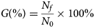
A treatment time of 15 min was chosen to compare the germination with untreated seeds. For each condition, 100 seeds were used, divided into four replicates of 25. In these tests, the seeds were placed between filter paper moistened with distilled water equal to 2.5 times the mass of the substrate used. These were stored in plastic boxes (capacity of 250 ml, dimension 11.0 × 11.0 × 3.5 cm3) which were placed in a germination chamber with a photoperiod of 12 h at 25°C. The number of radicles emerged, visible to the naked eye, was counted from the first to the 11th day, at the same time of day. The experiment was completely randomized. The percentage of cumulative germination (G), calculated for each day, was plotted as a function of time, in days (equation (2)).
 $$G\lpar \% \rpar = \displaystyle{{N_f} \over {N_0}} \times 100\% $$
where Nf is the cumulative number of seeds germinated in each day and N 0 is the number of sown seeds.
Statistical analysis
The experimental design used was completely randomized (DIC) for the analysis of variance, followed by the application of the Tukey (P < 0.05) averages Comparison Test, using Sisvar® Software, Lavras, Brazil.
Results and discussion
Seed morpho-anatomy
Morphologic analysis revealed that L. leucocephala seeds have structural characteristics of seeds which belong to the Fabaceae family, displaying a hard and smooth integument with fractures almost over its entire extension (Fig. 2A). On both sides of the seed, there is a region called a pleurogram (Fig. 2A) which is marked by a U-shaped fracture, whose opening faces the hilar region and micropyle (Fig. 2B).
Structural of the L. leucocephala seed presenting: (A) Pleurogram (Pl), (B) Region hilar (Hi) and micropyle (Mi), and (C) Cracks in the integumentary region (Ti).

The most accepted hypothesis on the function of the pleurogram comprises a hygroscopic valve, functioning like the hilum during the entry of water into the seed (Gunn, Reference Gunn, Polhill and Raven1981). In leucaena seeds, microcracks (Fig. 2B,C) and contour in the pleurogram (Fig. 2A) are important in the water absorption process, due to the proximity and communication with the hilar region, unlike what happens with other species within the family of Fabaceae. The probable origin of the pleurogram contour originates in the maturation process, as a result of the compressive stresses between structures of different degrees of stiffness and irregular pattern (Rodrigues-Junior et al., Reference Rodrigues-Junior, Faria, Vaz and Nakamura2014). These cracks play a key role in the water absorption mechanism. Most of the cracks present in the tegument, especially in the region of the pleurogram (Fig. 3A), extend through the first layer of the integument, the cuticle (CU) (Fig. 3B), consisting of epidermis formed by a layer of palisade, composed of thick-walled, radially elongated macrosclereid cells.
(A) Tegumentary region (Te) and (B) cross section of the seed showing the tegumentary region (Te), cuticule (Cu) and light line (ll). (C) Cell arrangement within the L. leucocephala seed: cuticle (Cu); line of light (11); macroesclereids (ma); osteo-esclereids (os) and parenchyma (pa).

This region of macrosclereid cells, when viewed by light microscopy, shows a light line, produced by neither chemical nor mechanical changes but caused by a modified molecular structure containing less water than the remainder of the cell wall (Fig. 3C). The presence of cellulose in the tegument's layers of tissues is indicated by the purple colouration. A similar result was obtained in Schizolobium parahyba, Senna multijuga and L. leucocephala seeds (Serrato-Valenti et al., Reference Serrato-Valenti, Cornara, Ghisellini and Ferrando1994; Voegele et al., Reference Voegele, Graeber, Oracz, Tarkowská, Jacquemound, Tureckova, Urbanová, Strnad and Leubner-Metzger2012; Rodrigues-Junior et al., Reference Rodrigues-Junior, Baskin, Baskin, Oliveira and Garcia2018). The light line has previously been reported as a region with callose (polysaccharide–glucose polymer) which is a sealant substance, an important factor in water permeability (Currier and Strugger, Reference Currier and Strugger1956; Bhalla and Slattery, Reference Bhalla and Slattery1984; Bevilacqua et al., Reference Bevilacqua, Fossati and Dondero1987).
Wettability tests
Plasma treatment of seeds had a significant effect on wettability. Contact angle measurements (Fig. 4) show that seeds before treatment had a hydrophobic surface (a mean contact angle of 95°). After treatment, the values of the contact angles decreased to values as low as 40° in the treatments of 15 min. Therefore, plasma caused a reduction in θ between 42% and 58%. According to De Gennes et al. (Reference De Gennes, Brochard-Wyart and Quéré2003) and Marmur (Reference Marmur and Mittal2009), the increase in surface wettability of the treated seeds may be related to chemical mechanisms of surface modification caused by the adsorption of radicals containing, for example, oxygen or nitrogen, making it more hydrophilic and resulting in higher water rate absorption (Stoffels et al., Reference Stoffels, Sakiyama and Graves2008; Bormashenko et al., Reference Bormashenko, Grynyov, Bormashenko and Drori2012). Results from the literature show that the treatment of seed surfaces by cold plasma improves the wettability level of the seeds, mainly for air plasma or atmospheres containing N2. It was observed that biological tissue plasma treatment affects only the outer layer at a nanoscale (France and Short, Reference France and Short1997; Kaminska et al., Reference Kaminska, Kaczmarek and Kowalonek2002; Mortazavi, and Nosonovsky, Reference Mortazavi and Nosonovsky2012; Bormashenko et al., Reference Bormashenko, Chaniel and Grynyov2013). Thus, a change from a hydrophobic state of the L. leucocephala seed surface in the untreated condition to hydrophilic states when treated, as observed in this work, indicates that this treatment increases the density of hydrophilic on the outer surface of the non-cellular layer (Fig. 4).
(A) Contact angle values on the L. leucocephala seed surface for different treatment conditions and (B) photographs of water droplets deposited on untreated seed surfaces and treated for 15 min.

Water path identification
Accompanying the markers (dyes) in the water, it was possible to identify the path of water entry in the seed coat (Fig. 5A). Initially the cuticle is gradually wet, especially in the central pleurogram, which has a high density of cracks and therefore greater capillary action (Fig. 5B).
Penetration of the dye in the seed illustrated by red arrows (A) seed cross section and (B) pleurogram centre region where the dye penetrates the cuticle. (C) Penetration of water through pleurogram cracks and (D) water reaches the layer of osteosclereid cells and embryo. Subsequently, the dye penetrates the upper sublayer of the palisade and reaches the layer of osteosclereid cells through the pleurogram crack (C). The water distributes to the osteoscleroid layer and then wets the lower part of the palisade and the mesophyll layers and the remaining endosperm, reaching the embryo (D). Due to the internal pressure caused by the osmotic pressure, the microcracks present on the surface of the integument will open after ~50 h, breaking the walls of the shell, which further potentiates the entry of water into the seed (Fig. 6).

Increased water absorption after tegument rupture. (A) Micrograph of the shell after 50 h, (B) before disruption and (C) after disruption.

The whole region below the cuticle was completely permeated by the dye (Fig. 6C) after 90 h of imbibition. Therefore, the water path in the seed results from the combination of tegument wettability, followed by absorption, by capillarity of the cracks and microchips and, thus, increasing the water potential from outside to inside the seed. The increased volume of water inside the seed causes swelling and increases the internal pressure, resulting in tegument rupture. Plasma treatment increases the wettability of the surface, enhancing the capillary effect in the cracks, thus producing a higher water potential than the untreated seeds. Higher water potential means greater driving force for water diffusion and therefore greater imbibition. This effect is illustrated in Fig. 7. There is an expressive increase of the thickness of the cuticle in the treated seed when compared to the untreated seed. Also, in the structure formed by macrosclereid cells, the light line in the plasma-treated seed disappeared. The strength of the light line was related to seed coat impermeability.
Seeds removed from the solution after 2 h of soaking: (A) untreated and (B) treated for 15 min.

According to Steinbrecher and Metzger-Leubner (Reference Steinbrecher and Metzger-Leubner2017) the level of physical dormancy depends on the species and habitat, and the anatomical structure of the seed includes impermeable layer(s). The methods of overcoming physical dormancy are more or less efficient according to the degree of breaking of these impermeable barriers.
Imbibition tests
In general, the percentage of imbibition increases with time (Fig. 8). Germination is a process that involves three main phases. Phase I, also called imbibition, is characterized by rapid absorption of water, followed by a plateau or marked reduction of absorption (Phase II) and ending with new absorption growth (Phase III), characterized by radicle protrusion. Phase II is the longest in the process, in which there is little variation in water absorption (Bewley and Black, Reference Bewley and Black1994). This behaviour was not observed in the curves of Fig. 8. The individual curves showed variable behaviour among the four treatment conditions. For untreated or treated samples for 3 min, there is marked absorption rate in the initial phase, followed by a decrease and again a rise in the absorption rate in the final phase. The first phase occurred until around 18 h and the second phase between approximately 18 and 40 h. The rapid growth after this phase was not characterized by Phase III, since there was no radicle protrusion. In this phase the germination process is diminished by the resistance offered by the upper part of the tegument, which have hydrophobic substances. After this phase, above 40 h, the water wets the osteosclereide layer, then wets the lower part of the palisade and the layer of steosclereides, in addition to the remaining endosperm. Associated with this is the gradual opening of the microcracks in the palisade, promoted by the swelling during imbibition.
Time-dependent soak for L. leucocephala seeds, for untreated and plasma-treated conditions, for 3, 9 and 15 min.

In the seeds treated for 9 and 15 min, a significant increase in the third phase, greater than the two previous conditions, is observed. In this case it is assumed that this increase is due to fracture of the integument caused by internal pressure during seed swelling. It is plausible to assume, therefore, that plasma treatment in L. leucocephala seeds increased the density of hydrophilic sites of the integument, promoting greater motoric force to overcome the impermeable barrier in the light line. The increase in internal pressure caused by the absorption of a larger volume of water was sufficient to produce propagation of the cracks in the integument, resulting in an abrupt increase in imbibition.
Electrical conductivity and pH
Exudation of substances by the seed can be monitored by determining EC and pH of the imbibition solution (Fig. 9). The pattern of EC of the leachate was monitored during imbibition (Fig. 9A). During imbibition, substances such as sugars, amino acids, organic acids, proteins and inorganic ions (K+, Ca2+, Mg2+ and Na+) (AOSA, 2002) may be extruded by the seeds, of which the ions, organic acids and sugars are largely responsible for the increase in acidification of the medium. The number of ions leaching in the imbibition solution indirectly reflects the degree of damage of the cell membranes resulting from the seed drying process. While cell membranes are restructured upon imbibition there is release of electrolytes, the amount of which is associated with membrane integrity. For all the experimental conditions used in this work, the pattern of EC indicates that the leachate mostly displayed a growth of net charge (NC), except for the time period between approximately 60 and 84 h, when NC remained constant or decreased. This means that charges are being exchanged between water and seeds. The pattern of the pH was more complex (Fig. 9B), because it depends on the H+/OH− ratio that makes up the NC and this ratio changes during imbibition, in the following sequence:
1. Initially, on contact with water, the surface of the seed captures H+ ions, resulting in the increase of EC due to OH− ions in the solution, raising the pH up to around 4 h (initial part of imbibition, Phase I). In the solution containing the 15-min treated samples, on the contrary, a reduction of the pH occurred. An explanation for this result can be given, assuming that the seeds treated for this duration already have these sites saturated with H+ ions, promoting the leaching of compounds that reduce the pH.
2. Subsequently, between approximately 4 and 60 h, when initiation and intensification of the leaching occur (period comprising Phase II), saturation of the H+ ion adsorption by the seed surface is attained, then the leaching of compounds occurs, promoting the generation of H+ ions in the solution, thereby reducing pH.
3. With the access of the water to the second layer of seed cells, in the interval between approximately 60 and 84 h, the release of other compounds that promote the formation of OH− and other ions occurs. With the increase in OH− ions in the solution, the pH increases. In this interval, the EC remains approximately constant or decreasing, indicating that in addition to OH− ions other ions are generated that reduce the EC.
(A) EC and (B) pH of leached water as a function of imbibition time.

Germination
In the present study we attempted to elucidated the seed coat morphology of Leucaena seeds to understand the water entry process and, hence, the germination process, which is determined by the interaction of two antagonistic forces: the increase in embryo growth potential and the decrease in the resistance of the covering layers (Steinbrecher and Metzger-Leubner, Reference Steinbrecher and Metzger-Leubner2017). Accumulated germination percentages, G (%), obtained in this study for untreated conditions and treated for 15 min are shown in Table 1.
Germination percentage of Leucaena seeds without or with treatment

Same letters resulted in similar statistics results to P < 0.05 Tukey test.
Although plasma treatment significantly increased seed imbibition, this was not sufficient to overcome dormancy. The maximum germination percentage changed only from 4% (untreated seeds) to 7% (treated seeds). A hypothesis for this fact may be the existence of another type of dormancy barrier for which the plasma conditions used here were not effective for overcoming. Indeed, tests carried out recently in our laboratory showed that maximum germination percentage of 95% for scarified seeds, much higher than the results reported in the present work. In these scarified seeds the imbibition weight was around 250% of the dry weight, while the maximum imbibition value found in seeds treated by plasma of the present work was 80%.
Financial Support
This work was partially supported by the Brazilian funding agencies FAPEMA and National Council for Scientific and Technological Development (CNPQ-306087/2013-8) and National Institute of Surface Engineering (CNPq-465423/2014-0).



