INTRODUCTION
Enteromyxosis in gilthead sea bream (GSB) (Sparus aurata) consists of severe catarrhal enteritis, which can cause a cachectic syndrome, including anaemia, leading to fish morbidity and mortality. The aetiological agent is the myxozoan parasite Enteromyxum leei, which penetrates and proliferates in the paracellular space between enterocytes following a posterior-anterior invasion pattern along the intestine. The parasite disrupts the epithelial organization and provokes epithelial desquamation, impairing nutrient absorption and triggering an intense local inflammatory response. In infected intestines, hypertrophied lamina propria-submucosa with inflammatory infiltrates is frequently found. In the intestinal lumen, desquamated epithelial cells together with parasite stages occur (Cuadrado et al. Reference Cuadrado, Marques, Diamant, Sitjà-Bobadilla, Palenzuela, Álvarez-Pellitero, Padrós and Crespo2008; Fleurance et al. Reference Fleurance, Sauvegrain, Marques, Le Breton, Guereaud, Cherel and Wyers2008; Cuadrado, Reference Cuadrado2009; Sitjà-Bobadilla and Palenzuela, Reference Sitjà-Bobadilla, Palenzuela, Woo and Buchmann2012).
The cellular effectors of the fish innate immune response play a key role in the defence against pathogens, as fish live in direct contact with the aquatic environment that may be rich in pathogens. The two main cellular mechanisms are phagocytosis and cytotoxicity. In particular, at the mucosal barriers, lymphoid elements constitute the basis for local and systemic interactions of the immune system (Salinas et al. Reference Salinas, Zhang and Sunyer2011). At the gastrointestinal level, the gut associated lymphoid tissue (GALT) involves a secretory component and two immune-cell compartments, namely the lamina propria-submucosa and the epithelium. In fish, these two compartments harbour dispersed leukocytic populations, mainly intraepithelial lymphocytes and submucosal granulocytes, plasma cells and macrophages (some of them with non-specific cytotoxic activity), which, however, show variable distribution patterns along the distinct intestinal segments (Rombout et al. Reference Rombout, Abelli, Picchietti, Scapigliati and Kiron2011). Parasite infections frequently trigger the recruitment of inflammatory cells at local or systemic sites. Their activation, with subsequent associated secretions, or induction of immune-relevant gene expression may affect parasite establishment and proliferation (Jones, Reference Jones2001; Álvarez-Pellitero, Reference Álvarez-Pellitero2008). In piscine myxosporosis the invoked inflammatory response often leads to an immunopathological condition, as occurs with the intestinal Enteromyxum spp. and Ceratomyxa shasta or with the branchial Henneguya ictaluri (Sitjà-Bobadilla and Palenzuela, Reference Sitjà-Bobadilla and Palenzuela2013).
Previous studies on blood and head kidney leukocytic populations of GSB demonstrated the existence of plasma cells and B cells, neutrophilic, basophilic and acidophilic granulocytes macrophages, melanomacrophages and thrombocytes in this species (Zuasti and Ferrer, Reference Zuasti and Ferrer1988; López-Ruiz et al. Reference López-Ruiz, Esteban and Meseguer1992; Meseguer et al. Reference Meseguer, López-Ruiz and Esteban1994a , Reference Meseguer, López-Ruiz and Esteban b ) and in other tissues, putative T cells and mast cells have been described (Noya and Lamas, Reference Noya and Lamas1996; Mulero et al. Reference Mulero, Sepulcre, Meseguer, García-Ayala and Mulero2007). Among these cell types, acidophilic granulocytes have been suggested to be the equivalent of mammalian neutrophils, being the predominant cell type involved in the immune response against bacterial or viral pathogens in GSB (Couso et al. Reference Couso, Castro, Noya, Obach and Lamas2001; Chaves-Pozo et al. Reference Chaves-Pozo, Pelegrin, García-Castillo, García-Ayala, Mulero and Meseguer2004, Reference Chaves-Pozo, Muñoz, López-Muñoz, Pelegrin, Ayala, Mulero and Meseguer2005; Dezfuli et al. Reference Dezfuli, Lui, Giari, Castaldelli, Mulero and Noga2012b ). Mast cells, showing functional and morphological similarities to mammalian mast cells, play a role in the mucosal immune response of GSB (Mulero et al. Reference Mulero, Sepulcre, Meseguer, García-Ayala and Mulero2007) and other teleosts (Reite and Evensen, Reference Reite and Evensen2006). Both aforementioned cell types are eosinophilic, magenta-red with Giemsa and can be differentiated by the G7 surface epitope and the histamine (HIS) content of the granules; thus, acidophilic granulocytes are G7+ HIS− and mast cells are G7− HIS+ (Sepulcre et al. Reference Sepulcre, Pelegrin, Mulero and Meseguer2002; Mulero et al. Reference Mulero, Sepulcre, Meseguer, García-Ayala and Mulero2007). Besides, in the E. leei infected intestinal mucosa of GSB, a significant expression of immunoglobulin M (IgM) and recruitment of IgM bearing cells (mostly plasma cells and some B cells) has been reported, which increased late after parasite exposure (Estensoro et al. Reference Estensoro, Calduch-Giner, Kaushik, Pérez-Sánchez and Sitjà-Bobadilla2012a ). In teleost lymphohaematopoietic organs, melanomacrophage centres (MMCs), which retain and process parasite-derived antigens during immune responses, are also associated to inflammation and their size and abundance can be modulated by myxosporean infections. Thus, head kidney and spleen, apart from their haematopoietic function, also serve as secondary lymphoid organs by scavenging foreign material and participating in the induction and elaboration of immune responses (Agius and Roberts, Reference Agius and Roberts2003; Álvarez-Pellitero, Reference Álvarez-Pellitero2008).
In order to shed some light on the host immune defence against enteromyxosis, the local and systemic cell responses were analysed in the current work. By means of light microscopy, histochemistry and immunohistochemistry, the distribution patterns of acidophilic granulocytes, mast cells and plasma cells/B cells in intestinal and lymphohaematopoietic tissues, and of splenic MMCs, were studied in E. leei experimentally infected GSB. This study aims to understand the contribution of these leukocyte types to the acute immune response of GSB against this parasite.
MATERIALS AND METHODS
Fish, experimental set up and sampling procedure
Naïve GSB were obtained from a commercial fish farm with no previous records of enteromyxosis. They were checked for the absence of the parasite by non-lethal PCR diagnosis as described in Estensoro et al. (Reference Estensoro, Benedito-Palos, Palenzuela, Kaushik, Sitjà-Bobadilla and Pérez-Sánchez2011) and, during 2 weeks, acclimated to the experimental conditions in fibre-glass tanks (UV-treated, 5 μm-filtered water at a mean temperature of 21·2±0·25 °C and 37·5‰ salinity). Fish were daily fed a commercial dry pellet diet (BioMar, Palencia Spain) at 1% of body weight. At the beginning of the experiment, fish (average initial weight 130·5 g) were placed in two 200-L tanks. The infection was performed by anal intubation as previously described (Estensoro et al. Reference Estensoro, Redondo, Álvarez-Pellitero and Sitjà-Bobadilla2010). Briefly, 20 GSB were intubated with 1 mL of E. leei infected-intestinal scrapings (recipient group, R) and 20 fish were intubated with the same volume of sterile phosphate buffer saline (control group, C) during 2 consecutive days. Seven fish from both the C and R groups were sampled at 15 (time point = t1) and 40 (time point = t2) days post intubation (dpi). They were starved for 2 days and killed by overexposure to benzocaine anaesthesia (3-aminobenzoic acid ethyl ester, 100 mg mL−1) (Sigma, St. Louis, MO, USA). Tissue portions of anterior intestine, middle intestine, posterior intestine, head kidney, spleen and thymus were taken, fixed in Bouin for 24 h and embedded in paraffin for histological processing, following standard histology procedures.
All efforts were made to minimize animal suffering and all experiments were carried out in accordance with national (Royal Decree RD1201/2005, for the protection of animals used in scientific experiments) and institutional regulations (CSIC, IATS Review Board) and the current European Union legislation on experimental animal handling. Minimum number of animals was used to produce statistically reproducible results.
Parasite diagnosis
Parasite prevalence and intensity of infection were evaluated by microscopic examination of 4 μm haematoxylin/eosin stained paraffin sections of the three intestinal segments. The intensity of infection of each intestinal segment was indicated according to a semi-quantitative scale ranging from 1+ to 6+ depending on the parasite stages found per microscope field at ×250 magnification (scaling, 0 = no parasite stages; 1+ = 1–5 parasite stages; 2+ = 6–10 parasite stages; 3+ = 11–25 parasite stages; 4+ = 26–50 parasite stages; 5+ = 51–100 parasite stages; 6+ >100 parasite stages).
IgM, G7 and HIS immunohistochemistry
Paraffin sections (4 μm thick) of anterior intestine, posterior intestine, head kidney, spleen and thymus were collected on Super-Frost-plus microscope slides (Menzel-Gläser) and allowed to dry overnight. Slides were deparaffinized, hydrated and the endogenous peroxidase activity was blocked by incubation in hydrogen peroxide (0·3% (v/v) for 30 min). Incubations were performed in a humid chamber at room temperature and all washing procedures consisted of successive 5 min immersions in TTBS (20 mm Tris-HCl, 0·5 m NaCl, 0·05% Tween 20, pH 7·2) and TBS (20 mm Tris–HCl, 0·5 m NaCl, pH 7·2). Slides were washed, blocked for 30 min either with 1·5% normal goat serum or with normal horse serum (VECTOR Laboratories, Burlingame, CA, USA). After washing, they were then incubated with the primary antibody for 1 h and washed again. Detailed information can be found in Table 1. Slides were incubated with a biotinylated secondary antibody (1 : 200), either goat anti-rabbit or horse anti-mouse (VECTOR Labs.), for one further hour. They were washed, incubated with the avidin–biotin–peroxidase complex (ABC) (VECTOR Laboratories) for 1 h and washed again. Bound peroxidase was finally revealed by adding 3,3′-diaminobenzidine tetrahydrochloride chromogen (Sigma) for 5 min and the reaction was stopped with deionized water. Eventually, tissue sections were counterstained with Gill's haematoxylin, dehydrated and mounted with di-N-butyl phthalate in xylene. Incubation of tissue sections with ABC alone served as control to discard the presence of endogenous biotin-binding proteins. Negative controls omitting the primary antibodies, the secondary antibody and the ABC, respectively, were carried out on all the tested tissues and were negative.
Antibodies used for the detection of gilthead sea bream (GSB) leukocytes in histological sections

In addition, four routine staining procedures were performed on some tissue sections, namely toluidine blue, Giemsa, alcian blue and PAS.
Image analysis and statistics
For all C and R fish sampled, 10 random digital fields from each tissue section were captured with an Olympus DP70 camera connected to a Leitz Dialux22 light microscope at ×400 magnification. Immunoreactive cells (IRCs) were detected and quantified using ImageJ software (open-source Java-based imaging program). The high density of G7-IRCs found in head kidney hampered their individual quantification and, thus, immunoreactive surface in renal sections was measured instead by selecting a threshold colour for immunoreactive cells and calculating their total area. In addition, splenic images at ×100 magnification covering the entire organ section were used to quantify the amount of MMCs and their surface.
Statistically significant differences in the IRC number, the immunoreactive surface and the MMC values between C and R fish tissues at both sampling times were analysed by one-way analyses of variance (ANOVA-I) followed by Student–Newman–Keuls test. Data which failed the normality or equal variance test were analysed with Kruskal–Wallis ANOVA-I on ranks followed by Dunn's method. If the differences between C and R groups at t1 and at t2 were not significant, data were pooled into C and R and Student t-tests were performed to detect differences between the two groups or instead, Mann–Whitney U sum tests were applied for non-normal distributed data. ANOVA-I was also performed to compare three groups when data were pooled and categorized according to the fish infection status as C (not exposed), exposed and not infected R (non-parasitized), and exposed and infected R (parasitized). See sample size details in Table 2.
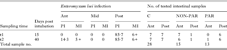
Enteromyxum leei infection in experimentally infected gilthead sea bream. Prevalence of infection (%) (PI) and mean intensity of infection (MI) are given for the three intestinal histological sections. Sample sizes for the control, unexposed (C), for the exposed, non-parasitized (NON-PAR) and for the parasitized (PAR) groups are given. Anterior intestine (Ant); Middle intestine (Mid); Posterior intestine (Post)

RESULTS
Enteromyxum leei infection
At 15 (t1) and 40 (t2) dpi prevalence of infection reached 85·7% in R GSB. At both times, all sampled fish except one were infected with a high intensity at the posterior intestine, and the observed parasite stages in all examined intestine sections ranged between proliferative stages with secondary cells and disporous sporoblasts with mature as well as immature spores. Details are summarized in Table 2. There were no significant differences between biometrical data of the sampled fish (control not exposed (C) vs recipient (R) and t1 vs t2) (average weights: C-t1 = 131·6 g; C-t2 = 168·0 g; R-t1 = 149·4 g; R-t2 = 149·0 g).
Granulocyte histochemistry
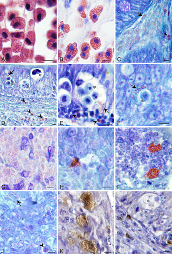
In haematoxylin/eosin and Giemsa stained sections, fuchsia-red coloured eosinophil/acidophil granulocytes were observed in all examined organs (Fig. 1A–J). They predominated in the lamina propria-submucosa (Fig. 1C and D) in both C and R intestines and degranulation close to intraepithelial parasite stages was best observed (Fig. 1E and F). Weakly stained, large eosinophil/acidophil granulocytes and smaller eosinophil/acidophil granulocytes densely packed with cytoplasmatic granules were distinguishable (Fig. 1C). Such cells were also found within blood vessels of different organs. No metachromasy was observed in toluidine-stained tissues, no granulocytes were stained with alcian blue, and no PAS positive granules were found (not shown).
Granulocyte heterogeneity in infected gilthead sea bream tissue sections: histochemistry (A: haematoxylin & eosin, B–J: Giemsa) and anti-histamine immunohistochemistry (K–L: haematoxylin). Enteromyxum leei stages are indicated by asterisks. (A, B) Eosinophil/acidophil granulocytes in the posterior intestine (Pi). (C) Abundant, large eosinophil/acidophil granulocytes (arrow) and scarce, small eosinophil/acidophil granulocytes (arrow) in lamina propria-submucosa of Pi. Note the diffuse granule content in large ones and the densely packed granules in the small ones. (D) Small eosinophil/acidophil granulocytes in infected Pi. Note their abundance in the lamina propria-submucosa (arrowheads) and some infiltration in the epithelium (arrow). (E) Degranulation (arrow) into a proliferative parasite stage with numerous secondary daughter cells in the Pi. Small eosinophil/acidophil granulocytes (arrowheads) are infiltrated in the epithelium. (F) Eosinophil/acidophil granules surrounding an epithelial parasite stage in the Pi. (G) Head kidney parenchyma with numerous large eosinophil/acidophil granulocytes. (H) One small eosinophil/acidophil granulocyte close to a blood vessel in head kidney. (I) Two eosinophil/acidophil granulocytes in the thymus. (J) Scarce eosinophil/acidophil granulocytes in the splenic parenchyma showing different granule densities: disperse granules (arrow), dense granules (arrowhead). (K) Large histamine IRCs in the lamina propria-submucosa of the anterior intestine. (L) Small histamine IRCs in the lamina propria-submucosa of Pi. Scale bars = 5 μm.

IgM-IRC distribution pattern
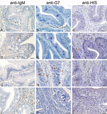
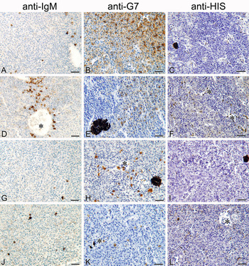
IRCs for the polyclonal antibody (Pab) anti-GSB IgM (Pab-IgM) were detected in intestines as well as in lymphohaematopoietic tissues of C and R fish (Figs 1–3). Their morphology was in most cases compatible with plasma cells for their voluminous cytoplasm (large cytoplasm/nucleus ratio) and in some occasions with B cells for their round shape and thin cytoplasm rim (low cytoplasm/nucleus ratio) (Supplementary online material 1A – in Online version only). In both cell types, the staining was located in the cytoplasm and different intensities of immunoreactivity were observed depending on the degree of cell differentiation. In intestines, IgM-IRCs were mainly located in the lamina propria-submucosa and occasionally infiltrated in the epithelium in close contact to the parasite (Fig. 2D and J; Supplementary online material 1E – in Online version only). On the epithelial basal lamina and in the hypertrophied lamina propria-submucosa of R intestines, high amounts of cells morphologically resembling lymphocytes were present, which were not immunoreactive (Supplementary online material 1G – in Online version only). In lymphohaematopoietic tissues, IgM-IRCs were found in the renal and thymic parenchyma and in the splenic white pulp (Fig. 1G–J), often close to blood vessels (Fig. 2D) and MMCs. In C tissues, IgM-IRCs were more abundant in lymphohaematopoietic tissues than in intestines and head kidney presented the highest IgM-IRC counts.
Leukocyte immunohistochemistry of gilthead sea bream intestinal sections counterstained with haematoxylin. Anterior intestine (A–F) and posterior intestine (G–L) sections of control, unexposed fish (A–C and G–I) and of Enteromyxum leei-exposed fish (D–F and J–L) were immunolabelled with the anti-IgM polyclonal antibody (Pab), with the anti-G7 monoclonal antibody or with the anti-histamine (anti-HIS) polyclonal antibody, which detect plasma cells/B cells, acidophilic granulocytes and mast cells, respectively. Scale bars = 20 μm.

Leukocyte immunohistochemistry of gilthead sea bream sections of lymphohaematopoietic organs counterstained with haematoxylin. Head kidney (A–F) and spleen (G–L) sections of control, unexposed fish (A–C and G–I) and of Enteromyxum leei-exposed fish (D–F and J–L) were immunolabelled with the anti-IgM polyclonal antibody (Pab), with the anti-G7 monoclonal antibody or with the anti-histamine (anti-HIS) polyclonal antibody, which detect plasma cells/B cells, acidophilic granulocytes and mast cells, respectively. Note the presence of immunoreactive cells in and around blood vessels (*). Scale bars = 20 μm.

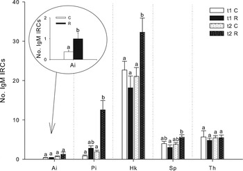
At t1, no statistically significant differences in IgM-IRCs between R and C fish were observed (Fig. 4). By contrast, at t2, a statistically significant increase in IgM-IRCs occurred in the posterior intestine and the head kidney of R fish compared with C, though more pronounced at the posterior intestine, in which a six-fold increase was detected. In R spleens, the IgM-IRC number was significantly higher at t2 than at t1. Though no significant differences in IgM-IRCs were detected at the anterior intestine for either of the two sampling times individually, when data were pooled into C and R groups, regardless of the time of infection, the number of IRCs was significantly higher in R than in C fish (inset Fig. 4). Interestingly, at t1, a pattern of IgM-IRC increase was recorded for both intestinal segments of R fish compared with their C group, in contrast with the decreasing trend observed in the lymphohaematopoietic tissues (head kidney, spleen and thymus).
Mean+s.e.m. of IgM immunoreactive cells (IRCs) in tissue sections of recipient (R) and control (C) gilthead sea bream at two sampling times: t1, 15 days post Enteromyxum leei intubation (dpi) and t2, 40 dpi. Pooled Ai data of t1 and t2 for C and R fish, respectively, are represented in the inset. For each tissue, letters indicate statistically significant differences (P<0·05) between groups. Anterior intestine (Ai), posterior intestine (Pi), head kidney (Hk), spleen (Sp), thymus (Th).

G7-IRC distribution pattern
The monoclonal antibody (Mab) against the G7 molecule (Mab-G7) labelled acidophilic granulocytes in all the examined intestinal and lymphohaematopoietic tissues of C and R GSB (Figs 1–3). These leukocytes had a rounded eccentric nucleus and a voluminous granular cytoplasm (large cytoplasm/nucleus ratio), in which the immunolabel was observed by light microscopy. Their shape was round in the spleen (Fig. 3H), while in the other tissues they presented heterogeneous morphologies (Fig. 2P, Q and R). Different degrees of immunoreactivity were mainly observed in head kidney (Fig. 3B). In intestinal sections, acidophilic granulocytes predominated in the lamina propria-submucosa but also infiltrated in the epithelium (Fig. 2H; Supplementary online material 1F – in Online version only). In head kidney, the highest amounts of acidophilic granulocytes formed clusters that extended over the parenchyma (Fig. 3B). By contrast, acidophilic granulocytes in the spleen were scattered throughout the organ (Fig. 3H) as in the thymic parenchyma, in which scattered acidophilic granulocytes were scarcely found (Fig. 1K; Supplementary online material 1B – in Online version only).
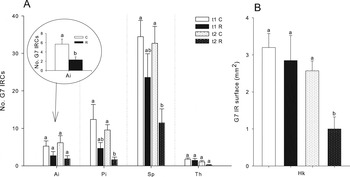
Statistically significant decreases in the number of G7 immunoreactive granulocytes of R fish were only found at t2 in the posterior intestine, head kidney and spleen (Fig. 5). However, such a decreasing trend of the acidophilic granulocytes was found in all tissues at both infection times. Although no significant differences were observed at any sampling point at the anterior intestine, a significantly lower acidophilic granulocyte number was detected in R fish with respect to C fish for pooled data of both samplings (inset Fig. 5).
Mean+s.e.m. of G7 immunoreactive cells (IRCs) (A) or immunoreactive surface (B) in tissue sections of recipient (R) and control (C) gilthead sea bream at two sampling times: t1, 15 days post Enteromyxum leei intubation (dpi) and t2, 40 dpi. Pooled Ai data of t1 and t2 for C and R fish, respectively, are represented in the inset. For each tissue, letters indicate statistically significant differences (P<0·05) between groups. Anterior intestine (Ai), posterior intestine (Pi), head kidney (Hk), spleen (Sp), thymus (Th).

HIS-IRC distribution pattern
Immunoreactive mast cells for the Pab against HIS (Pab-HIS) were present in all the studied tissues of C and R fish (Figs 1 and 2). Two morphological types of mast cells were immunolabelled by the Pab-HIS, regarding their size. The small-sized type (4–7 μm) showed a round to oval eccentric nucleus and a small cytoplasm, which was densely immunolabelled (Fig. 1L). The large-sized type presented a similar nuclear appearance, but an extremely larger cytoplasm (10–15 μm cell size), in which a more dispersed and granular immunolabel was observed (Fig. 1K). Intermediate sizes of immunoreactive mast cells could be observed mainly in lymphohaematopoietic tissues (Fig. 3G, F, I and L; Supplementary online material 1C – in Online version only). Notably, the large mast cell type was mainly observed at the anterior intestine (Fig. 2F). At the posterior intestine, by contrast, both morphological types coexisted, and scarce intermediate stages were found.
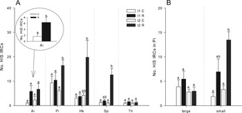
A similar increasing pattern of mast cells in R compared with C fish was detected in all tissues at the two sampling times, though the increase was statistically significant only at t2 in the posterior intestine and the spleen (Fig. 6). The increase of HIS-IRCs in the head kidney of R fish at t2 was only significant compared with R fish at t1, but not with respect to C. At the anterior intestine, a significant increase in large-sized mast cells was detected in R compared with C, after pooling data of both infection times. At the posterior intestine, the two size types of mast cells were quantified separately due to their high amount. No variations in the number of the large type were found due to infection, whereas significant increase was found in the small type in R fish at t2.
Mean+s.e.m. of histamine (HIS) immunoreactive cells (IRCs) in tissue sections of recipient (R) and control (C) gilthead sea bream at two sampling times: t1, 15 days post Enteromyxum leei intubation (dpi) and t2, 40 dpi. (A) Pooled Ai data of t1 and t2 for C and R fish, respectively, are represented in the inset. Anterior intestine (Ai), posterior intestine (Pi), head kidney (Hk), spleen (Sp), thymus (Th); (B) HIS-IRC types by their size at the Pi. For each tissue and cell type, letters indicate statistically significant differences (P<0·05) between groups.

MMCs distribution pattern
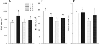
The size of the MMCs was analysed in splenic sections and no significant differences between C and R fish groups were found (Fig. 7, Supplementary online material 2 – in Online version only). The same happened for the measurements on the number of MMCs per mm2 and for the percentage of splenic surface occupied by MMCs.
Mean+s.e.m. of melanomacrophage centres (MMCs) in splenic sections of recipient (R) and control (C) gilthead sea bream at two sampling times: t1, 15 days post Enteromyxum leei intubation (dpi) and t2, 40 dpi. (A) MMC size in μm2; (B) Average count of splenic MMCs per mm2; (C) Percentage of splenic surface occupied by MMCs. Different letters indicate statistically significant differences (P<0·05) between groups.

Leukocyte populations vs parasitic status
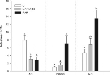
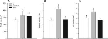
When data for acidophilic granulocytes in intestinal segments were pooled into C, NON-PAR and PAR, no differences between NON-PAR and PAR groups was found, but both groups showed a significantly lower number of acidophilic granulocytes compared with C fish (Fig. 8). Intestinal plasma cells/B cells, by contrast, were significantly higher in PAR than in C and NON-PAR intestines (Fig. 8). The number of intestinal mast cells was significantly higher only in PAR tissues compared with C counts, though a certain increase of mast cells was also found in NON-PAR (Fig. 8). A significant increase of the splenic surface occupied by MMCs was detected in NON-PAR fish compared with C and PAR, when fish were pooled by the infective status at the posterior intestine (Fig. 9).
Mean+s.e.m. of immunoreactive cells (IRCs) (acidophilic granulocytes (AG), plasma cells and B cells (PC/BC) and mast cells (MC)) in intestinal sections of control (C) unexposed gilthead sea bream and recipient gilthead sea bream inoculated with Enteromyxum leei. Data of anterior and posterior intestines of two samplings (15 and 40 days post intubation) are pooled and recipient fish, split into parasitized (PAR) and non-parasitized (NON-PAR) intestinal segments. Different letters indicate statistically significant differences between groups (P<0·05).

Mean+s.e.m. of melanomacrophage centres (MMCs) in splenic sections of control (C) and recipient gilthead sea bream inoculated with Enteromyxum leei. Data of two samplings (15 and 40 days post inoculum) are pooled and recipient fish, split into parasitized (PAR) and non-parasitized (NON-PAR) fish at the posterior intestine. (A) MMC size in μm2; (B) Average count of splenic MMCs per mm2; (C) Percentage of splenic surface occupied by MMCs. Different letters indicate statistically significant differences between groups (P<0·05).

DISCUSSION
Enteromyxum spp. entail a threat for several aquaculture-reared fish species and the lack of available therapeutic treatments and vaccines point out the need to broaden the knowledge on the immune factors that may participate in host protection (Sitjà-Bobadilla and Palenzuela, Reference Sitjà-Bobadilla, Palenzuela, Woo and Buchmann2012). Most available data on GSB immune response to enteromyxosis have been gathered from long-term experimental infections by cohabitation with E. leei-infected fish or exposure to E. leei-contaminated effluent (>100 days exposure) (Cuesta et al. Reference Cuesta, Muñoz, Rodríguez, Salinas, Sitjà-Bobadilla, Álvarez-Pellitero, Esteban and Meseguer2006b , Reference Cuesta, Salinas, Rodríguez, Muñoz, Sitjà-Bobadilla, Álvarez-Pellitero, Meseguer and Esteban c ; Sitjà-Bobadilla et al. Reference Sitjà-Bobadilla, Diamant, Palenzuela and Álvarez-Pellitero2007, Reference Sitjà-Bobadilla, Calduch-Giner, Saera-Vila, Palenzuela, Álvarez-Pellitero and Pérez-Sánchez2008; Davey et al. Reference Davey, Calduch-Giner, Houeix, Talbot, Sitjà-Bobadilla, Prunet, Pérez-Sánchez and Cairns2011; Estensoro et al. Reference Estensoro, Benedito-Palos, Palenzuela, Kaushik, Sitjà-Bobadilla and Pérez-Sánchez2011, Reference Estensoro, Calduch-Giner, Kaushik, Pérez-Sánchez and Sitjà-Bobadilla2012a , Reference Estensoro, Redondo, Salesa, Kaushik, Pérez-Sánchez and Sitjà-Bobadilla b , Reference Estensoro, Jung-Schroers, Álvarez-Pellitero, Steinhagen and Sitjà-Bobadilla2013). In the current study, the relatively fast progression of the E. leei infection by the anal route resulted in high prevalence and intensity of infection as early as 15 dpi, confirming previous findings on this experimental infection route (Estensoro et al. Reference Estensoro, Redondo, Álvarez-Pellitero and Sitjà-Bobadilla2010). Recipient (R) fish harboured a well-established infection of proliferative as well as sporogonic parasite stages, but the disease outcome was not yet apparent, since neither mortalities nor macroscopic signs of the disease were recorded.
The infection route not only influenced the infection level (intensity and prevalence), but it also determined the onset of the host immune response. This becomes particularly evident in regard to the IgM local response, when the current results are compared with data from a previous trial, in which GSB were exposed to the parasite by effluent (Estensoro et al. Reference Estensoro, Calduch-Giner, Kaushik, Pérez-Sánchez and Sitjà-Bobadilla2012a ). In that previous study, a detailed analysis of IgM dynamics was performed, showing a positive correlation between IgM transcription levels and IgM-IRC abundance, and only after 133 days of exposure to the parasite, a significant increase of IgM expression occurred at the posterior intestine. By contrast, the current experimental infection triggered a significant recruitment of plasma cells/B cells in PAR fish much earlier (40 dpi), when a six-fold IgM-IRC increase was recorded at the target site of the parasite, the posterior intestine, and also weaker at the anterior intestine. Such higher number of intestinal plasma cells/B cells, either from local synthesis, blood derived or both, contributed to the mucosal inflammatory response. At this time point (t2), renal and splenic plasma cells/B cells had significantly increased, probably as a sign of haematopoietic proliferation and of the maturation and initiation of the adaptive immune mechanisms.
The initial decreasing trend in lymphohaematopoietic tissues at t1, may suggest an early mobilization of IgM-IRCs from the systemic reservoirs to the local infection site, where such a cell type increased at this time point, but not significantly. Interestingly, this faster infection course seems to approach the infection dynamics and immune response of the GSB–E. leei model to those of the more pathogenic turbot-Enteromyxum scophthalmi model. In this species, an IgM-IRC increase in spleen and head kidney of R fish at 20 and 40 dpi was associated with the initiation of a humoral immune response, which was followed by a subsequent migration of IgM-IRCs to the infected intestines and finally, by lymphohaematopoietic depletion owing to immunodepression (Bermúdez et al. Reference Bermúdez, Vigliano, Marcaccini, Sitjà-Bobadilla, Quiroga and Nieto2006). In regard to IgM as well as to the mucosal IgT, the participation of mucosal-systemic and cellular–humoral interactions in the immune response of fish has been reported previously (Álvarez-Pellitero, Reference Álvarez-Pellitero2008; Jorgensen et al. Reference Jorgensen, Heinecke, Skjodt, Rasmussen and Buchmann2011; Olsen et al. Reference Olsen, Kania, Heinecke, Skjoedt, Rasmussen and Buchmann2011; Salinas et al. Reference Salinas, Zhang and Sunyer2011; Ordas et al. Reference Ordas, Castro, Dixon, Sunyer, Bjork, Bartholomew, Korytar, Kollner, Cuesta and Tafalla2012). Although the contribution of natural antibodies before the induction of an adaptive immune response has been recognized in teleosts, the involvement of innate-like lymphocytes is still far from being understood (Whyte, Reference Whyte2007; Álvarez-Pellitero, Reference Álvarez-Pellitero2008; Gómez and Balcázar, Reference Gómez and Balcázar2008; Magnadottir et al. Reference Magnadottir, Gudmundsdottir, Gudmundsdottir and Helgason2009; Hu et al. Reference Hu, Zhu, Xiang and Shao2011). Remarkable is, however, the phagocytic capacity detected for B cells in some fish species suggesting their evolutionary relationship with macrophages and their possible role in innate immune functions (Li et al. Reference Li, Barreda, Zhang, Boshra, Gelman, Lapatra, Tort and Sunyer2006; Overland et al. Reference Overland, Pettersen, Ronneseth and Wergeland2010; Sunyer, Reference Sunyer2012). However, the possible contribution of IgM-IRCs to the innate immune response of GSB against enteromyxosis deserves further study. Additionally, Ig-binding to Fc receptors and phagocytosis of immune complexes and plasma cells have been previously associated in fish to the presence of few IgM+ macrophages and neutrophils (Pettersen et al. Reference Pettersen, Bjerknes and Wergeland2000; Bermúdez et al. Reference Bermúdez, Vigliano, Marcaccini, Sitjà-Bobadilla, Quiroga and Nieto2006; Grove et al. Reference Grove, Tryland, Press and Reitan2006), though such cells with phagosomal-like structures were not identified in the current study.
The involvement of innate defence mechanisms in the immune response of fish to parasite infections has been studied in several host–parasite models (Álvarez-Pellitero, Reference Álvarez-Pellitero2008). In GSB, lymphocytes, monocyte/macrophages and acidophilic granulocytes are considered non-specific cytotoxic cells (NCCs) (Meseguer et al. Reference Meseguer, Esteban and Mulero1996; Cuesta et al. Reference Cuesta, Esteban and Meseguer2005). In particular, an important role is attributed to the cellular effectors involved in the intestinal inflammatory response against E. leei (Fleurance et al. Reference Fleurance, Sauvegrain, Marques, Le Breton, Guereaud, Cherel and Wyers2008; Cuadrado, Reference Cuadrado2009; Estensoro et al. Reference Estensoro, Redondo, Álvarez-Pellitero and Sitjà-Bobadilla2010) and responsible for the variations detected in the cytotoxic, phagocytosis and respiratory burst activities and peroxidase content during enteromyxosis (Cuesta et al. Reference Cuesta, Muñoz, Rodríguez, Salinas, Sitjà-Bobadilla, Álvarez-Pellitero, Esteban and Meseguer2006b , Reference Cuesta, Salinas, Rodríguez, Muñoz, Sitjà-Bobadilla, Álvarez-Pellitero, Meseguer and Esteban c ; Sitjà-Bobadilla et al. Reference Sitjà-Bobadilla, Calduch-Giner, Saera-Vila, Palenzuela, Álvarez-Pellitero and Pérez-Sánchez2008; Estensoro et al. Reference Estensoro, Benedito-Palos, Palenzuela, Kaushik, Sitjà-Bobadilla and Pérez-Sánchez2011). Such innate mechanisms were also engaged in the response of GSB to bacterial and viral threats (Reyes-Becerril et al. Reference Reyes-Becerril, López-Medina, Ascencio-Valle and Esteban2011b ; Chaves-Pozo et al. Reference Chaves-Pozo, Guardiola, Meseguer, Esteban and Cuesta2012; Dezfuli et al. Reference Dezfuli, Lui, Giari, Castaldelli, Mulero and Noga2012b ), as well as to bioactive compounds (Cuesta et al. Reference Cuesta, Cerezuela, Esteban and Meseguer2008; Reyes-Becerril et al. Reference Reyes-Becerril, Ascencio-Valle, Tovar-Ramírez, Meseguer and Esteban2011a ; Cabas et al. Reference Cabas, Liarte, Garcia-Alcazar, Meseguer, Mulero and García-Ayala2012; Águila et al. Reference Águila, Castillo-Briceno, Sánchez, Cabas, García-Alcázar, Meseguer, Mulero and García-Ayala2013). Indeed, acidophilic granulocytes are professional phagocytes of GSB (together with macrophages) and are considered the functional equivalents of mammalian neutrophils as they can be rapidly recruited from head kidney and are the most abundant circulating granulocytes (Sepulcre et al. Reference Sepulcre, Pelegrin, Mulero and Meseguer2002; Chaves-Pozo et al. Reference Chaves-Pozo, Muñoz, López-Muñoz, Pelegrin, Ayala, Mulero and Meseguer2005). They contribute to the respiratory burst activity in tissues and blood and contain in their granules reactive oxygen intermediates (Couso et al. Reference Couso, Castro, Noya, Obach and Lamas2001; Sepulcre et al. Reference Sepulcre, Pelegrin, Mulero and Meseguer2002), antimicrobial peptides (piscidin 3) (Mulero et al. Reference Mulero, Noga, Meseguer, García-Ayala and Mulero2008) and lysosomal enzymes (peroxidase and acid phosphatase) (Meseguer et al. Reference Meseguer, López-Ruiz and Esteban1994a ), which can all be intracellularly (into phagosomes) or extracellularly secreted to degrade pathogens. Acidophilic granulocytes also express the pro-inflammatory cytokine interleukin-1β, the inflammatory prostanoid COX-2 (Chaves-Pozo et al. Reference Chaves-Pozo, Pelegrin, García-Castillo, García-Ayala, Mulero and Meseguer2004; Sepulcre et al. Reference Sepulcre, López-Castejón, Meseguer and Mulero2007), and antigen-presenting major histocompatibility complex II alpha chain (Cuesta et al. Reference Cuesta, Esteban and Meseguer2006a ), thus suggesting that acidophilic granulocytes participate in the regulation of inflammation and link innate and adaptive immune mechanisms. Our results showed that this paramount leukocyte for the defence of GSB decreased in all examined tissues of R fish (PAR and NON-PAR, except in thymus), which may explain at least part of the limited protection of this species against the E. leei. In accordance, a decrease of the acidophilic granulocyte percentage (and consequent decrease in respiratory burst, phagocytic activity and leukocyte peroxidase) in the head kidney of E. leei-infected GSB was detected (Cuesta et al. Reference Cuesta, Salinas, Rodríguez, Muñoz, Sitjà-Bobadilla, Álvarez-Pellitero, Meseguer and Esteban2006c ). On the other hand, the high serum peroxidase content found after E. leei exposure supports leukocyte mobilization into the circulatory system (Cuesta et al. Reference Cuesta, Muñoz, Rodríguez, Salinas, Sitjà-Bobadilla, Álvarez-Pellitero, Esteban and Meseguer2006b ). Nevertheless, immunosuppressive effects of some fish parasites included the inhibition of NCC proliferation, of their phagocytic activity and the induction of NCC apoptosis. Moreover, immunomodulation by some parasites interferes with cytokine transcription to facilitate their survival (Sitjà-Bobadilla, Reference Sitjà-Bobadilla2008). The observed significant and almost ubiquitous decrease of acidophilic granulocytes during acute E. leei infection in GSB deserves further investigation.
Fish mast cells are mainly eosinophilic granulocytes actively involved in bacterial and parasite clearance at mucosal sites through degranulation of antimicrobial peptides and enzymes such as lysozyme (Reite, Reference Reite1998; Murray et al. Reference Murray, Leggiadro and Douglas2007; Corrales et al. Reference Corrales, Mulero, Mulero and Noga2010). The cytoplasmic granules of GSB mast cells participating in the innate mucosal defence may contain HIS, piscidin 3 and likely piscidins 1 and 2 (Mulero et al. Reference Mulero, Sepulcre, Meseguer, García-Ayala and Mulero2007, Reference Mulero, Noga, Meseguer, García-Ayala and Mulero2008). Our results show HIS-containing mast cells with different morphologies among the granular infiltrates in the intestines of GSB, but also in lymphohaematopoietic tissues, and these leukocytes presented an opposite distribution pattern to the distribution of acidophilic granulocytes during the trial. Even if HIS-containing granules are present in mature mast cells, different sizes of HIS-IRCs might be associated to different developmental cell stages, thus the small ones being the ‘young’ cells proliferating in response to the parasite threat and being recruited from lymphohaematopoietic tissues into infection sites. The large-sized mast cells would therefore correspond to a more constant tissue resident population of ‘older’ mast cells. Furthermore, Giemsa staining also revealed the existence of two types of acidophilic granular cells by their size and granule density. Generally speaking, fish mast cells are considered a heterogeneous cell population. It is accepted that mast cell precursors leave haematopoietic organs and, via blood circulation, reach mucosal sites in which maturation takes place locally (Reite, Reference Reite1996, Reference Reite1998). The presence of eosinophilic granular cells (EGCs), likely mast cells, among the intestinal inflammatory infiltrates of E. leei-infected species has been previously reported (Sitjà-Bobadilla et al. Reference Sitjà-Bobadilla, Diamant, Palenzuela and Álvarez-Pellitero2007; Álvarez-Pellitero et al. Reference Álvarez-Pellitero, Palenzuela and Sitjà-Bobadilla2008; Fleurance et al. Reference Fleurance, Sauvegrain, Marques, Le Breton, Guereaud, Cherel and Wyers2008; Estensoro et al. Reference Estensoro, Redondo, Álvarez-Pellitero and Sitjà-Bobadilla2010; Katharios et al. Reference Katharios, Rigos and Divanach2011), as well as in response to other parasitic diseases (Álvarez-Pellitero, Reference Álvarez-Pellitero2008). Many authors have claimed EGCs to be mammalian mast cell equivalents for their functional and morphological similarities (Reite, Reference Reite1996; Reite and Evensen, Reference Reite and Evensen2006; Mulero et al. Reference Mulero, Sepulcre, Meseguer, García-Ayala and Mulero2007; Gómez and Balcázar, Reference Gómez and Balcázar2008; Dezfuli et al. Reference Dezfuli, Giari, Squerzanti, Lui, Lorenzoni, Sakalli and Shinn2011b , Reference Dezfuli, Lui, Giari, Castaldelli, Mulero and Noga2012b ). Apparently, different EGC1 and EGC2 types were involved in the response of both sparids GSB and Diplodus puntazzo to E. leei, though in the current study neither PAS positive nor different sized granules according to this description could be found (Álvarez-Pellitero et al. Reference Álvarez-Pellitero, Palenzuela and Sitjà-Bobadilla2008; Estensoro et al. Reference Estensoro, Redondo, Álvarez-Pellitero and Sitjà-Bobadilla2010). In the current results, the absence of PAS, but also toluidine and alcian blue positive granules reported for mast cells from other teleosts, may have been influenced by the different fixative we used, by the staining procedure itself, or by interspecific differences (Noya and Lamas, Reference Noya and Lamas1996; Reite, Reference Reite1996, Reference Reite1998; Rocha and Chiarini-Garcia, Reference Rocha and Chiarini-Garcia2007). However, the number of EGC1 decreased, whereas the number of EGC2 increased in R D. puntazzo (Álvarez-Pellitero et al. Reference Álvarez-Pellitero, Palenzuela and Sitjà-Bobadilla2008), in coincidence with the detected acidophilic granulocyte decrease and mast cell increase in R GSB. While the term EGC refers to the staining properties of mononuclear granule-containing cells, it encompasses functionally different cell types, at least acidophilic granulocytes and mast cells. In any case, a remarkable intraspecific heterogeneity among the EGCs of GSB seems obvious.
The large EGC type with dispersed granules seems to correspond to acidophilic granulocytes, the predominant type in head kidney, but also to some intestinal mast cells. The small and densely packed EGC type seems to correspond exclusively to the mast cell population being recruited into the parasitized intestine. Both types of granular cells studied were found infiltrated in the intestinal epithelium in close contact with the parasite and degranulation could be observed. Interestingly, intraepithelial acidophilic granulocytes, mainly present in unexposed GSB, have not been observed in previous studies. Zones of extracellular IgM immunostaining and plasma cells surrounding parasite stages were also observed. Thus, the involvement of all three leukocyte populations in the GALT inflammatory reaction against the E. leei seems evident and, furthermore, this is apparently supported at systemic level since the distribution patterns of these leukocytes at the posterior intestine and at the head kidney show parallel changes. Circulating granulocytes observed in tissue vessels supports this. Accordingly, the perivascular position of many leukocyte types is associated to their regulatory function during inflammatory responses (Mekori, Reference Mekori2004; Mulero et al. Reference Mulero, Sepulcre, Meseguer, García-Ayala and Mulero2007; Dezfuli et al. Reference Dezfuli, Giari, Lui, Squerzanti, Castaldelli, Shinn, Manera and Lorenzoni2012a ). Some examples of cellular innate responses to piscine myxosporeans are documented, such as the neutrophil and eosinophil-like infiltration during gill inflammation in channel catfish infected with Henneguya spp., whose degranulation within lesions was associated with further host tissue damage (Lovy et al. Reference Lovy, Goodwin, Speare, Wadowska and Wright2011). Furthermore, the time-dependent succession of granulocytes and lymphocytes in the oedemata surrounding Myxobolus pendula branchial cysts was described in creek chub (Martyn et al. Reference Martyn, Hong, Ringuette and Desser2002). Other studies on leukocyte contribution during piscine parasitoses involve tapeworms and acanthocephalans (Dezfuli et al. Reference Dezfuli, Castaldelli, Bo, Lorenzoni and Giari2011a , Reference Dezfuli, Giari, Squerzanti, Lui, Lorenzoni, Sakalli and Shinn b , Reference Dezfuli, Giari, Lui, Squerzanti, Castaldelli, Shinn, Manera and Lorenzoni2012a , Reference Dezfuli, Lui, Giari, Castaldelli, Shinn and Lorenzoni c , Reference Dezfuli, Lui, Pironi, Manera, Shinn and Lorenzoni2013; Hansen et al. Reference Hansen, Trees, Bah, Hetzel, Martin, Bain, Tanya and Makepeace2011). It should be noted that HIS may have an ambivalent effect on respiratory burst activity of professional fish phagocytes, thus suggesting a role for mast cells in regulating phagocyte activity. Mast cells may also regulate the inflammatory response by inducing vasodilatation and leukocyte chemotaxis and activation (Matsuyama and Iida, Reference Matsuyama and Iida1999, Reference Matsuyama and Iida2001; Mulero et al. Reference Mulero, Sepulcre, Meseguer, García-Ayala and Mulero2007).
Our results show for the first time the presence of acidophilic granulocytes and mast cells in thymus of GSB. It is worth mentioning the absence of changes in the distribution patterns of the studied leukocytes in this organ during the whole trial. In agreement with the main role of this organ in T cell proliferation and maturation (Bowden et al. Reference Bowden, Cook and Rombout2005), it does not seem to play a relevant role in granulocyte or plasma cell kinetics during the mucosal response. Nevertheless, the presence of thymus IgM-IRC was relatively high and constant, similar to the IgM-IRC content in spleen, and higher than in intestines of control (C) fish (about 4 and 10 times of posterior and anterior intestine levels, respectively). IgM-producing cells in the teleost thymus have led several authors to suggest the involvement of this organ in humoral immunity of fish (Tian et al. Reference Tian, Xie, Zhang, Xu, Yao and Nie2009). Alternatively, IgM-bearing macrophages might be the IRCs we detected, since thymic macrophages also participate in the clearance of immature self-reactive thymocytes during the development of functional T cells (Bowden et al. Reference Bowden, Cook and Rombout2005). At this point, a remark should be made on the absence of specific markers for GSB T cells, as occurs for several teleosts. IgM negative, lymphocyte-resembling cells in E. leei-infected intestines of GSB were previously considered putative T cells (Estensoro et al. Reference Estensoro, Calduch-Giner, Kaushik, Pérez-Sánchez and Sitjà-Bobadilla2012a ), agreeing with observations made in E. scophthalmi-infected turbot intestines (Bermúdez et al. Reference Bermúdez, Vigliano, Marcaccini, Sitjà-Bobadilla, Quiroga and Nieto2006). In the latter species, cross-reactivity with a heterologous anti-human CD3ε was found (Vigliano et al. Reference Vigliano, Losada, Castello, Bermúdez and Quiroga2011), which unfortunately did not cross-react with GSB in spite of its wide interspecific cross-reactivity (unpublished data).
The role of MMCs in the fish immune response as primitive germinal centres is related to antigen retention and processing, and thus involved in maintaining humoral memory (Vigliano et al. Reference Vigliano, Bermúdez, Quiroga and Nieto2006). The significant increase in the percentage of splenic surface occupied by MMC in NON-PAR fish vs PAR and C fish could be related to larger number of MMC rather than to their size increase. An increase in MMCs was already reported in the spleen of E. leei-infected GSB (Fleurance et al. Reference Fleurance, Sauvegrain, Marques, Le Breton, Guereaud, Cherel and Wyers2008), of E. scophthalmi-infected turbot (Sitjà-Bobadilla et al. Reference Sitjà-Bobadilla, Redondo, Bermúdez, Palenzuela, Ferreiro, Riaza, Quiroga, Nieto and Álvarez-Pellitero2006; Ronza et al. Reference Ronza, Coscelli, Losada, Bermúdez and Quiroga2013) and in the intestine of Enteromyxum fugu-infected tiger puffer (Tun et al. Reference Tun, Ogawa and Wakabayashi2002). This increase was associated with the inflammatory response and in some cases with the presence of engulfed parasite stages in melanomacrophages. Focal development of MMCs was also detected in response to several infections, including myxosporean infections (Agius and Roberts, Reference Agius and Roberts2003). It is tempting to suggest that a fast melanomacrophage proliferation and activation in the spleen may protect GSB from parasite invasion, but little evidence is still available.
The current results suggest the involvement of acidophilic granulocytes, mast cells, plasma cells, B cells and melanomacrophages in the acute immune response of GSB to enteromyxosis at both lymphohaematopoietic and intestinal levels. Some morphological differences were detected between acidophilic granulocytes and mast cells, which presented an opposite response to the parasite infection. Several links have been drawn between humoral and cellular mechanisms (leukocyte derived humoral factors like serum peroxidase), as well as between innate and adaptive immune responses (antigen presenting phagocytic and cytotoxic acidophilic granulocytes; mucosal-systemic cooperation). The participation of such cell types outlines once more the artificial nature of such boundaries (Álvarez-Pellitero, Reference Álvarez-Pellitero2011; Criscitiello and de Figueiredo, Reference Criscitiello and De Figueiredo2013). In addition, immune evasive, immunosuppressive or even immunomodulatory effects of the parasite on such cellular effectors are poorly understood and deserve further study.
SUPPLEMENTARY MATERIAL
To view supplementary material for this article, please visit http://dx.doi.org/10.1017/S0031182013001789.
ACKNOWLEDGEMENTS
The authors thank R. del Pozo for assistance in fish maintenance and samplings, J. Monfort and L. Rodríguez for histological processing and J. Pertusa for ImageJ assessment.
FINANCIAL SUPPORT
This work was funded by MICINN through project AGL2009-13282-C02-01, and by the ‘Generalitat Valenciana’ (projects PROMETEO 2010/006 and ISIC 2012/003). I. E. received a PhD FPI fellowship from MEC.