Superior-inferior or upstairs-downstairs ventricles describes the anomaly in which the ventricles are situated one above the other, rather than the usual side-by-side arrangement. When the ventricles are positioned in this abnormal fashion on top of one another, then the ventricular septum adopts a horizontal position.Reference Anderson1, Reference Freedom, Culham, Moes, Freedom, Culham and Moes2 Most of the hearts thus far reported with this arrangement have been characterized by unexpected ventricular and great arterial relationships for the given segmental connections, with twisted or criss-crossing atrioventricular junctions.Reference Freedom, Mawson, Yoo, Benson, Freedom, Mawson, Yoo and Benson3 Less commonly, a superior-inferior relationship of the ventricles is present without a twisted nature of the atrioventricular junction, as if the apex of the ventricles is simply tilted upwards.Reference Anderson1 Almost always, superior-inferior ventricles are associated with cardiovascular pathology such as discordant atrioventricular connections, ventricular septal defects, straddling atrioventricular valves, ventricular hypoplasia, obstruction of the ventricular outflow tracts, and abnormal ventriculo-arterial connections.Reference Freedom, Culham, Moes, Freedom, Culham and Moes2–Reference Seo, Yoo, Ho, Lee and Anderson8 Due to the complexity of these anomalies, a biventricular repair is often not possible, or else is achieved only after extensive surgery.Reference Peirone, Freedom, Yoo, Freedom, Yoo, Mikailian and Williams7
In their Angiographic Textbooks of Congenital Heart Disease, Freedom and colleagues reported three unrelated children with yet another form of superior-inferior ventricles, which they named “topsy turvy hearts”.Reference Freedom, Culham, Moes, Freedom, Culham and Moes2, Reference Freedom, Mawson, Yoo, Benson, Freedom, Mawson, Yoo and Benson3 In its literal sense, this phrase means a state of inversion, with the top-side pointing the other way. Indeed, in this condition, it is the entire cardiac mass as a block, rather than only the ventricles, which is rotated clockwise along the axis from apex to base, leaving the right ventricle and right atrium in a superior spatial relationship to their left-sided counterparts, while the great arteries are displaced inferiorly and posteriorly into the distal mediastinum. To the best of our knowledge, no documentation of this exceedingly rare form of superior-inferior cardiac malposition has been published in the peer-reviewed medical literature. We present here two examples of topsy-turvy hearts detected by fetal echocardiography, and describe the postnatal consequences of the peculiar anomaly.
Presentation of the cases
Two consecutive fetuses conceived by healthy parents, who were first degree cousins, were referred to our fetal programmes in mid-gestation with virtually identical cardio-vascular findings. As with the initial description by Freedom et al.,Reference Freedom, Culham, Moes, Freedom, Culham and Moes2 both of the fetuses exhibited a superior-inferior relationship of the cardiac chambers in the setting of usual atrial arrangement, left-sided heart, and concordant atrioventricular and ventriculo-arterial connections. The true nature of the cardiovascular anomalies of the first fetus was only recognized retrospectively, after comparing the fetal echocardiogram with the correctly interpreted prenatal findings of the second sibling.
In the first case, the mother, 24 years old and carrying her first child, was referred for fetal echocardiography at 19 gestational weeks subsequent to detection of a discrepancy in the size of the ventricle at a routine obstetrical ultrasound scan. The fetus also had a single umbilical artery, and a small pericardial effusion. On detailed fetal echocardiography, the structures of the left heart were smaller when compared to the right side, the aortic arch was thought to be interrupted, and the left pulmonary artery branch was not seen. The couple decided to terminate the pregnancy after being counselled that the child would require extensive surgery to the aortic arch, and that a functionally biventricular outcome would be uncertain due to the suspected degree of left heart hypoplasia. An autopsy was declined. The karyotype was normal and male. Fluorescent in situ hybridization for 22q11.2 showed no deletion or duplication of this segment. Following their first pregnancy, the couple was counselled regarding their consanguinity, and the possibility that the cardiac condition may be associated with a single gene disorder with autosomal recessive mode of inheritance, and thus could carry a risk of recurrence as high as 25% for future pregnancies.
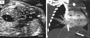
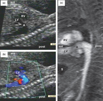
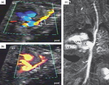
Their second pregnancy, conceived some months later, was initially uncomplicated. On the fetal echocardiogram at 18 weeks gestation, the heart was seen in an almost horizontal position, as if the arterial outlets, and with it the cardiac base, were pulled inferiorly towards the diaphragm by a shortened descending aorta. The normally-connected atriums and ventricles were situated in a superior-inferior spatial relationship, suggesting that the heart was also rotated clockwise along its long axis when viewed from the apex (Fig. 1a). The atrial and ventricular septums were in a virtually horizontal plane. Due to the abnormal orientation of the heart, the cardiac four-chamber view was obtained from an oblique coronal plane, rather than the standard position that is close to an axial plane. The inferior, morphologically left ventricle gave rise to a short ascending aorta which coursed horizontally and posteriorly (Fig. 2). The aortic arch was unusually low in the thorax, closer to the diaphragm than to the thoracic inlet. As a consequence, the brachiocephalic arteries were markedly elongated (Fig. 3a). The pulmonary trunk was also located in an unusually posterior and inferior position, angling towards the posterior aspect of the diaphragm. The arterial duct was short and wide. The intracardiac anatomy was essentially normal. Because of an unusually low position of the aortic arch, the possibility of significant compression of the airways was discussed at prenatal counselling.
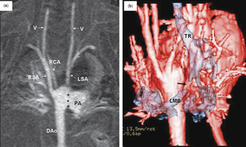
The parents opted to continue the pregnancy, and a female child was born with a normal karyotype, and a normal fluorescent in situ hybridization test for 22q11.2 deletion or duplication. The cardiovascular findings and consequences suspected prenatally were confirmed after birth by cross-sectional echocardiography, magnetic resonance imaging (Figs 1b, 2c, 3c, and 4a) contrast-enhanced computerized tomography of the chest (Fig. 4b) and at the time of surgery. The postnatal course was complicated by significant gastroesophageal reflux, with recurrent aspiration requiring fundoplication. Trapping of air in the left lung produced chronic respiratory distress, in consequence of near total occlusion of the main stem of the left bronchus. This was related to the elongated course of the left bronchus beneath the downwardly displaced aortic arch. Bronchial and tracheal decompression was ultimately achieved at one year of age by surgical translocation of the aortic arch into a more cephalad position, and by enlargement of the left bronchus with a patch, with eventual stenting using a 23 millimetre Genesis stent. The short and widely patent arterial duct (Fig. 4a) was closed during the surgical procedure. The child is now 3 years old, asymptomatic from the cardiac stance, but requires regular treatment and physiotherapy for repeated chest infections and reactive airways.
The unusual superior-inferior arrangement of the atriums and ventricles in our second patient with the interventricular septum (*) in an abnormal horizontal plane. Figure 1a shows the fetal oblique coronal echocardiogram, while Figure 1b is the postnatal contrast-enhanced magnetic resonance image. Abbreviations: A = ascending aorta; ant = anterior; DAo = descending aorta; LA = left atrium; LV = left ventricle; PA = pulmonary trunk; S = stomach; RA = right atrium; RV = right ventricle; post = posterior.

The left ventricular outflow tract is inferior displaced, supporting an abnormal shape intrathoracic aorta. The short “ascending” aorta is directed in a horizontal anterior-posterior direction to reach the aortic arch in the inferior-posterior mediastinum just above the diaphragm. Figure 2a is the fetal oblique coronal echocardiogram, Figure 2b shows the colour Doppler image, and Figure 2c is the postnatal contrast enhanced magnetic resonance image. Abbreviations: A = ascending aorta; ant = anterior; DAo = descending aorta; LA = left atrium; LV = left ventricle; PA = pulmonary trunk; S = stomach; RA = right atrium; RV = right ventricle; post = posterior.

There is marked elongation of the left subclavian artery (arrows), shown by fetal colour Doppler (a), fetal power Doppler (b) and postnatal contrast enhanced magnetic resonance image (c). Abbreviations: A = ascending aorta; ant = anterior; DAo = descending aorta; LA = left atrium; LV = left ventricle; PA = pulmonary trunk; S = stomach; RA = right atrium; RV = right ventricle; post = posterior.

The postnatal contrast-enhanced magnetic resonance image reconstructed in a coronal plane (a) illustrates the elongated brachiocephalic vessels originating from the posterior aspect of the left-sided aortic arch in the lower mediastinum. There is a large window-like patent arterial duct (asterisks). The elongated right (RSA) and left subclavian (LSA) arteries continue as the vertebral arteries (V) in the neck. Note that the right common carotid artery (RCA) is seen in the medial aspect of the right subclavian artery. The left common carotid is out of plane and not seen. The composite 3-dimensionally reconstructed computed tomographic image of the heart (b) shows the aorta (red) and airways (blue) seen from behind, illustrating the elongated trachea (TR) and left bronchus (LMB), which is compressed (arrow) by the distally displaced aortic arch.

Discussion
We describe here a cardiac malformation, to the best of our knowledge unique in the history of fetal diagnosis, characterized by a superior-inferior spatial relationship of the atriums and ventricles, and by downward displacement of the aortic arch and pulmonary trunk into the distal thorax, with markedly elongated brachiocephalic arteries. This rare condition has previously been detected after birth in three unrelated cases, and was named by Freedom and colleaguesReference Freedom, Culham, Moes, Freedom, Culham and Moes2, Reference Freedom, Mawson, Yoo, Benson, Freedom, Mawson, Yoo and Benson3 the topsy-turvy heart.
The spatial orientation of the heart in our two affected siblings may appear at a first glance similar to that seen in so-called criss-cross hearts. While there is, indeed, a similar superior-inferior relationship of the ventricles, the structure of the atrioventricular connections differs markedly between topsy-turvy and criss-cross hearts. The essential feature of the criss-cross variants is the twisted nature of the atrioventricular junctions, with loss of the usual parallel alignment of the atrioventricular connections.Reference Hery, Jimenez, Didier, van Doesburg, Guerin, Fouron and Davignon5, Reference Peirone, Freedom, Yoo, Freedom, Yoo, Mikailian and Williams7 Thus, in criss-cross hearts, the cardiac chambers and great arterial trunks are oriented as if the heart is twisted by a hand placed on the cardiac apex. In most cases, twisting is such as to place the right ventricular inlet superiorly, and the left ventricular inlet inferiorly. The extreme cases, with a higher degree of twisting, may have a side-by-side orientation at the ventricular apex, with only the inlet portions being located in supero-inferior position. The atrial and ventricular septums typically show an angulated or curved configuration. In contrast to criss-cross or twisted hearts, there are cases in which the ventricles have a superior-inferior relationship with a curved ventricular septum, but in which the parallel axes of the atrioventricular valves are maintained. Anderson described this group of hearts, showing exclusively a supero-inferior ventricular relationship, as a consequence of ventricular tilt.Reference Anderson1
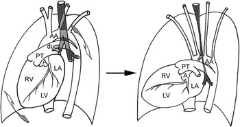
Our two cases reported herein differ from both these criss-cross hearts, and from superior-inferior ventricles with ventricular tilt, suggesting yet another potential mechanism for upstairs-downstairs ventricles. There is an approximately 90 degree clockwise rotation of the entire heart as a block around the cardiac long axis as seen from the apex, without any twisting of the atrioventricular connections, and no ventricular tilt (Fig. 5). As a consequence, the right-sided chambers assume a superior position relative to the chambers of the left heart, with the atrial and ventricular septums being oriented in a horizontal plane, and with the atrioventricular valves retaining their normal parallel axes. Rotation of the whole heart brings the great arterial trunks posteriorly towards the diaphragmatic surface, while elongation of the brachiocephalic arteries maintains the perfusion of the upper body. The major impact of the abnormally low position of the aortic arch, left-sided arterial duct, and pulmonary trunk is on the trachea and bronchuses, which become elongated and stretched. The potential for significant compression of the airways, therefore, can be predicted when this abnormality is identified during fetal echocardiography.
Illustrations of the normal heart (left panel) and the topsy-turvy heart (right panel) in steep left anterior oblique views. The left panel shows the normal concordant atrioventricular and ventriculo-arterial connections, with the expected spatial relationship of the cardiovascular segments. In the right panel, we show rotation of the entire heart through 90 degrees in clockwise fashion along its apex-base axis, producing an unexpected upstairs-downstairs positioning of the cardiac chambers. Despite their abnormal spatial relationship, the atriums, ventricles and great arteries are normally connected, without any twisting of their junctions, and without any major cardiac defect. The great arterial trunks are displaced posteriorly towards the diaphragm, resulting in elongation of the brachiocephalic arteries. A = ascending aorta; AA = aortic arch; LA = left atrium; LV = left ventricle; PT = pulmonary trunk; RV = right ventricle

As we have discussed in our introduction, criss-cross or twisted hearts are almost always associated with major cardiovascular pathology. In contrast, despite the bizarre external appearance of topsy-turvy hearts, none of the 3 cases reported by Freedom and his colleagues,Reference Freedom, Culham, Moes, Freedom, Culham and Moes2, Reference Freedom, Mawson, Yoo, Benson, Freedom, Mawson, Yoo and Benson3 nor our 2 cases, showed any major cardiac defects, apart from a short and wide persistently patent arterial duct. We speculate that abnormal cardiac embryogenesis is the primary pathogenesis of the criss-cross hearts with twisted atrioventricular connections, while the topsy-turvy hearts are the consequence of simple rotation of the entire heart, which otherwise develops normally. The parental consanguinity in our cases suggests that the topsy-turvy heart is genetically orchestrated, and is controlled by a single gene with an autosomal recessive mode of inheritance.
Based on our experiences, we suggest that a superior-inferior ventricular relationship can be the consequence of twisting of the heart along its long axis to produce the so-called criss-cross variant, horizontal right- or leftwards tilting of the ventricles to give the supero-inferior arrangement, or rotation of the whole heart as a block around the long axis to give the topsy-turvy heart. The main clinical significance of this last rarely encountered pathology is downward displacement of the aortic arch, the left-sided arterial duct and the left pulmonary artery, with elongation and severe compression of the left bronchus. The fact that our two siblings were of different gender, but born to first-degree cousins, suggests a recessive pattern of inheritance at least for some cases with topsy-turvy hearts, with a risk of recurrence of 25% for future pregnancies.
Acknowledgement
We thank Mrs. Eul Kyung Kim for making the diagrams shown in Figure 5.