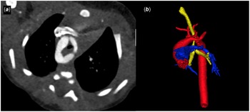
A vascular ring is a congenital malformation reflecting persistence, to varied extent, of the bilaterally symmetrical arterial structures which, during cardiac development, encircle the trachea and oesophagus. Such rings are extremely rare, accounting for no more than 1% of congenital heart abnormalities. Reference Tarmahomed and Umapathi1 Persistence to produce a double arch is the most common variant, accounting for just over half of reported cases, Reference Tarmahomed and Umapathi1 with several patterns reported in the literature. The pattern associated with ligamentous atresia of the distal left arch Reference Hung, Fu, Wei, Tsai and Chen2 is the focus of our report.
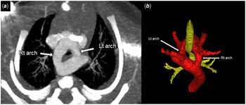
The patterns with luminal patency of both arches are readily recognised by echocardiography, but more definitively when using CT (Fig 1). The variant with a ligamentous atresia of the distal left arch is more difficult to diagnose. In the presence of such ligamentous atresia, the double arch can be mistaken on transthoracic echocardiography as a right aortic arch with mirror-imaged branching. We show the subtle differences in the transthoracic echocardiographic images of a right arch with mirror-imaged branching compared to suspected double arch with ligamentous atresia in movie clips 1 and 2. When there is a right arch with mirror-imaged branching, the left brachiocephalic trunk turns leftward. It then branches into left subclavian and left common carotid arteries, remaining anterior to the trachea. In the variant with ligamentous atresia of the distal left arch, in contrast, after giving off the left common carotid artery, a segment of the left arch extends posteriorly to give off the left subclavian artery. This can, on occasion, give the impression of mirror-imaged branching. These variants must be distinguished, since the form with ligamentous atresia can produce more prominent symptoms due to airway and oesophageal compression, and thus warrant surgical treatment. Whereas the right arch with mirror-imaged branching and a left-sided arterial duct can form a loose ring, the arrangement may not always require surgery, this depending on symptoms and the extent of airway compression. Here, we present six cases with ligamentous atresia of the distal left arch (Table 1). We emphasise the role of computed tomographic angiography in permitting timely and efficient diagnosis and treatment.
A double aortic arch showing complete patency of both right and left arches as seen on CT (panel A) and three-dimensional reconstruction (panel B). Note the stenotic trachea (yellow) due to the vascular compression.

Clinical presentation and echocardiographic findings of six patients with double aortic arch with ligamentous atresia of distal left arch

Results
In our series, half of the patients were diagnosed with right aortic arch or double aortic arch prenatally, with only half being symptomatic at the time of diagnosis. Our third patient, for example, was diagnosed during fetal scanning as having a right arch with a left-sided arterial duct. The first postnatal echo was again reported as showing a right arch with mirror-imaged branching, with the possibility of a vascular ring being dismissed. The fetal images, however, had been convincing for presence of a vascular ring with aortic arch on the right side of the trachea and ductus on the left side, completing the ring. Cardiac CT was obtained, which showed evidence of the double arch with ligamentous atresia of the distal left component. Careful review of the postnatal transthoracic echocardiograms then showed subtle features of the double aortic arch, which initially had been missed. The next three patients were then diagnosed correctly. Having made the diagnosis of the presence of ligamentous atresia within the double arch, we proceeded to early surgery, irrespective of the presence or absence of symptoms. Of our six patients, one was lost to follow-up. All the others have undergone surgical correction, with the findings confirmed intra-operatively. They all underwent division, via a lateral thoracotomy, of a ductal ligament and the ligament representing the atretic distal left arch.
Discussion
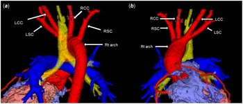
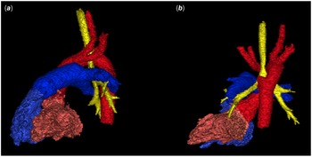
Remodelling of the arteries of the initial symmetrical arteries of the pharyngeal arches during cardiac development normally produces a single arch, which tracks in front of the trachea, crossing the left bronchus. Reference Hikspoors, Kruepunga, Mommen, Köhler, Anderson and Lamers3 The arch has three main branches. The first is the brachiocephalic trunk, which divides to become right subclavian and right common carotid arteries. This is followed by the left common carotid and then the left subclavian arteries. So-called vascular rings reflect failure of regression of the right-sided components of the initially symmetrical arteries extending through the pharyngeal arches. In consequence of the persistence of the right-sided components, an arch is also found on the right side, which crosses the right bronchus to track to the right of the trachea. Reference Hanneman, Newman and Chan4,Reference Weinberg, Allen, Driscoll, Shaddy and Feltes5 The persisting right and left arches then surround the trachea and oesophagus, joining together to form the descending aorta. The arrangement can promote stridor and dysphagia by virtue of the compression thus produced (Fig 1). Should the right arch persist with regression of the left-sided dorsal components, the result is a right arch with mirror-imaged branching (Fig 2). Reference Glew and Hartnell6 This variant needs to be differentiated from the variants of the double arch with ligamentous atresia of some of its parts (Figs 3-6). Reference Hung, Fu, Wei, Tsai and Chen2
Three-dimensional reconstruction of CT images of a right arch with mirror-imaged branching, with panel A showing the posterior view and panel B showing the anterior view.
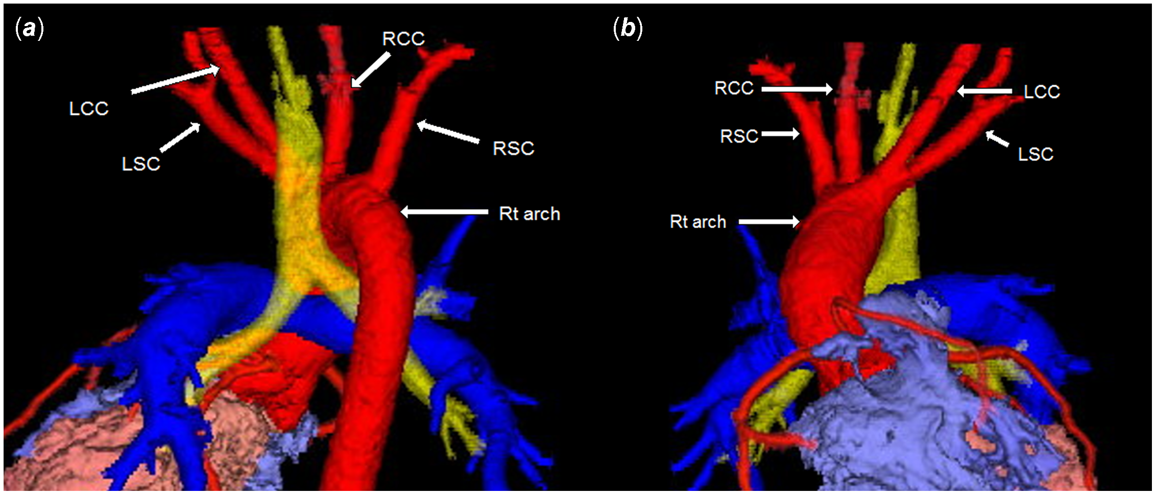
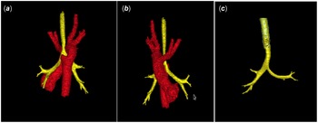
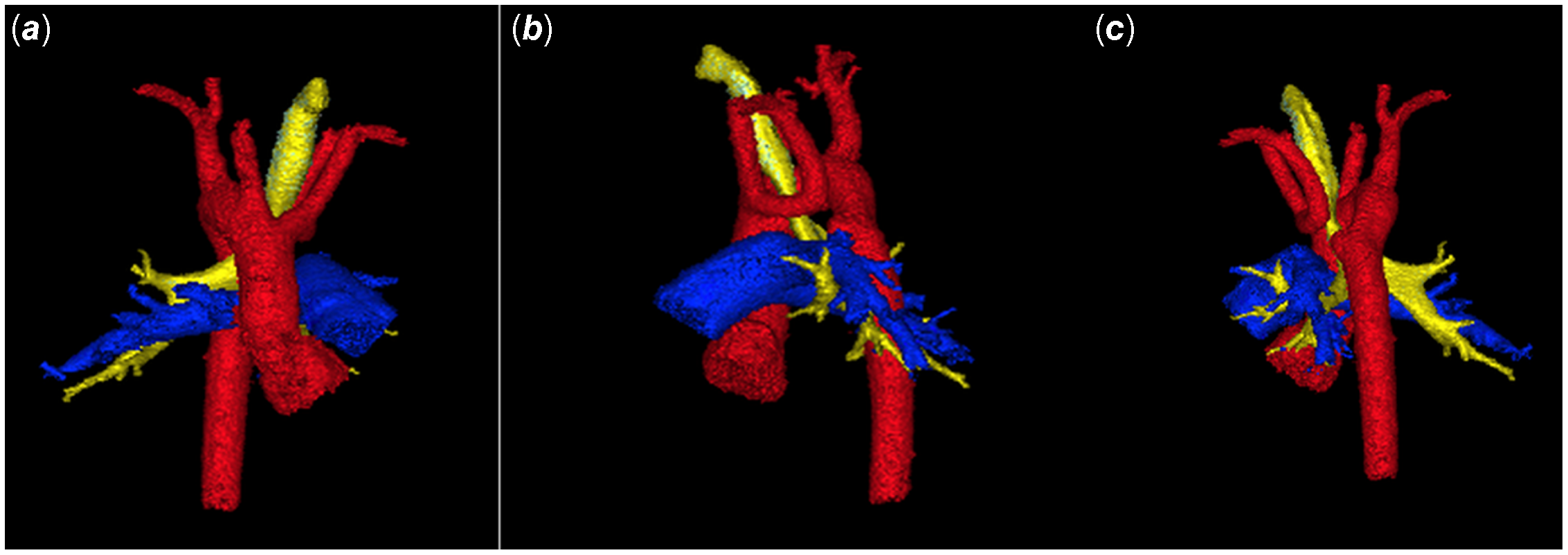
Three-dimensional reconstruction showing the features of a double aortic arch with ligamentous atresia of the distal left arch, including the tethering of the left subclavian artery, as seen in panels A and B, which predisposed to tracheal compression (panel C).
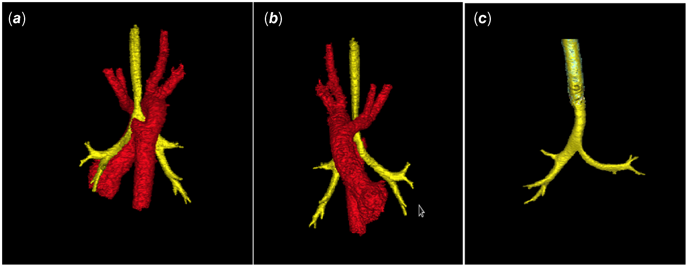
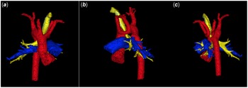
Another three-dimensional reconstruction of a left arch with ligamentous atresia of distal left arch, with panel A showing the anterior view, panel B the lateral view, and panel C the posterior view.
Panel A shows the computerised tomographic image of a double aortic arch with ligamentous atresia of distal left arch. The three-dimensional reconstruction shown in panel B, as viewed from the left side, shows the stenotic trachea (yellow), with ligamentous atresia distal to the origin of the left subclavian artery with a prominent diverticulum representing the remnant of the dorsal arch.

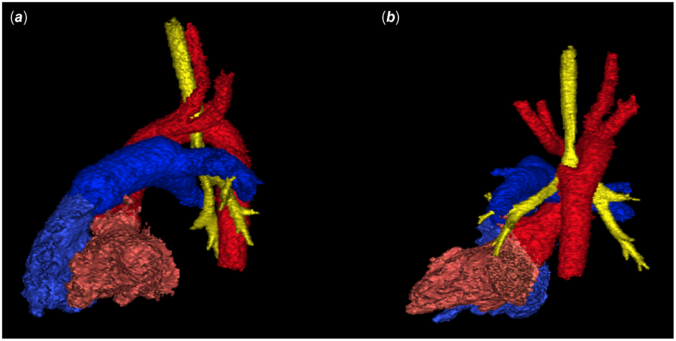
An additional three-dimensional reconstruction of left arch with ligamentous atresia of distal left arch shows the lateral view in panel A, and the posterior view in panel B.
All the variants of the double arch Reference Hanneman, Newman and Chan4 are well explained on the basis of the hypothetical model proposed by Edwards. Reference Edwards7 The hypothetical arrangement also provides the explanation for the right aortic arch with mirror-imaged branching. The latter arrangement is usually associated with congenital cardiac malformations, including tetralogy of Fallot, common arterial trunk, tricuspid atresia, and transposition. It is the consequence of regression of the left dorsal aorta distal to the origin of the seventh intersegmental artery, such that the left fourth arch forms the proximal component of the left subclavian artery, rather than the definitive left aortic arch. The right arch, of course, is defined on the basis that it crosses over the right bronchus. Reference Glew and Hartnell6 A double arch is formed when there is persistence of the arteries of both fourth pharyngeal arches, with each lateral arch giving rise to separate carotid and subclavian arteries. Reference Hanneman, Newman and Chan4,Reference Li, Wen and Liang8,Reference Priya and Nagpal9 Usually, only one of the arteries of the ultimate pulmonary arch persists as the arterial duct. The duct itself, furthermore, usually remodels to become the arterial ligament. Reference Hikspoors, Kruepunga, Mommen, Köhler, Anderson and Lamers3 It follows that remodelling of some component of the double arch can also produce a ligamentous, and hence an atretic, segment.
The double arch, when patent on both sides, usually produces symptoms because of the compression of the trachea-oesophageal pedicle. Such symptoms of compression and stridor can also be produced by the variants with ligamentous atresia, but the symptoms are typically less severe, or even absent. Reference Priya and Nagpal9 Although echocardiography is typically used as the initial tool for evaluation of a suspected vascular ring, Reference Priya, Thomas, Nagpal, Sharma and Steigner10 there are many factors that limit its efficacy. In the first place, it is operator dependent and can present technical problems, especially in small children. Reference Priya, Thomas, Nagpal, Sharma and Steigner10 Second, there is difficulty in visualisation of the posterior branching of the double aortic arch because of interference from anterior structures, such as the overlying bony thorax. Reference Priya, Thomas, Nagpal, Sharma and Steigner10 Additionally, echocardiography only provides cross-sectional imaging in two dimensions, limiting visualisation of complex anatomy and the spatial relationships between the component parts. Where echocardiography may suggest the presence of a right arch with mirror-imaged branching, or suspicion of a double aortic arch with ligamentous atresia of distal left arch (movie clips 2 and 3), reconstruction of computed tomographic datasets of the aortic arch, airway, and oesophagus is quite helpful. It is important to image the ductus or infer the location of its ligament as well the atretic ligament of the left arch to accurately plan the surgery. It can illustrate the compressed trachea, presence of ductal flow, diverticulum/ampulla on the superior aspect of left pulmonary artery, diverticulum from the descending aorta (remnant of the dorsal left arch or Kommerell’s diverticulum), downward tethering of the left subclavian artery (Figs 3-6), and perhaps calcified ligaments in older patients. Our experience serves to validate all these points. When using echocardiography, we initially made correct diagnoses in only three of our cases. With the addition of our reconstructed tomographic datasets, we were able to correctly identify the location of the atretic segment in all cases. The reconstructions also revealed the extent of narrowing of the airways, a feature essential for optimal planning of treatment, and the approach should surgical intervention be deemed necessary.
All children with right aortic arch should have a CT at some point to evaluate for airway compression. When using anaesthesia, the airways should always be interpreted with regard to the mode of ventilation (with positive pressure reducing the accuracy of assessment) and if inconclusive, performance of bronchoscopy may be indicated. In our institution, CT studies for arch anomalies are generally performed without anaesthesia and do not pose a problem with airway assessment.
In conclusion, accurate diagnosis of a double aortic arch with ligamentous atresia of distal left arch is critical for prompt surgical planning and correction. The appropriate treatment relieves discomfort and the other problems related to tracheoesophageal compression, such as stridor, dysphagia, gastroesophageal reflux, and chronic cough. The potential presence of a double arch with ligamentous atresia should always be suspected when unexplained dysphagia is found in the setting of a right aortic arch. Reference Priya, Thomas, Nagpal, Sharma and Steigner10 Even when the vascular ring is recognised using echocardiography, subsequent computed tomographic interrogation is recommended better to visualise the abnormal vessels and the compressed structures so as optimally to plan for surgery. Reference Mądry, Karolczak, Myszkowski and Zacharska-Kokot11 Late diagnosis, and delayed surgery, can lead to chronic issues with the airways, problems that can be avoided with appropriate timely imaging and for better counselling of the patients to monitor for symptoms of airway and oesophageal compression.
Financial support
This research received no specific grant from any funding agency, commercial, or not-for-profit sectors.
Conflicts of interest
None.