Background
Cor triatriatum sinister is a very rare congenital anomaly in which the left atrium is partitioned into two chambers by a fibromuscular membrane. A communication between the two chambers may exist in the form of fenestrations.Reference Van Praagh and Corsini 1 The clinical presentation of these patients varies from being totally asymptomatic to advanced heart failure symptoms. 2 , Reference Yaroglu Kazanci, Emani and McElhinney 3 Failure to diagnose this condition early in life may lead to pulmonary hypertension and right ventricular failure. We report a case of cor triatriatum sinister with a restrictive fenestration, decompressing vertical vein, pulmonary hypertension, and severe tricuspid regurgitation as a result of severe right ventricular dilation and dysfunction.
Case report
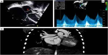
A 3-year-old girl who was born at term with no perinatal issues had her first onset of respiratory symptoms at 2–3 months of age. She was diagnosed with asthma, and was started on asthma medications. She has had several hospital re-admissions for her respiratory illness. Lately, she had increased respiratory symptoms that were not relieved by her asthma medications. She was referred to her local emergency department where a chest X-ray showed severe cardiomegaly and pulmonary oedema. The patient was transferred to our institution where an echocardiogram revealed the following: cor triatriatum sinister (Fig 1a) with a very small restrictive fenestration between the pulmonary venous chamber – that is, the proximal chamber – and the distal chamber with a mean gradient of 9 mmHg (Fig 1b); a decompressing vertical vein inserting into the innominate vein; a massive right atrium; intact interatrial septum; right ventricle and pulmonary artery dilation; evidence of significant pulmonary hypertension; and severely depressed right ventricular function with severe tricuspid regurgitation. A CT angiography scan confirmed the echocardiographic findings (Fig 1c) with a prominent cor membrane and a decompressing vertical vein. She was immediately taken to the operating room for resection of the cor membrane.
( a ) Preoperative echocardiogram showing the cor membrane (red arrow) in the left atrium. ( b ) Preoperative echocardiogram showing the mean gradient across the fenestration in the cor membrane. ( c ) Preoperative CT scan showing the cor triatriatum membrane (arrow sign) in the left atrium with the decompressing vein (star sign) draining into the innominate vein.

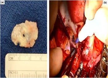
In the operating room, via median sternotomy and after opening the pericardium, there was massive right atrial and right ventricular dilatation with externally very poor right ventricular function. The main pulmonary artery was dilated and hypertensive. There was a large, left-sided ascending vertical vein, which drained to a dilated innominate vein. Given the severe right atrial dilatation, cardiopulmonary bypass was initially instituted with a single right atrial venous cannula. This was later converted to bicaval cannulation. Intracardiac examination was carried out under cardiopulmonary bypass and deep hypothermia with a brief period of deep hypothermic circulatory arrest at 18°C for cor membrane excision. The atrial septum was intact. There was a thick cor triatriatum membrane with two very small fenestrations in the membrane (Fig 2a); four pulmonary veins were seen draining to the proximal cor chamber. The distal chamber contained the mitral valve and left atrial appendage. The tricuspid valve annulus was markedly dilated. There appeared to be relatively normal tricuspid valve leaflets and subvalvar apparatus. The repair consisted of resection of the cor membrane, ligation of the ascending vertical vein (Fig 2b), and tricuspid valve annuloplasty using a De Vega technique. After repair, the pulmonary artery pressures were approximately one-third systemic with a central venous pressure of 6 mmHg and left atrial pressure of 7 mmHg. The patient was weaned from bypass on epinephrine, milrinone infusions, and inhaled nitric oxide. An intraoperative transoesophageal echocardiogram showed unobstructed pulmonary venous drainage to the mitral valve and trivial tricuspid regurgitation. There continued to be severely depressed right ventricular function. The cardiopulmonary bypass time was 113 minutes, with a cross-clamp period of 42 minutes, and circulatory arrest period of 5 minutes. The chest was closed in the usual manner, and the patient tolerated the procedure remarkably well.
( a ) An intraoperative picture of the resected membrane with a small fenestration in the centre. ( b ) An intraoperative picture showing the decompressing vertical vein (black arrow) draining into the innominate vein.

Postoperatively, the patient was started on enteral sildenafil with weaning of nitric oxide. She was extubated on postoperative day 3, and was transferred out of the cardiovascular ICU on postoperative day 7. An echocardiogram performed 1 day later revealed moderately depressed right ventricular function with trivial tricuspid regurgitation. She was discharged home on postoperative day 10. An echocardiogram at 1 month follow-up showed normal right ventricular function and normal pulmonary artery pressures.
Discussion
The first successful surgical repair for cor triatriatum was performed by Dr Lewis in 1956. 4 Van Praagh defined the anomaly in 1969 as being a rare congenital malformation in which the left atrium is partitioned into two chambers, an anterior chamber and a posterior chamber, by a fibromuscular membrane. The anterior chamber contains the mitral valve and appendage, whereas the posterior one contains the pulmonary veins. The communication between both chambers can be large, small, or absent.Reference Van Praagh and Corsini 1 The lesion can be asymptomatic and discovered incidentally or can be present along with symptoms such as dyspnoea, arrhythmia, heart failure signs, or even cyanosis. It may present in any age group depending on the degree of obstruction. It can be misdiagnosed as bronchial asthma or primary pulmonary hypertension. It may present as an isolated cardiac lesion, similar to our case here, or in association with other lesions such as ventricular septal defect, tetralogy of Fallot, transposition of the great arteries, or complete atrioventricular septal defect.Reference Van Praagh and Corsini 1 – Reference Yaroglu Kazanci, Emani and McElhinney 3 , 5 Echocardiography is the primary method for diagnosis; however, cardiac catheterisation, CT scanning, and MRI may be used.Reference Nassar and Hamdan 6 The standard treatment for this condition is surgery with excision of the membrane and repair of any associated lesions. Other treatment modalities have been reported, such as medical therapy for non-obstructed lesions and percutaneous balloon angioplasty of the fenestration, 7 but are generally reserved for patients not suitable for surgical intervention. Repairing the tricuspid valve regurgitation associated with this condition is somewhat controversial. Despite not being a durable repair method in many cases, the De Vega technique was used in this particular patient to help manage her immediate postoperative period. Having severe tricuspid regurgitation with severe right ventricular dysfunction after coming off bypass would have not helped the haemodynamics and would have posed a great risk for being on extracorporeal life support in the postoperative period. Furthermore, the tricuspid valve annulus was reduced from 25 to 20 mm after repair. De Vega repair may theoretically limit the growth of a part of the annulus but not the whole valve. The early-, mid-, and long-term outcomes of surgical repair of the cor membrane are extremely good with a very low risk for re-intervention. Survival is highly dependent on the associated lesions. 2 , Reference Yaroglu Kazanci, Emani and McElhinney 3 , 5 Pulmonary hypertension and ventricular dysfunction are usually reversible with this condition when surgical correction is performed early in life. 8 The late presentation and diagnosis of this patient, despite having a very restrictive communication, is explained by having a persistent levoatrial cardinal vein that functioned as a decompressing channel for the left atrium. 9 , 10
This case was unusual in its presentation in a toddler with a severely restrictive fenestration, a decompressing vertical vein, and an intact interatrial septum; the treatment was successful as demonstrated by normalisation of ventricular function and pulmonary artery pressure.
Acknowledgements
Permission was granted by the parents to publish the case report.
Authors’ contributions: all authors participated in creating content for the manuscript, editing, and provided final approval for submission. No undisclosed authors contributed to the manuscript.
Financial Support
This research received no specific grant from any funding agency or from commercial or not-for-profit sectors.
Conflicts of Interest
The author(s) declare no potential conflicts of interest with respect to the research, authorship, and/or publication of this article.