Introduction
One of the challenges in reproductive biology is to understand how intraovarian and/or extraovarian factors influence the maturation of oocytes during follicular development. Oocyte maturation is defined as a reinitiation and completion of the first meiotic division and its subsequent progression to metaphase II. In this regard, nuclear and cytoplasmic maturation are mainly regulated by microenviromental factors in the follicles and also, apparently, by follicular size, quality (Carolan et al., Reference Carolan, Lonergan, Monget, Monniaux and Mermillod1996), donor's age (Khatir et al., Reference Khatir, Carolan, Lonergan and Mermillod1997) and LH surge (Lonergan et al., Reference Lonergan, Gutiérrez-Adán, Rizos, Pintado, De la Fuente and Boland2003). To understand the mechanism of nuclear and cytoplasmic maturation in the oocyte, it has been necessary to reproduce in vitro the composition of the natural biological fluids. For this purpose, a great number of incubation media that included exogenous chemicals and biological preparations have been described (Ikeda et al., Reference Ikeda, Azuma, Hashimoto and Yamada1999; Bu et al., Reference Bu, Xie, Tao, Wang and Xia2004; Sutton-McDowall et al., Reference Sutton-McDowall, Gilchrist and Thompson2005).
Our understanding of the role of heparin in mammalian fertilization began in the 1980s (Valencia et al., 1980). Moreover, the presence of heparin has been reported in ooplasm (Reyes et al., Reference Reyes, Ramírez and Delgado2004), follicular fluid (Ax & Bellin, Reference Ax and Bellin1988) and uterine fluid (Foley et al., Reference Foley, Griffin, Zuzel, Aparicio, Bradbury, Bird, Clayton, Jenkins, Scott, Rajah and McNichol1978) participating in events leading to in vitro fertilization (IVF). Recently, several carbohydrates, including d-mannose, fucoidan, dextran sulphate, d-galactose, N-acetyl-d-galactosamine, d-fucose, sialic acid and the glycoprotein fibronectin were shown to inhibit (by 40–90%) spermatozoa penetration of in vitro matured cumulus–oocyte complexes (COCs) (Tanghe et al., Reference Tanghe, Van Soom, Duchateau, Nauwynck and De Kruif2004). However, heparin was the only carbohydrate that significantly increased the penetration rate.
Heparin interferes with the regulation of intracellular signals during oocyte maturation. For instance, after insemination, phases 1 and 3 of the transient calcium signal are inhibited by preloading oocytes (metaphase I) with 5 mM heparin (Russo et al., Reference Russo, Kyozuka, Antonazzo, Tosti and Dale1996). In addition, when growing pig oocytes are cultured in a medium conditioned by COCs, heparin suppresses cumulus expansion in both EGF- and FSH-treated cultures. However, it does not prevent germinal vesicles breakdown (GVBD) stimulated by hormones (Downs, Reference Downs1989). On the other hand, Fenton et al. (Reference Fenton, Dentine and Ax1993) observed that treatment with 0.1 mg/ml heparin for 24 h induces cumulus cell expansion in bovine COCs in the absence of fetal bovine serum. Fetal bovine serum inhibits this expansion, perhaps due to binding of serum proteins with heparin before addition of the COCs to the culture (Fenton et al., Reference Fenton, Dentine and Ax1993).
Heparin increases sperm capacitation, motility and the acrosome reaction (Valencia et al., Reference Valencia, Wens, Merchant, Reyes and Delgado1984; Chamberland et al., Reference Chamberland, Fournier, Tardif, Sirard, Sullivan and Bailey2001). It also induces changes in chromatin, initiating sperm nuclei decondensation in more that seven animal species (Sanchez-Vazquez et al., 1998). Therefore, the primary goal of this study was to evaluate the possible effect of heparin on bovine oocyte chromatin. We determined if heparin acts as an oocyte maturation factor and if the heparin-treated oocytes can be fertilized. A secondary goal of this study was to verify the heparin effect on maturation of oocytes incubated in simplified salt and culture medium systems that eliminate excessive variability. This could lead to a better understanding of the heparin effect.
Material and methods
All reagents were purchased from Sigma Chemical Company unless otherwise noted.
Cell preparation
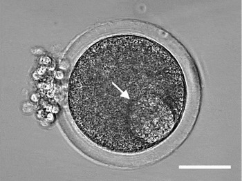
Bovine ovaries were obtained from a local abattoir (Atlixco, Pue, Mexico) where the dimensions were measured prior to transportation on ice to the laboratory, which took less than 1 h. Ovaries with corpora lutea or morphological abnormalities were excluded. Follicles were measured with a micrometer and classified according to diameter in: (1) small = 2–5 mm; (2) medium = 6–10 mm; and large = 11 mm. Within 1 h after slaughter, oocytes were aspirated from ovarian antral follicles (6 < /alt − text mm in diameter) using a 22 gauge needle and a 3 ml syringe. The oocytes were collected in 1 ml of the following salt medium used by Keyhani & Storey (Reference Keyhani and Storey1973): 113 mM KCl, 12.5 mM KH2PO4, 2.5 mM K2HPO4, 2.5 mM MgCl2 and 20 mM Tris, pH 7.4, containing 100 U/ml of penicillin and 100 μg/ml of streptomycin. In all cases, the granulosa-enclosed oocytes were freed from the surrounding cells by vortexing at low velocity for 15–30 s. Thus, the ova with normal morphology utilized in our experiments are completely denuded or had only a small amount of granulosa cells and the germinal vesicle was clearly visible (Fig. 1). With the aid of a stereoscopic microscope (Nikon SMZ-800), the collected oocytes were placed in a glass multiwell slide with 0.2 ml of the incubation salt medium per well where they were washed three times.
Control bovine oocyte. Phase contrast image of an oocyte with normal morphology and germinal vesicle clearly visible (arrow). Bar = 50 μm.

Heparin effect on oocyte in vitro maturation
For each of five replicates, 15–20 collected oocytes were randomly assigned to each treatment. The cells were placed in an organ culture dish containing 1 ml of salt medium or 1 ml of HEPES-buffered Tissue Culture Medium 199 (TCM 199) containing 4 mM NaHCO3, 100 U/ml penicillin and 100 μg/ml streptomycin. They were incubated at 37.5 °C in a humidified atmosphere of 5% CO2. Control denuded ova were maintained in the presence of each incubation medium alone for 24 h. Using MetaMorph imaging software (MetaMorph Universal Imaging Corporation) the total surface area (μm2), volume (μm3), perimeter (μm) and diameter (μm) were measured at 0 and 24 h.
Heparin-treated oocytes were incubated 24 h with 1.0, 6.5 or 65.0 mg/ml heparin in salt medium or TCM 199. To determine the effects of these treatments on ova maturation during the incubation time, images were captured with time-lapse photography (MetaMorph) every 10 min for 24 h. The effect of heparin on oocyte in vitro maturation was measured as the times of GVBD and formation of the first polar body (PB). The endpoint of GVBD was taken when the nuclear envelope was completely dispersed in the cytoplasm. The time for first PB formation time was taken when the pinching off of the PB body was completed.
Sperm preparation and in vitro fertilization
Spermatozoa were isolated by removing the entire epididymis and repeatedly washing it with 0.9% NaCl followed by perfusion with the same solution via the spermatic artery. Spermatozoa were obtained from the cauda region by procedures previously described (Carranco et al., Reference Carranco, Reyes, Magdaleno, Huacuja, Hernández, Rosado, Merchant and Delgado1983). To diminish interdonor variability, pools of sperm cells were obtained from five different animals and used for individual experiments. These samples were washed by centrifugation at 700 g, resuspended and diluted if necessary to obtain a final concentration 106 spermatozoa/ml in TCM 199. Spermatozoa were capacitated in TCM 199 supplemented with 10 μg/ml heparin and 3 mg/ml BSA fraction V for 1.5 h at 37.5 °C.
For IVF, oocytes were matured in TCM 199 with 0 (control), 1.0 and 6.5 mg/ml heparin. After first PB formation, they were washed three times in the same medium. Five replicates of 10–15 oocytes were placed in an IVF dish containing 1 ml of TCM 199. A 10 μl aliquot of prepared sperm suspension was added to each fertilization dish to obtain a final concentration of 0.1 × 10⁶ spermatozoa/ml. Dishes were incubated at 37.5 °C with 5% CO2 in air and maximum humidity. The formation of pronuclei (PN) and the first cleavages (embryos that developed to the 2-cell stage) were evaluated 24 and 72 h post-fertilization respectively under a phase contrast microscope (Nikon E-600, Técnica en Laboratorios).
Fluorescent probes
DNA structure
To observe DNA structure after oocyte maturation, the control and heparin-treated oocytes were washed three times with 1 ml salt medium, placed in a chamber slide with 0.2 ml of 0.005% quinacrine mustard and incubated for 20 min at 37.5 °C. Quinacrine mustard is a cationic acridine dye with a high affinity for A–T rich regions of DNA. The oocytes were then placed in a 0.2 ml chamber slide with salt medium and viewed with a confocal microscope (Nikon C1, Técnica en Laboratorios) equipped with ion-argon and helio-neon lasers. Fluorescence was detected in 2 μm sections upon excitation with argon and helio-neon lasers at 488 and 633 nm, respectively.
Metaphase II oocytes
After maturation, the control and heparin-treated oocytes were washed with salt medium and stained 10 min with 100 nM SYBR14 (Molecular Probes Inc.) in 1 ml of salt medium. SYBR14 is a membrane-permeant nucleic acid stain. The washed cells were examined in 2 μm sections with the confocal microscope described above.
Statistical analysis
Each heparin-treated oocyte experiment was repeated five times and data were expressed as means ± standard errors (SE). The differences of maturation times between experimental groups were analyzed using the Mann–Whitney Rank Sum Test. The per cent differences in oocyte DNA fragmentation, PN formation and first cleavages were analyzed using the chi-squared test. All analyses were performed with SigmaStat v2.0 software (Jandel Corporation). Differences of p < 0.05 were taken as significant.
Results
The length of the ovaries was 29.9 ± 5.1 mm (n = 65), which was significantly greater (p < 0.05) than the width, 19.2 ± 7.0 mm. The morphometric para-meters of 512 oocytes were as follows: total sur-face area 19,895.0 ± 348.0 μm2, volume 2,116,000.0 ± 54,589.0 μm3, perimeter 562.6 ± 8.2 μm and diameter 158.8 ± 1.8 μm. There were no differences after 24 h of incubation in salt medium or TCM 199.
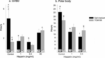
For control oocytes, the time of GVBD was 4.7 ± 1.1 h in salt medium, which was significantly longer than in TCM 199 (p < 0.05, Fig. 2a). Heparin at both 1.0 and 6.5 mg/ml significantly reduced the time of GVBD in salt medium to about 1.5 h (p < 0.05 each compared with control). These values were similar to those for TCM 199 without or with heparin (Fig. 2a).
Heparin effects on (a) germinal vesicle breakdown and (b) polar body formation. Bovine oocytes were incubated in the presence of 1.0, 6.5 and 65.0 mg/ml heparin in salt medium and TCM 199. Each bar represents the mean (±SE) hours when GVBD occurred (a) and PB separation was completed (b). The number of oocytes for each group, n, is shown at the bottom of each bar. The means were compared by the Mann–Whitney Rank Sum Test. Treatment means that are labelled with the same letters (a–g) differed significantly (p < 0.05).

The time of first PB formation in controls was 22.0 ± 1.1 h in salt medium and 18.0 ± 1.2 h in TCM 199 (Fig. 2b). Heparin at 1.0 and 6.5 mg/ml significantly reduced the time for PB formation to 11.0 ± 1.1 and 9.0 ± 0.8 h respectively in salt medium. The time of PB formation in TCM 199 was reduced from 17.7 ± 1.2 h in the control oocytes to 12.1 ± 1.7 h in oocytes treated with 6.5 mg/ml heparin (p < 0.05; Fig. 2b).
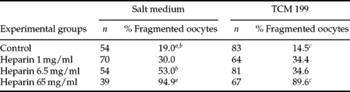
After maturation, staining of nuclei by quinacrine mustard showed that control oocytes in salt medium and TCM 199 had 19.0% and 14.5% chromatin fragmentation, respectively (Table 1). Heparin at 1.0 and 6.5 mg/ml appeared to increase fragmentation, but the values were not significantly greater than the controls incubated in either salt medium or TCM 199. However, 6.5 mg/ml heparin in salt medium significantly increased fragmentation (p < 0.05).
Heparin effects on oocyte DNA fragmentation

The data were compared by chi-squared test. Per cents with the same letter (a–c) differ significantly (p < 0.05).
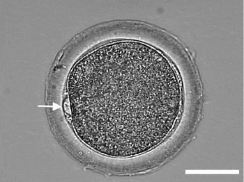
The first PB formation (Fig. 3) was the most reliable morphological indicator of oocyte maturation. With the SYBR14 stain, we observed metaphase II after the PB was completely separated from the oocyte in both media (Fig. 4). Metaphase II nuclei were present in 43–64% of the oocytes treated with 1.0 or 6.5 mg/ml heparin. These values were not significantly different from the control groups.
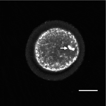
Polar body formation. Phase contrast microscopy of an oocyte treated 11 h with 1.0 mg/ml of heparin in salt solution. The arrow shows the polar body just before extrusion. Treatment with 6.5 mg/ml heparin in salt medium and TCM 199 gave similar results. Bar = 50 μm.

Metaphase II oocyte. Confocal microscope image of a metaphase II nucleus (arrow) for an in vitro matured bovine oocyte after 14 h with 1.0 mg/ml heparin in salt medium. The DNA was labelled with SYBR14. Similar results were obtained with 6.5 mg/ml heparin. Bar = 50 μm.

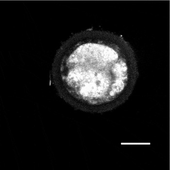
Heparin at 65.0 mg/ml was detrimental to oocyte maturation. At this concentration, the oocytes in both incubation media had significantly increased chromatin fragmentation (Fig. 5, Table 1). Approximately 90–95% of the oocytes did not form PBs due to chromatin fragmentation after GVBD occurred. This indicates that heparin at 65.0 mg/ml damaged the DNA to the point of interfering with maturation.
Oocyte DNA fragmentation by heparin. Confocal image of an oocyte with fragmented DNA after incubation for 24 h with 65.0 mg/ml heparin in salt medium and staining with quinacrine mustard. The same heparin treatment in TCM 199 gave similar results. Bar = 50 μm.

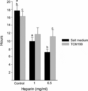
The heparin effect on the maturation of the oocyte nucleus was evident when we used the salt medium for the incubations. The latency time between GVBD and first PB formation for control oocytes was 17.7 ± 1.3 h in salt medium (Fig. 6). This time was significantly reduced (p < 0.05) when the oocytes were incubated in 1.0 and 6.5 mg/ml heparin. The latency in TCM 199 was 16.2 ± 1.2 h and this time was significantly reduced only by 6.5 mg/ml heparin (Fig. 6).
Heparin effects on latencies between GVBD and PB formation. The oocytes were treated with heparin (1.0 and 6.5 mg/ml) in salt medium and TCM 199. Each bar represents the mean (±SE) latency hours between the GVBD and PB separation. The means were compared by the Mann–Whitney Rank Sum Test. Treatment means labelled with the same letter (a, b, c) differed significantly (p < 0.05).

Both control and heparin-treated oocytes (1.0 and 6.5 mg/ml) were fertilized in TCM 199 (Table 2). The per cents of PN formation and cleavage of IVF oocytes were similar for control and heparin-treated oocytes. However, 6.5 mg/ml heparin treatment caused a significant reduction in PN formation and in cleavage when compared to treatment with 1 mg/ml.
IVF of bovine oocytes matured with heparin

The data were compared by chi-squared test. Per cents with the same letter (a, b) differ significantly (p < 0.05).
Discussion
Many culture media have been developed for recovering and culturing oocytes. Some of these have compounds that are toxic for oocyte development (Aárabi et al., Reference Aárabi, Roussel and Candler1997). Despite the known toxicity of Tris buffer used in the salt medium in previous studies, this buffer gave us good results and preserved oocyte viability (Reyes et al., Reference Reyes, Ramírez and Delgado2004). In the present study, the controls clearly demonstrate that bovine oocytes can mature and maintain physical dimensions and integrity in this salt solution. This could be due to the beneficial effect of the high concentration of potassium ions (135.5 mM). This is consistent with the current use of the potassium simplex optimized media for the preservation of the spermatozoa, oocytes, blastomeres and embryos (Bhuiyan et al., Reference Bhuiyan, Cho, Jang, Park, Kang, Lee and Hwang2004; Nedambale et al., Reference Nedambale, Dinnyes, Groen, Dobrinsky, Tian and Yang2004; McGinnis et al., Reference McGinnis, Zhu, Lawitts, Bhowmick, Toner and Biggers2005).
Many studies have shown that heparin plays an important role in oocyte maturation and fertilization. Fenton et al. (Reference Fenton, Dentine and Ax1993) reported that heparin stimulates COC expansion, increases production of hyaluronic acid by COCs and allows normal GVBD with decreased cAMP production. In addition, the cAMP reduction induces the resumption of meiosis in most mammalian oocytes during nuclear maturation (Homa et al., Reference Homa, Webster and Russell1991). Furthermore, when 1.0 mg/ml heparin was included in fertilization medium for pig oocytes, it increased the cleavage rates and subsequent development of blastocysts (Kim et al., Reference Kim, Lee, Hyun, Nam, Lee, Jeong, Kim, Kim, Kang, Lee and Hwang2005). At 0.5 mg/ml, it increased the rates of cleavage and blastocyst formation in IVF bovine oocytes (Jang et al., Reference Jang, Lee, Kang and Hwang2003). Finally, Li et al. (Reference Li, Seidel and Squires2004) showed that incubation of oocytes in 10.0 μg/ml heparin before or after intracytoplasmic sperm injection benefited pronuclear formation, cleavage rate and the number of cells in the blastocysts.
Follicular fluid that accumulates in the antrum during maturation of ovarian follicles contains high amounts of glycosaminoglycans such as chondroitin, hyaluronic acid and heparin-like compounds (McGaughey, Reference McGaughey1975). Gebauer et al. (Reference Gebauer, Lindner and Amsterdam1978) found that rat ovarian slices in vitro synthesize heparin-like compounds. This also occurs in vivo as measured by incorporation of intraperitoneally administered 35SO42− in sexually mature rats. They found that the heparin-like compounds are synthesized mainly by the granulosa and theca cells and are released into the follicular antrum, suggesting a process of active secretion. Heparin inside oocytes occurs in several species, from invertebrates to mammals (Reyes et al., Reference Reyes, Ramírez and Delgado2004). Although the presence of glycosaminoglycans may be associated with the mechanism of ovulation (Yoshimura & Wallach, Reference Yoshimura and Wallach1987), it is possible that these compounds also play a role in the initiation of nuclear decondensation of spermatozoa deposited into the female reproductive tract (Kawakami et al., Reference Kawakami, Arai, Oishi, Hori and Tsutsui2000).
In vitro oocyte maturation is influenced largely by the culture media and conditions employed (Eckert &Niemann, Reference Eckert and Niemann1995). To eliminate excessive variability in the present study, the cultures were prepared in simplified salt medium and in TCM 199. Thus, we observed the effect of heparin on GVBD and PB formation without any natural or introduced factors that might influence the maturation of the oocytes. In both in vitro and in vivo conditions, spontaneous GVBD in mammals occurs after follicular release. For bovine oocytes, the first PB develops and the metaphase II nucleus appears in approximately 20 h (Hyttel et al., Reference Hyttel, Greve and Callesen1989; Sirard et al., Reference Sirard, Florman, Leibfried-Rutledge, Barnes, Sims and First1989; Homa, Reference Homa1995). In our study, heparin reduced the time of GVBD and PB formation with respect to the controls by 2–3 h and 11–16 h respectively in salt medium. The most effective concentration of heparin for oocyte maturation was 1.0 mg/ml. At this concentration, 43–64% of the oocytes reached metaphase II and DNA structure was fragmented in only 30–34% (Table 1). The increased DNA fragmentation at 6.5 mg/ml of heparin in salt medium was higher than that in TCM 199 (53.0 versus 34.0%). This effect could be due to the Tris buffer in the salt medium. At high heparin concentrations (65.0 mg/ml), there was increased DNA fragmentation in both culture mediums. Russo et al. (Reference Russo, Kyozuka, Antonazzo, Tosti and Dale1996) reported that preloading oocytes with 5 mM heparin (65.0 mg/ml) inhibited the calcium transients (phases 1 and 3) and the polar body extrusion in ascidian oocyte maturation. In our experiments, we observed chromatin fragmentation and no PB formation when the oocytes were treated with that concentration, demonstrating that it damages DNA and partially inhibits the maturation process. Currently, we do not know the intracellular concentration of heparin in bovine oocyte.
In TCM 199, only the 6.5 mg/ml heparin concentration had a significant effect on oocyte maturation. The reduced effect with 1.0 mg/ml could be due to the binding of TCM 199 components to heparin, causing inhibition of the stimulant effect on oocyte maturation.
Due to the high rate of DNA fragmentation of oocytes matured in the salt medium, we did not test the ability of these oocytes to become fertilized. For those matured in TCM 199 with 1.0 mg/ml heparin, the percentages of PN formation and cleavage of IVF oocytes were similar to controls. These results showed that heparin treatment did not interfere with the IVF process, at least at 1.0 and 6.5 mg/ml. However, these results are lower than previously reported (Choi et al., Reference Choi, Takagi, Kamishita, Wijayagunawardane, Acosta, Miyazawa and Sato1998), probably because we used TCM 199, which is a protein-free medium (Eckert & Niemann, Reference Eckert and Niemann1995).
Some of the oocytes obtained from large-sized normal follicles (11 < /alt − text mm) were surrounded by compact layers of follicle cells, while some from medium follicles (6–10 mm) were completely denuded. Nevertheless, when the denuded oocytes from large- and medium-sized follicles were used, the results of treatment with heparin were the same. Thus, there may not be any relationship between follicle size and the degree of the oocyte maturation. In any case, we have clearly established that, with the exception of the presence of first polar body, there is no oocyte morphometric parameter that serves as a maturation index.
Based on our results, we conclude that heparin plays an important role in the rearrangement of the oocyte chromatin and it acts as a maturation factor, at least in vitro. This is consistent with our results of reorganization of sperm nuclei chromatin in seven different species (Sánchez-Vázquez et al., Reference Sánchez-Vázquez, Reyes, Ramírez, Merchant-Larios, Rosado and Delgado1998). For oocyte maturation, it remains to be determined if heparin acts on one or several steps of the complex network of signal transduction pathways (Cook & Aikawa, Reference Cook and Aikawa1973; Saiga & Kinoshita, Reference Saiga and Kinoshita1976; Sato et al., Reference Sato, Ishibashi and Koide1987).
Acknowledgements
We are grateful to M.V.Z. Juan Manuel Ayestaran Nava and M.V.Z. Eduardo Cabrera Bautista of the Rastro Municipal de Atlixco, Puebla, México, for the bovine gametes.