Introduction
Acute mastoiditis denotes a suppurative inflammation within the mastoid air cell system of the temporal bone. The condition typically arises as a complication of acute otitis media through local propagation via the aditus ad antrum connecting the middle ear to the mastoid. Conventional teaching suggests causative organisms to be predominantly those colonising the upper respiratory tract, such as Streptococcus pneumoniae, Staphylococcus aureus, group A streptococci, H influenzae, as well as P aeruginosa.Reference Cassano, Ciprandi and Passali1
The routine infant pneumococcal conjugate vaccine (PCV) programme was first introduced in the UK in 2006 with a 7-valent vaccine (PCV7), which was replaced with the 13-valent vaccine (PCV13) in 2010. Studies have found the introduction of the vaccine to be highly protective in reducing invasive pneumococcal infection in both children and adults by up to 50 per cent.Reference Miller, Andrews, Waight, Slack and George2, Reference Waight, Andrews, Ladhani, Sheppard, Slack and Miller3 In the United States, there have been reports of reducing numbers of middle ear fluid cultures growing S pneumoniae in young children with acute otitis media since this introduction.Reference Tamir, Roth, Dalal, Goldfarb and Marom4 Interestingly, Attlmayr et al. previously summarised the microbiological results of patients presenting with mastoiditis at Alder Hey Children’s Hospital for the years 2003 and 2012, covering a pre-vaccination period. At that time, the most common micro-organisms responsible for acute mastoiditis included Streptococcus pyogenes (group A), S pneumoniae and P aeruginosa.Reference Attlmayr, Zaman, Scott, Derbyshire, Clarke and De5
In this study, we aim to assess the latest microbiological profile of patients with acute mastoiditis in a major UK paediatric tertiary centre.
Material and methods
A retrospective observational study was undertaken at Alder Hey Children’s Hospital, Liverpool, UK between January 2017 and September 2022. Inclusion criteria involved all patients with a coded diagnosis of mastoiditis requiring hospital admission. Institutional approval was granted prior to data collection.
Patient demographics, length of stay, biochemical markers including maximal C-reactive protein (CRP), culture and sensitivity analysis (obtained from pus swabs, pus cultures or blood cultures), and interventions were recorded for analysis. Data was stored securely on the hospital database. Patients were followed up for a minimum of nine months for evidence of complications and recurrence.
The primary outcome was the identification of the type of pathogen responsible for mastoiditis infections within this cohort of children. Secondary outcomes included length of hospital stay, 30-day mortality, biochemical markers of inflammation including CRP and white blood cell count (WBC), complications and the need for surgery. These secondary outcomes were stratified by the identified causative pathogen, when available.
Data was collected and analysed using Microsoft Excel (Microsoft, Redmond, Washington, USA) and Statistical Package for the Social Sciences (IBM SPSS, Armonk, New York, USA).
Results and analysis
A total of 108 patients were admitted with 112 episodes of mastoiditis; 61 males and 47 females. Recurrent presentations were recorded in three male patients and one female patient. The median age on admission was 2 years old (aged 4 months–14 years old), and the average length of stay was 4.6 days (range 0–40; median 3). Study patient characteristics cross-referenced by the more common organisms isolated on microbiological analysis are summarised in Table 1. Mixed skin flora was not included in the summary table due to the likelihood of contamination.
Patient characteristics with comparison between most common organisms identified on microbiological analysis. Categorical data are summarised as N (%); GAS: group A streptococci; LOS: Length of stay; CRP represented as average; Age and LOS are represented as medians

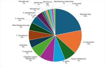
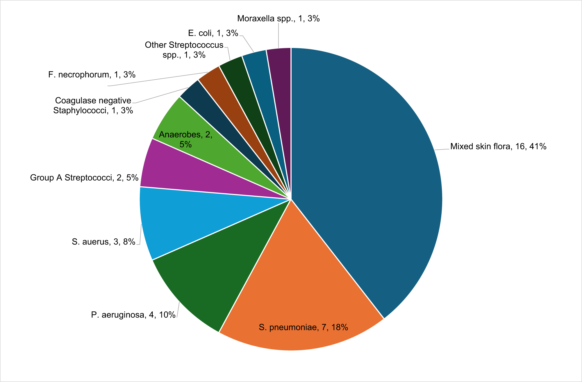
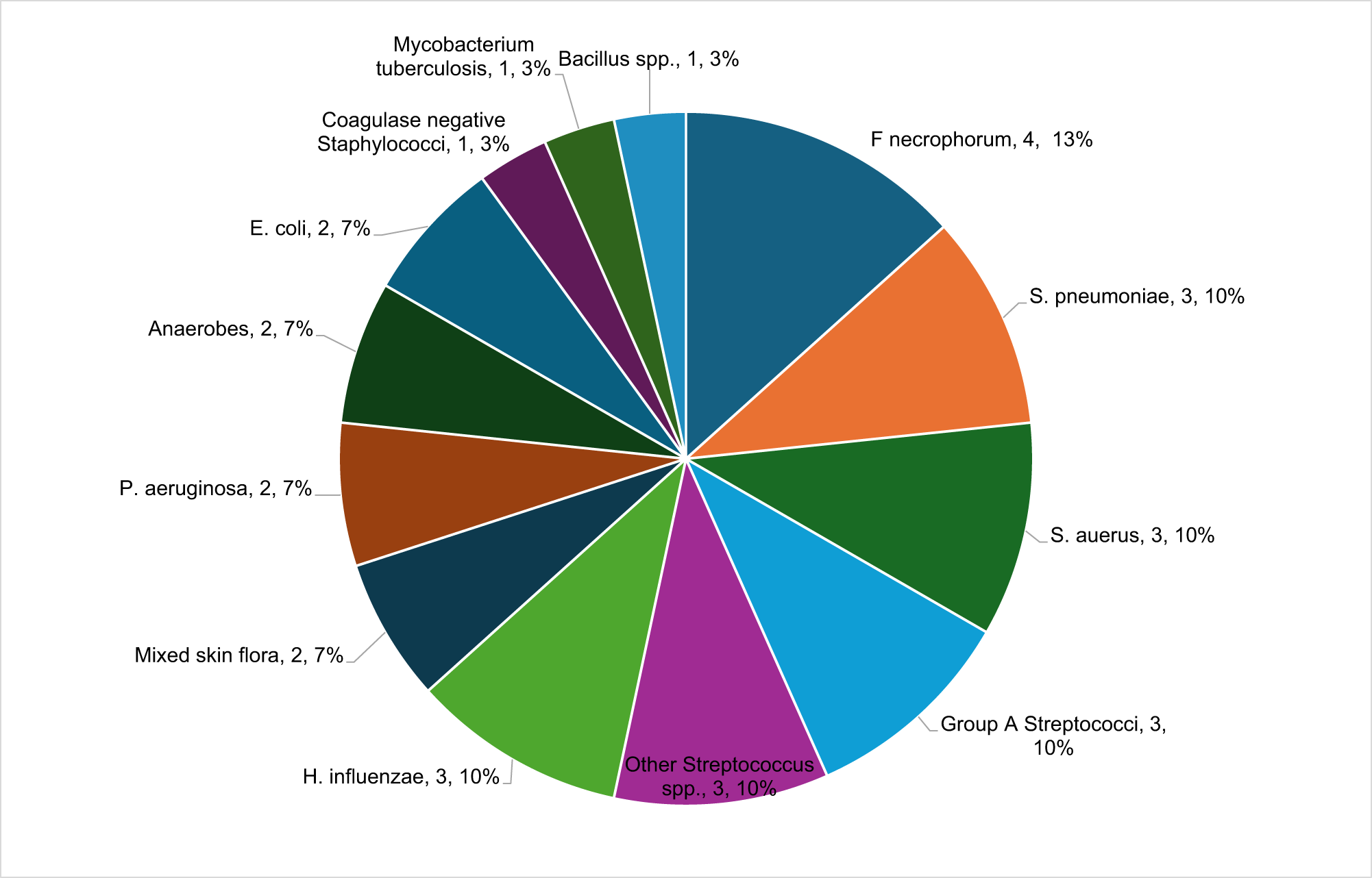
In 87 cases (78 per cent), samples (ear swabs, pus cultures or blood cultures) were obtained and sent for microbiology. Of those, 50 cases had 58 positive samples, and 82 organisms were isolated (summarised in Figure 1). After excluding mixed skin flora as a likely contaminant (n = 18, 22 per cent), the most commonly isolated organisms from all samples were Streptococcus spp., isolated in 22 cases; 12 (15 per cent) S pneumoniae, 6 (7 per cent) group A streptococci, 1 (1 per cent) Streptococcus intermedius, 1 (1 per cent) Streptococcus oralis, 1 (1 per cent) Streptococcus constellatus and 1 (1 per cent) Streptococcus massiliensis. The second most common organisms were Staphylococcus spp., isolated in 13 cases; 6 (7 per cent) S aureus and 7 (9 per cent) coagulase-negative staphylococci. P aeruginosa was isolated in six cases (7 per cent), Fusobacterium necrophorum in five cases (6 per cent), anaerobes in four cases (5 per cent), H influenzae in four cases (5 per cent) and Escherichia coli in three cases (4 per cent). Other organisms isolated once only were Haemophilus parainfluenzae, Prevotella spp., Moraxella spp., Bacillus spp., Micrococcus spp., Acinetobacter baumannii and Mycobacterium tuberculosis (one each). Figures 2 and 3 provide a summary of positive cultures by site of specimen.
Eighty-two organisms isolated from 58 microbiological samples.

A total of 39 organisms were isolated from 31 ear swabs.

A total of 30 organisms were isolated from 18 mastoid pus samples.

Sensitivity data was available in 34 of the 58 positive samples. The most common antibiotic was cefotaxime which was tested in 18/34 samples and was effective in 100 per cent of cases tested. This was followed by amoxicillin which was effective in 94 per cent of cases and tested in 16/34 samples. Clarithromycin was tested in 13/34 samples and was effective in 93 per cent of cases. A total of 8/34 (24 per cent) samples were found to have antibiotic-resistant strains of bacteria. This included an S aureus resistant to clarithromycin and two cases of H influenzae resistant to co-trimoxazole. Ciprofloxacin resistance was encountered in four cases, organisms included S pyogenes (which additionally was resistant to metronidazole), S pneumoniae and two cases of P aeruginosa.
Blood cultures were obtained in 56 cases and were positive in 9 (16 per cent). The most commonly isolated organisms were coagulase-negative staphylococci in five cases, with one case positive for A baumannii from the same sample; all were thought to be contaminants on consultant microbiologist review. This was followed by two cases of S pneumoniae, which in one case was associated with positive H influenzae from the same sample. There was one case of group A streptococci and one positive for Micrococcus spp., which was thought to be a contaminant based on expert microbiologist opinion.
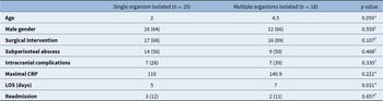
After excluding mixed skin flora isolates, a total of 18 cases had multiple organisms isolated from specimens which were obtained during the same presentation, while 25 patients had a single organism isolated from specimens (Table 2). There was a significant difference between the length of stay for each group (median 7 days for multiple organisms, median 5 days for single isolated organisms, p = 0.03), but no significant difference was detected across other variables (surgical intervention, subperiosteal abscess, intracranial complications or readmission). When multiple culture samples were obtained from the same child, only 2/87 cases had positive cultures from more than one sample. In both cases, the bacteria cultured from the positive samples were different. One case reported S pyogenes as positive from a blood culture, whilst the ear swab from the same patient reported S aureus. The second case demonstrated S pneumoniae from pus cultured during mastoidectomy and Bacillus sp. from a sample of granulation tissue sent for culture.
Patient characteristics subdivided by single vs multiple organism isolation. Categorical data are summarised as N (%); LOS: Length of stay; CRP represented as average; Age and LOS represented as median

* Mann–Whitney U test.
† Fisher’s exact test – 1-sided.
A total of five children in our study were found to have concurrent cholesteatoma. Of these, four had culture results available; P aeruginosa was isolated in two children, with S aureus, Prevotella. sp. and E coli isolated in the other two children.
Intracranial complications were recorded in 18 patients (median age = 4.5 years), 15 (83 per cent) of which had positive cultures. There were 4 cases positive for S pneumoniae (27 per cent), 4 S aureus (27 per cent), 3 group A streptococci (20 per cent), 3 anaerobes (20 per cent), 1 S intermedius (7 per cent), 1 S oralis (7 per cent), 1 F necrophorum (7 per cent), 1 E coli (7 per cent), 1 H parainfluenzae (7 per cent), 1 Prevotella spp. (7 per cent) and 1 Bacillus spp. (7 per cent). In most cases, cultures were positive from a combination of ear swabs and mastoid pus/intracranial source; three patients had ear swabs only, two of which were positive for S pneumoniae, while one yielded mixed skin flora. None of these patients had positive blood cultures.
As per the local hospital guidelines, patients were treated with intravenous cefotaxime and metronidazole unless allergic. In the event of an allergy, intravenous clarithromycin was used as an alternative. Mortality at 30 days across all groups was zero.
Discussion
Our study confirms that, despite the introduction of the pneumococcal vaccine across the UK, the majority of acute mastoiditis is secondary to Streptococcus spp. Moreover, we identified that there is a high incidence of mastoiditis-related complications (subperiosteal abscess and intracranial complications) in patients with Fusobacterium-positive cultures, followed by mixed anaerobes. Surgical intervention was required in all patients with F necrophorum, H influenzae and positive anaerobic cultures.
Notably, the high incidence of abscess and intracranial complications reported in the present study is likely to be that Alder Hey Children’s Hospital is a large tertiary referral centre for paediatric ENT and neurosurgery, therefore these cases are usually referred from other centres.
Our data aligns with the findings of previously published data regarding causative pathogens responsible for mastoiditis infection. Studies conducted globally consistently showed Streptococcus spp. as the most prevalent, with many of those studies identifying S pneumoniae as the most isolated bacterium.Reference Laulajainen-Hongisto, Saat, Lempinen, Markkola, Aarnisalo and Jero6-, Reference Mierzwiński, Tyra, Haber, Drela, Sinkiewicz and Puricelli10
Although the data focusing only on the post-pneumococcal vaccine era is limited, Roddy et al. identified no difference in the proportion of paediatric mastoiditis cases caused by S pneumoniae before and after the introduction of the PCV7 in the US in 2000.Reference Roddy, Glazier and Agrawal11 Moreover, in a prospective study conducted between 2010 and 2011, Giannakopoulos et al. identified that 81 per cent of cases of acute mastoiditis were attributed to S pneumoniae.Reference Giannakopoulos, Chrysovergis, Xirogianni, Nikolopoulos, Radiotis and Lebessi12 However, Tamir et al. observed an inverse relationship between the increasing rate of immunised children and the proportion of mastoiditis cases with confirmed S pneumoniae-positive cultures.Reference Tamir, Roth, Dalal, Goldfarb and Marom13
Interestingly, our study has identified a comparable proportion of mastoiditis cases attributable to Streptococcus spp., when compared to the pre- and intra-pneumococcal conjugate vaccine era data from our institute. In the retrospective analysis of mastoiditis cases presenting to Alder Hey Hospital between 2003 and 2012, Attlmayr et al. identified that both S pyogenes (6/32 patients) and S pneumoniae (5/32 patients) were the most commonly isolated pathogens, collectively comprising 34 per cent of all identified pathogens.Reference Attlmayr, Zaman, Scott, Derbyshire, Clarke and De5 Paradoxically, our data demonstrated an increased number of S pneumoniae infections in the post-pneumococcal conjugate vaccine era (24/85 (28 per cent) versus 5/32 (16 per cent)). This could be partially explained by an increase of the serotype 19A in pneumococcal mastoiditis cases following the introduction of PCV7, which did not decline after PCV13 was introduced, as previously reported by Koutouzis et al.Reference Koutouzis, Michos, Koutouzi, Chatzichristou, Parpounas and Georgaki14 Other possible aetiologies for the continuing presence of S pneumoniae include shifting of bacterial serotypes after vaccine introduction, increasing resistance secondary to inadequate antibiotic use and also the low-to-late use of antibiotics in acute otitis media.Reference Goldberg-Bockhorn, Hurzlmeier, Vahl, Stupp, Janda and von Baum15, Reference Koelman, Brouwer and van de Beek16
The findings from this study suggest that in cases of mastoiditis, empirical treatment should focus on targeting organisms frequently colonising the upper respiratory tract. However, resistance patterns are likely to differ between regions/nations, therefore, local microbiological advice should always be sought, and antibiotic therapy should be guided by microbiology sampling, where possible. Importantly, antibiotic stewardship is vital to ensure prevention against resistance, and responsible prescribing of broad-spectrum antibiotics is highly encouraged. Moreover, given the high incidence of mastoiditis-related complications as demonstrated in this study in patients identified as infected with F necrophorum, H influenzae or mixed anaerobic bacteria, caution is advised when the specific pathogens are identified, and the responsible clinicians should carefully consider further investigations and aggressive treatment. However, in a majority of cases, these cultures were taken during surgery, raising the question of whether these bacteria may also be present in cases where surgery was not performed.
The study’s design being both retrospective and observational lends itself susceptible to selection bias. Microbiological samples were more likely to be obtained from the more severely unwell patients and those requiring surgical intervention, therefore, the estimated incidence of these organisms could be subjected to bias. In the case of conservatively managed mastoiditis (with an intact tympanic membrane), the likelihood of being able to obtain a microbiological diagnosis is low. Some of the specimens were swabs from the ear canal, which means the bacteria grown may not be directly responsible for mastoiditis. The use of nasopharyngeal swabs as a surrogate marker for acute otitis media infections in cases with an intact tympanic membrane has been explored in the literature, with high negative predictive value rates quoted between 68–97 per cent.Reference van Dongen, van der Heijden, van Zon, Bogaert, Sanders and Schilder17–Reference Yatsyshina, Mayanskiy and Shipulina19 Additionally, serotyping was not performed, limiting our ability to identify specific bacterial strains. In cases where multiple pathogens were detected, determining the causative pathogen becomes challenging.
Conclusion
Mastoiditis infections have predominantly gram-positive, facultative anaerobic microbiology. Despite the introduction of the pneumococcal vaccine, S pneumoniae remains the most prevalent identified pathogen. From this sample of data, empirical treatment with cefotaxime and metronidazole gives adequate antibiotic cover. Microbiology culture and sensitivity, wherever possible, are essential to ensure effective antibiotic therapy, and further studies are required in order to definitively evaluate the effect of the pneumococcal vaccination scheme on the bacteriology of mastoiditis.
Funding statement
This research did not receive any specific grant from any funding agencies in the public, commercial or not-for-profit sectors.
Competing interests
The authors declare none.
Ethical standards
The study was conducted in accordance with the Declaration of Helsinki and institutional approval was granted prior to data collection. The requirement for informed consent was waived because of the retrospective design.