Leucoraja erinacea, the little skate, is a member of the order Rajiformes (skates) sensu Naylor et al. (Reference Naylor, Caira, Jensen, Rosana, White and Last2012a), one of the most diverse and well-defined monophyletic groups within Batomorphii, including almost half of all batoid fishes (ca.290 valid living species in 38 genera). Skates are benthic batoids characterised by a series of derived morphological traits. Although morphological analyses have retrieved conflicting phylogenetic positions for Rajiformes within Batomorphii (e.g., Aschliman et al. Reference Aschliman, Nishida, Miya, Inoue, Rosana and Naylor2012a; Naylor et al. Reference Naylor, Caira, Jensen, Rosana, White and Last2012a, Reference Naylor, Caira, Jensen, Rosana, Straube, Lakner, Carrier, Musick and Heithausb; Bertozzi et al. Reference Bertozzi, Lee and Donnellan2016), molecular data have recovered skates as the most plesiomorphic order within batomorphs (e.g., Aschliman et al. Reference Aschliman, Claeson, McEachran, Carrier, Musick and Heithaus2012b; Last et al. Reference Last, White, de Carvalho, Séret, Stehmann and Naylor2016; ‘Chondrichthyan Tree of Life' project https://sharksrays.org/).
Skates are becoming increasingly important model organisms for understanding the early development of neurocranial traits, such as those related to inner ear morphology, within a phylogenetic framework. Ultimately, this will cast new light on the origin and transformations of the neurocranium and its associated structures in the common ancestor of elasmobranch fishes (e.g., Maisey Reference Maisey2001, Reference Maisey2004; Maisey & Lane Reference Maisey and Lane2010). Skates such as the little skate (Leucoraja erinacea) or the winter skate (Leucoraja ocellata) are increasingly being used as model organisms in a range of studies addressing their development, renal physiology, immunology, neurobiology, regeneration processes, and ecology (e.g., Stolte et al. Reference Stolte, Galaske, Eisenbach, Lechene, Schmidt-Nielson, Boylan, Antkowiak, Patel and Niermann1977; Elger et al. Reference Elger, Hentschel, Litteral, Wellner, Kirsch, Luft and Haller2003; Maxwell et al. Reference Maxwell, Fröbisch and Heppleston2008; Gillis et al. Reference Gillis, Dahn and Shubin2009; DiSanto Reference DiSanto2015; Criswell et al. Reference Criswell, Coates and Gillis2017). Additionally, a skate genome project is currently under development with several resources already publicly available (Wyffels et al. Reference Wyffels, King, Vincent, Chen, Wu and Polson2014). These skates also are readily available from embryonic stages to adults, because they can be easily raised within the egg cases in the lab. Identifying developmental traits of the sensory system such as the inner ear of Leucoraja erinacea thus complements the already available information about this skate, making it an ideal model organism in developmental studies of elasmobranch fishes.
Cartilaginous (chondrichthyan) and ray-finned bony (actinopterygian) fishes have diverse sensory systems including mechanoreception, vision, chemoreception, electroreception, and an electrically sensitive magnetic sensory system finely attuned to their aquatic environments (e.g., Roberts Reference Roberts, Hodgon and Mathewson1978; Braun & Grande Reference Bleckman, Hoffman and Hamlett2008; Ladich & Schulz-Mirbach Reference Ladich and Schulz-Mirbach2016). The vestibular system, as one of several mechanosensory organs, is important in aquatic vertebrates and is responsible for equilibrium and balance (Retzius Reference Retzius1881), but represents a poorly understood system. Therefore, it is likely to yield crucial information relevant to comparative neuroanatomical studies in an evolutionary context.
Essentially, all gnathostomes share common features of the otic system (Maisey Reference Maisey2001), such as the presence of the third horizontal canal, antero-posteriorly asymmetrical canals and vestibular chambers, widely curved semicircular canals (SCCs), an utricular chamber and macula, a crista acustica neglecta, distinct saccular–lagenar chambers, and calcium carbonate polycrystalline otoliths. In aquatic gnathostomes such as chondrichthyans and actinopterygians, the auditory system is composed of two portions that have been traditionally referred to as the pars superior, consisting of the SCCs and an otolithic element (called utriculus in actinopterygians), and the pars inferior, consisting of two otolithic organs (called sacculus and lagena in actinopterygians) (Carlström Reference Carlström1963; Platt & Popper Reference Platt, Popper, Tavolga, Popper and Fay1981; Kasumyan Reference Kasumyan2004). Published accounts focused on the SCCs (bony fishes: Rayner Reference Rayner1952; Grande & Bemis Reference Grande and Bemis1998; Maisey Reference Maisey1999; Giles et al. Reference Giles, Rogers and Friedman2016; cartilaginous fishes: Maisey Reference Maisey1983, Reference Maisey1985, Reference Maisey1987; Coates et al. Reference Coates, Gess, Finarelli, Criswell and Tietjen2017) and semicircular ducts (bony fishes: Grande & Bemis Reference Grande and Bemis1998; cartilaginous fishes: Maisey Reference Maisey2001; Evangelista et al. Reference Evangelista, Mills, Siebeck and Collin2010). Several characters of the inner ear differentiate elasmobranchs from extant holocephalans and osteichthyans (Maisey Reference Maisey2001), such as the isolation of the posterior semicircular canal (PSC) from the other SCCs (Retzius 1881), the single connection of the PSC with the vestibular region, the rounded shape of PSC (Retzius 1881; de Beer Reference De Beer1931; Schaeffer Reference Schaeffer1981), the separation of the saccular and utricular regions, an elaboration and enlargement of the crista neglecta, a chondrification of the medial capsular wall, the occurrence of a perilymphatic fenestra and a crus between the anterior and external canals, an out-of-plane and curved orientation of the external canal, a single canal between the anterior and external canals with the vestibular region, and exogenous sand grains within the endolymphatic duct and sacculus. According to Maisey (Reference Maisey2001), derived features of elasmobranchs include the presence of an endolymphatic (parietal) fossa and a horizontally lateral otic process, a closure of the embryonic metotic fissure, the anterior ampulla located between or in front of the postorbital process, and the occipital block wedged between the otic capsules (e.g., Schaeffer Reference Schaeffer1981). All of these characters reveal the complex anatomy of the membranous system in cartilaginous fishes. In osteichthyans, however, no apomorphic characters of the otic region have been recognised and it has been suggested that the labyrinth of extant bony fishes is highly conservative and represents the anatomical pattern of early chondrichthyans (Maisey & Lane Reference Maisey and Lane2010).
To obtain fundamental insights into the evolution and functional morphology of the vertebrate inner ear, the comparative anatomy of taxa from early diverging groups are the key to our understanding of the evolution of the vertebrate sensory system (Fay & Popper Reference Fay and Popper2000; Ladich & Schulz-Mirbach Reference Ladich and Schulz-Mirbach2016). Since the in-depth studies of Hyrtl (Reference Hyrtl1873), hundreds of papers focusing on the anatomy of the vestibular system have been published, mainly focusing on terrestrial vertebrates (e.g., Clack et al. Reference Clack, Ahlberg, Finney, Dominiquez, Robinson and Ketcham2003; Spoor et al. Reference Spoor, Garland, Krovitz, Ryan, Silcox and Walker2007; Lebrun et al. Reference Lebrun, De León, Tafforeau and Zollikofer2010; Benoit et al. Reference Benoit, Lehmann, Vatter, Lebrun, Merigeaud, Costeur and Tabuce2015; Pfaff et al. Reference Pfaff, Martin and Ruf2015; Clack Reference Clack, Clack, Fay and Popper2016; Clack & Anderson Reference Clack, Anderson, Clack, Fay and Popper2016; Ekdale Reference Ekdale2016; Grohé et al. Reference Grohé, Tseng, Lebrun, Boistel and Flynn2016b; Coutier et al. Reference Coutier, Hautier, Cornette, Amson and Billet2017; Pfaff et al. Reference Pfaff, Nagel, Gunnell, Weber, Kriwet, Morlo and Bastl2017a, Reference Pfaff, Czerny, Nagel and Kriwetb). Detailed anatomical descriptions of the ear region of sharks, skates, and rays have already been used to contrast morphological differences related to body shape (cylindrical vs. flat: Cuvier Reference Cuvier1799; Weber Reference Weber1820; Cuvier & Valenciennes Reference Cuvier and Valenciennes1828; reviewed in Breschet Reference Breschet1838; Leydig Reference Leydig1852; Norris Reference Norris1929; Hasse Reference Hasse1873a, Reference Hasseb), to present morphological information of inner ear structures (e.g., deBeer 1931; Holmgren Reference Holmgren1940; Jollie Reference Jollie1971; Corwin Reference Corwin and Johari1978; Maisey Reference Maisey1983, Reference Maisey1985, Reference Maisey1987, Reference Maisey2001; Pradel et al. Reference Pradel, Langer, Maisey, Geffard-Kuriyama, Cloetens, Janvier and Tafforeau2009; Evangelista et al. Reference Evangelista, Mills, Siebeck and Collin2010; Coates et al. Reference Coates, Gess, Finarelli, Criswell and Tietjen2017; Schnetz et al. Reference Schnetz, Kriwet and Pfaff2017), to review phylogenetic aspects of inner ear evolution (Maisey Reference Maisey2001), and also to investigate phonoreception (e.g., Ladich & Popper Reference Ladich, Popper, Manley, Fay and Popper2004; Casper & Mann Reference Casper and Mann2006; Maisey & Lane Reference Maisey and Lane2010), including an ontogenetic study of the Thornback Ray, Raja clavata (Corwin Reference Corwin1983). As a step towards expanding this research and as a baseline for future studies focusing on the SCC sensory system, we present here the first description of the early development of the otic region in Leucoraja erinacea.
1. Material and methods
We conducted observations on five ontogenetic stages of Leucoraja clavata. These stages were defined following Maxwell et al. (Reference Maxwell, Fröbisch and Heppleston2008) and comprise the prenatal stages 29 (unsexed; Natural History Museum, London (BMNH) 2018.9.26.5; Associated 3D data in MorphoMuseumM (M3 Id): M3#376_2018.9.26.5), 31 (unsexed; BMNH 2018.9.26.4; M3 Id: M3#370_2018.9.25.4), and 32 (male; BMNH 2018.9.26.3; M3 Id (skel. lab.): M3#369_2018.9.25.3; memb. lab.: M3#372_2018.9.25.3), the post-hatching stage 34 (male; BMNH 2018.9.26.2; M3 Id: M3#368_2018.9.25.2), and one adult specimen (male; BMNH 2018.9.26.1; M3 Id: M3#367_2018.9.25.1) with a total length of 52cm. Specimens were obtained euthanised and formalin fixed from Woods Hole Marine Biological Laboratory, Woods Hole, Maryland (USA), following standard protocols, under approved procedures and with required scientific permits.
1.1. Anatomical terminology of the vestibular system
Following the Federal Committee on Anatomical Terminology (Reference Gardiner, Hueter, Maruska, Sisneros, Casper, Mann, Demski, Carrier, Musick and Heithaus1998) and Sipla & Spoor (Reference Sipla, Spoor, Thewissen and Nummela2008), within the otic system, the term ‘semicircular canals' refers to the bony labyrinth or skeletal labyrinth, whereas the ‘semicircular ducts/tubes' represent the membranous ducts inside these bony canals. The closed membranous system is composed of fluid-filled ducts/tubes (SCDs: semicircular ducts; ASD: anterior semicircular duct; PSD: posterior semicircular duct; LSD/HSD: lateral/horizontal semicircular duct) and surrounded by the skeletal labyrinth with its three SCCs (ASC: anterior semicircular canal; PSC; LSC/HSC: lateral/horizontal semicircular canal) and the sensory organs (utriculus, sacculus, and lagena) (e.g., Hyrtl Reference Hyrtl1845, Reference Hyrtl1873; Retzius 1881). The skeletal labyrinth is separated from the membranous labyrinth by perilymphatic fluid acting as a shock absorber (e.g., Torrey Reference Torrey1962; Maisey Reference Maisey2001). While the SCDs of gnathostomes are responsible for detecting angular acceleration, the utriculus, sacculus, and lagena are three sensory areas that detect linear movements during locomotion (e.g., de Burlet Reference De Burlet, Bolk, Göppert, Kallius and Lubosch1934; Tester et al. Reference Tester, Kendall and Milisen1972).
1.2. Computed tomography (CT) scanning
Leucoraja specimens were scanned in the Imaging and Analysis Centre, Natural History Museum, London. The adult was scanned on the Metris X-Tek HMX ST 225 CT scanner, while stages 29–34 (one specimen per stage) were scanned on the ZEISS Xradia 520 Versa CT scanner. These smaller specimens were washed in dehydrating solutions consisting of still water with increasing amounts of ethanol (30%, 50%, 70% of ethanol mixed with distilled water) and stained with phosphotungstic acid (PTA) (0.1% wt/vol in 70% ethanol) for soft tissue visualisation, for three days, then washed in pure 70% ethanol for one day, and scanned while immersed in 70% ethanol. For scanning settings see Table 1.
3D measurements of the neurocranium and skeletal labyrinth of Leucoraja erinacea. CT scan settings of the investigated specimens. Abbreviations: NCmin=minimum width of the neurocranium; NCmax=maximum length of the neurocranium; NACmax=maximum distance between the nasal capsules; OtRmax=maximum width of the otic region; ASCh=height of the anterior semicircular canal; PSCh=height of the posterior semicircular canal; HSCh=height of the horizontal semicircular canal; HSCw=width of the horizontal semicircular canal; HSCd = diameter of the horizontal semicircular canal; VESTl=length of the vestibulum.

1 3D reconstruction of skeletal labyrinth was not possible.
1.3. Three-dimensional (3D) visualisation
3D reconstructions and visualisations were performed using Drishti 2.6.2 (http://anusf.anu.edu.au/Vizlab/drishti/). The ‘clip function' was employed to visualise internal structures, while the ‘mop carve function' was used to erase some structures, such as the external gill elements, in order to expose the head and otic region (e.g., stage 31). Additionally, 3D models of the skeletal labyrinth of all stages and the membranous system of stage 32 were reconstructed with Amira 6.0. based on manual segmentation of the tiff stacks (supplementary Fig. 1 available at https://doi.org/10.1017/S1755691018000993). The reconstructed Polygon File Format (PLY) are published in M3 (Lebrun & Orliac Reference Lebrun and Orliac2016). Inventory numbers of associated data are found in Section 1.
1.4. 3D measurements
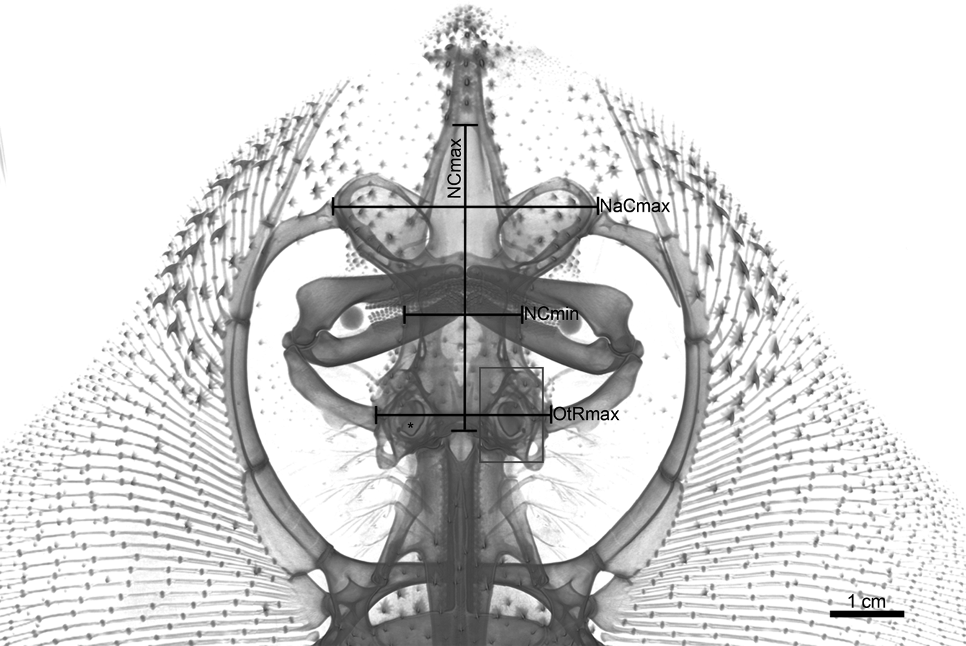
3D measurements were taken on the isosurfaces of the 3D models of the neurocranium and the virtually reconstructed skeletal labyrinths generated with Amira 6.0. Anatomical terms of the skull of Leucoraja erincaea follows Bustamate et al. (Reference Bustamate, Lamilla, Concha, Ebert and Benett2012). Measurements of the neurocranium are defined as (Fig. 1; Table 1):
• Minimum width of the neurocranium (NCmin): shortest distance between the supraoccipital ridges; transverse plane.
• Maximum length of the neurocranium (NCmax): between the anterior margin of the anterior fontanelle and the parietal depression of the occipital region; midsagittal plane.
• Maximum distance between the nasal capsules (NaCmax): between the outermost lateral edges of the nasal capsules; transverse plane.
• Maximum width of the otic region (OtRmax): between the outermost lateral edges of the otic capsules; transverse plane.
Leucoraja erinacea, 3D volume-rendered reconstruction, adult specimen (BMNH 2018.9.26.1). 3D measurements of the neurocranium with otic region. Abbreviations: NCmin=minimum width of the neurocranium; NCmax=maximum length of the neurocranium; NaCmax=maximum length between the nasal capsules; OtRmax=maximum width of the otic region. Grey box=otic region; asterisk=otolith-like structure (OLS) in the left otic region.

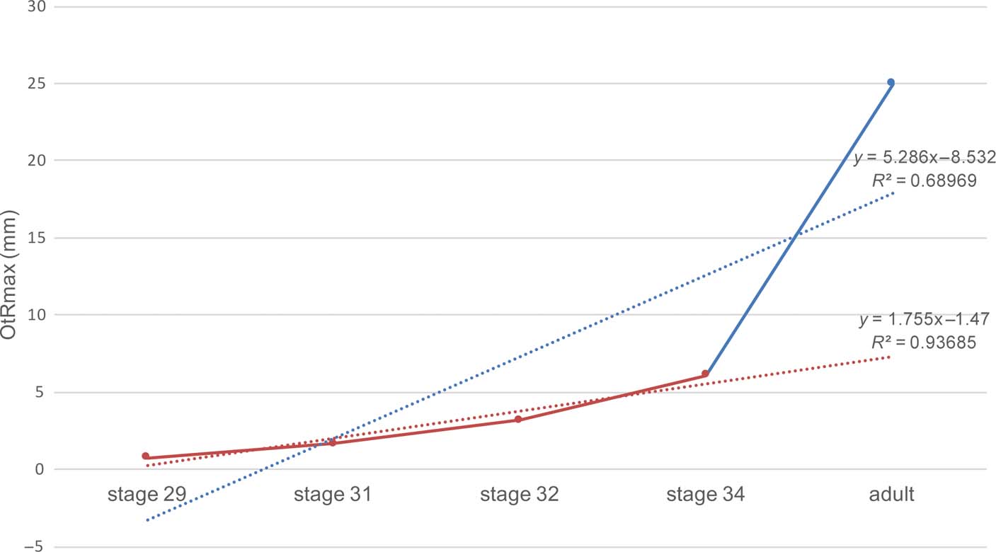
Differences in OtRmax among the different Leucoraja stages were visualised and analysed with a linear regression line using Microsoft Office Excel 14.1.0. Due to the large developmental gap between stage 34 and the adult specimen, we also fit a regression line between stage 29 and 34.
Measurements of the skeletal labyrinth are modified from Pfaff et al. (Reference Pfaff, Martin and Ruf2015):
• Height of the SCCs (ASCh, PSCh, HSCh): maximum height, expressed as the distance from the surface of the vestibulum to the inner wall of the skeletal canal taken at 50% of the distance between the ampulla of the canal and the crus commune.
• Width of the SCCs (ASCw, PSCw, HSCw): maximum width between the inner walls of the skeletal canal of each SCC, measured at 50% of the height of the corresponding canal at a right angle to canal height.
• Diameter of the SCCs (ASCd, PSCd, HSCd): diameter taken at 50% of the length of the corresponding skeletal labyrinth canal.
• Length of the vestibulum (VESl): maximum distance between the most dorsal point of the skeletal labyrinth and the most ventral point of the vestibulum.
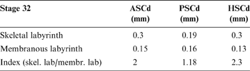
Anatomical distinction between the skeletal and membranous labyrinth were measured as two separate diameters of each SCC at 50% length. An index was calculated as their quotient (diameter of the skeletal SCC/diameter of the membranous SCD) with Microsoft Office Excel 14.1.0 (Table 2).
Diameter of the skeletal and membranous labyrinth of stage 32 at 50% SCCs length. Abbreviations: ASCd=diameter of the anterior semicircular canal; PSCd=diameter of the posterior semicircular canal; HSCd=diameter of the horizontal semicircular canal.

2. Results
2.1. Model of growth
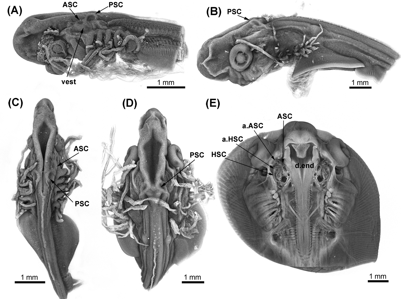
The growth model of the ear region was investigated using measurements at the respective ontogenetic stages (for a detailed list, see Table 1; Figs 2, 3; supplementary Fig 1). We were not able to fully reconstruct the vestibular system in stages 29 and 31 (supplementary Fig. 1) due to their early stage of development, even though they were discernible with Drishti (Fig. 2a, b).
Leucoraja erinacea, early ontogenetic stages, with vestibular system. Lateral view: (A) stage 29 (BMNH 2018.9.26.5); (B) stage 31 (BMNH 2018.9.26.4). Dorsal view: (C) stage 29; (D) stage 31; (E) stage 32 (BMNH 2018.9.26.3). Abbreviations: a.Asc=ampulla of anterior semicircular canal; a.HSC=ampulla of horizontal semicircular canal; ASC=anterior semicircular canal; d.end=ductus endolymphaticus; PSC = posterior semicircular canal; HSC=horizontal semicircular canal; vest=vestibulum.

Leucoraja erinacea, maximum length (mm) of the complete otic region (OtRmax) between investigated ontogenetic stages. Regression line (red): stage 29 (BMNH 2018.9.26.5) through stage 34 (BMNH 2018.9.26.2). Regression line (blue): stage 29 through adult stage (BMNH: 2018.9.26.1).

2.2. Morphological comparison between membranous and skeletal labyrinth (stage 32)
The skeletal labyrinth encloses the membranous system, with its three SCDs, and an utriculo–lagenar chamber. Precise anatomical differences are seen between the membranous and skeletal labyrinths (Fig. 4; Table 2) and calculated as an index (Table 2). In particular, we see a clear distinction of the membranous system and skeletal system in the SCCs and SCDs, the sacculus, and the utriculus. The connection between ASD and PSD represented by the crus commune is not observable in the membranous system, nor is any association between HSD and the sacculus detectable.
Leucoraja erinacea, 3D volume-rendered reconstruction of the vestibular system of stage 32 (BMNH 2018.9.26.3). (A) Skeletal labyrinth. (B) Membranous labyrinth. (C) Membranous labyrinth in the cavity of the skeletal labyrinth. PSC=posterior semicircular canal; ASC=anterior semicircular canal; HSC=horizontal semicircular canal.

2.3. SCCs
In the earliest stage available for this study (stage 29), the ASD, PSD, and the vestibulum are already developed and distinct, whereas the HSD and the endolymphatic duct cannot yet be detected (Fig. 2a, b; supplementary Fig 1). The PSD is rotated into a more dorsal position than the ASD (Fig. 2a–d), which is horizontally oriented. In stage 31, all three SCDs are present, dorso-ventrally flattened, and partly tilted laterally. The membranous system of stage 32 represents the anatomical morphology of the adult vestibular system (Fig. 2e). All three SCDs are circular in cross section, with a common canal connecting the ASD and HSD; however, the PSD is separated. The posterior canal duct connects the sacculus and PSD (Fig. 4b). Anteriorly, the endolymphatic duct runs dorsally and joins the parietal fossa (Fig. 4a). The position of the sensory maculae and cristae are not detectable.
2.4. Endolymphatic organ
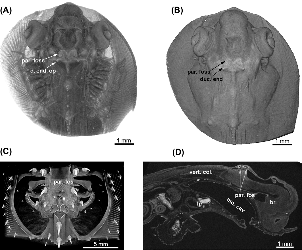
In stage 29 and 31, endolymphatic foramina are identifiable Fig. 5a, b; asterisks), but the endolymphatic organ is not yet developed. The parietal fossae appear as two highly mineralised hook-shaped structures in stage 32 and 34 (Fig. 5c, d). The shape of those hooks changes remarkably during ontogeny: two regular shaped and massive hook-like structures in a nearly horizontal plane (stage 32; Figs 5c, 6a, b) change into two fragile and irregularly shaped remnants (adult stage; Fig. 6c). First found at the anterior margins of the fossae, these rotate in a more occipital direction during ontogeny (adult stage; Figs 5e, 6c). The openings of the endolymphatic ducts appear at the posterior edge of the hook-like structures (Fig. 6b). Neither the course of the endolymphatic duct, the endolymphatic pouch, the muscle of the parietal fossa, nor the fenestra ovalis can be reconstructed in the investigated specimens (Fig. 6d).
Leucoraja erinacea, semi-transparent 3D volume-rendered images of all investigated specimens (dorsal view). (A) Stage 29 (BMNH 2018.9.26.5). (B) Stage 31 (BMNH 2018.9.26.4). (C) Stage 32 (BMNH 2018.9.26.3). (D) Stage 34 (BMNH 2018.9.26.2). (E) Adult (BMNH 2018.9.26.1). Red shading=3D reconstruction of the right vestibular system; asterisk=endolymphatic foramen; par foss=parietal fossa.

Leucoraja erinacea, parietal fossa. (A) Semi-transparent volume image of Stage 34 (BMNH 2018.9.26.2). (B) Isosurface image with the hook-like parietal fossa and endolymphatic foramen. (C) Semi-transparent image of the adult stage (BMNH 2018.9.26.1) with the slender parietal fossa. (D) Phosphotungstic acid-stained sagittal section of stage 34. Abbreviations: br.=brain; duc. end=ductus endolymphaticus; duc. end. op=opening of ductus endolymphaticus; hr=heart; mo. cav=mouth cavity; p. foss=parietal fossa; vert. col.=vertebral column.

2.5. Otoliths
During the ontogenetic development of Leucoraja erinacea, otoliths (mineralised homologues of actinopterygian otoliths) appear in the otic region. In the earliest investigated stage 29, mineralised otoliths are seemingly not yet developed, but in stages 32 and 34, two identifiable otoliths appear. The largest otolith (Fig. 1; asterisk), which we assume to be homologous to the sagitta in actinopterygians, is situated in the saccular chamber and possesses a small process. In stages 32 and 34, the sagitta appears in the vestibulum as a spoon-like structure with a small process, whereas in the lagena, the lapillus is bean-shaped. Unlike in actinopterygians, a third otolith is not discernible in the micro-CT scans, probably due to its low mineralisation in the investigated stages. This needs to be tested further.
3. Discussion
During the ontogeny of Leucoraja erinacea, the ear region shows two phases of growth (Fig. 3). From stage 29 (pre-hatching) to stage 34 (post-hatching), the size of the otic region changes isometrically (Fig. 3; red regression line) and then during the last phase of ontogenetic development, the size changes exponentially (Fig. 3; blue regression line). However, this last part of development might be an artefact (e.g., edge effect) due to missing stages between stage 34 (post-hatching) and the adult stage.
Early in the ontogeny of Leucoraja erinacea, the ASD and PSD are distinct and developed further than the HSD. Comparisons of L. erinacea with other elasmobranchs are hampered by the lack of ontogenetic studies in other species. With an isolated and circular PSC duct, which is already developed in the earliest investigated stage 29, the anatomy of the vestibular system of L. erinacea presents the characteristic morphology for elasmobranch fishes (e.g., de Beer Reference De Beer1931; Maisey Reference Maisey2001; Evangelista Reference Evangelista, Mills, Siebeck and Collin2010; Maisey & Lane Reference Maisey and Lane2010). Importantly, this represents a derived morphological trait in gnathostomes (Schaeffer Reference Schaeffer1981; contra Stensiö Reference Stensiö and Jorge1950, Reference Stensiö1963). However, the gross anatomy of the skeletal labyrinth in subadult stages of L. erinacea does not reflect the shape of the membranous system as was indicated for Squalus and Notorynchus (Maisey Reference Maisey2001). A crus commune is present in the skeletal labyrinth of L. erinacea between ASC and PSC (Fig. 4), which is not found in the underlying membranous system between ASD and PSD. This crus commune was assumed to be generally absent in fossil and modern elasmobranchs (Maisey Reference Maisey2004), but is seen in extant chimaeroids, osteichthyans, lampreys, osteostracans, and galeaspids (Miles Reference Miles, Miles, Patterson and Greenwood1973; Maisey Reference Maisey2001; Maisey & Lane Reference Maisey and Lane2010; Davis et al. Reference Davis, Finarelli and Coates2012; Coates et al. Reference Coates, Gess, Finarelli, Criswell and Tietjen2017). Overall, the membranous labyrinth of batoids appears more specialised than in sharks (Maisey Reference Maisey2001). To interpret anatomical changes in the evolutionary development of L. erinacea, visualisation of the membranous system in early stages should be the focus. Investigating functional-morphological adaptations using the skeletal labyrinth of batoids as an indicator of velocity and acceleration to elucidate the inhabitation of different zones of the ocean (see, e.g., Corwin Reference Corwin and Johari1978), might be problematic. Detecting phylogenetic signals in the skeletal anatomy of the ear region, as in sharks (Maisey Reference Maisey2001), is beyond the scope of this study.
Mineralised structures in the vestibulum and sacculus (otoliths; Fig. 1) appear early in the ontogeny of Leucoraja erinacea. Personal observations in other batoids show that these unambiguously display three otoliths, similar to the condition in actinopterygians. However, we were only able to detect two of these elements in L. erinacea. This most likely represents an artefact because otoliths contain high amounts of organic matter in batoids (Carlström Reference Carlström1963) and are highly vulnerable to fixation and handling of specimens.
It has been shown that the otic region in elasmobranchs, and especially in batomorphs, does not only contain the vestibular system, but also a sound-conducting mechanism (endolymphatic organ), involving the complex anatomy of the endolymphatic duct with its external aperture (‘fenestra ovalis'), the PSD and the sacculus, the loop or endolymphatic pouch (held within the parietal fossa), an internal passage, and an additional muscle of the parietal fossa (Scarpa Reference Scarpa1800; Ayers Reference Ayers1892; Norris Reference Norris1929; Tester et al. Reference Tester, Kendall and Milisen1972). An additional sensory structure (macula neglecta; macula acustica neglecta according to Retzius (1881)) is located in the posterior canal duct at the conjunction to the sacculus (Fay et al. Reference Fay, Kendall, Popper and Tester1974; Barber et al. Reference Barber, Yake, Clark and Pungur1985; Corwin Reference Corwin1989; Gardiner et al. Reference Gardiner, Hueter, Maruska, Sisneros, Casper, Mann, Demski, Carrier, Musick and Heithaus2012). This non-otolithic system determines the sound direction in elasmobranchs (Tester et al. Reference Tester, Kendall and Milisen1972; Corwin Reference Corwin1977; Fänge Reference Fänge1982) and is specialised toward low-frequency semi-directional phonoreception (Banner Reference Banner and Cahn1967; Corwin Reference Corwin, Tavolga, Popper and Fay1981; Bleckman & Hoffman Reference Bleckman, Hoffman and Hamlett1999; Maisey & Lane Reference Maisey and Lane2010). It is suggested that sound waves enter the PSD via the fenestra ovalis connecting the otic chamber and the parietal fossa and displace the cupula with its sensory cells lying over the macula neglecta (Tester et al. Reference Tester, Kendall and Milisen1972; Corwin Reference Corwin1977). The fenestra ovalis is covered with a small membrane, acting as a tympanic membrane analogous of higher vertebrates (Daniel Reference Daniel1934, p. 272). The macula neglecta in elasmobranchs shows a graduation from a single batch with a variety of oriented cells in rays to two separate patches composed of two cell groups oriented in opposite directions in carcharinid sharks, which might be related to increase auditory sensitivity (Corwin Reference Corwin1977, Reference Corwin and Johari1978, Reference Corwin1989; Gardiner et al. Reference Gardiner, Hueter, Maruska, Sisneros, Casper, Mann, Demski, Carrier, Musick and Heithaus2012). According to Corwin (Reference Corwin1989), the morphology of the auditory system of Leucoraja erinacea is indicative of type I classification with a small ventrally located sacculus and posterior canal duct, a small macula neglecta, and small lateral perilymphatic fluid space. However, this type of vestibular system characterises sharks that feed by pursuit predation (Tester et al. Reference Tester, Kendall and Milisen1972), whereas the macula neglecta in skates, rays, and sedentary sharks is small (Corwin Reference Corwin1989). This might explain why we were not able to visualise the macula neglecta in the sacculus of L. erinacea.
Even though we were not able to visualise the soft tissue of the endolymphatic canal duct in the parietal fossa (Fig. 6d), the overall shape and size of the parietal fossa changes remarkably during ontogeny (Figs 5, 6). During ontogenetic development, the size of the calcified part of the fossae is reduced in its length relative to the neurocranium and has an irregular and unusual shape. In the adult stage the calcified part is restricted to the posterior cranial margin of the fossa (Fig. 6c), whereas the fenestra ovalis is closed dorsally by soft tissue, which, as suggested above, may be comparable to the tympanic membrane in tetrapods and, thus, might be important for hearing. Endolymphatic foramina at the posterior margin of the parietal fossa are present in subadult stages, which become smaller relative to the neurocranium during development. The parietal fossa appears early in the ontogeny of Leucoraja erinacea, which might represent a heterochronic development of the otic region. We were not able to identify any exogenous material in either the endolymphatic duct or in the sacculus of the adult, although previous studies indicate an intake of sediment particles into the sacculus of prenatal batomorphs through the endolymphatic duct (Norris Reference Norris1929).
4. Conclusions
Ontogenetic studies of the vestibular system of sharks, skates, and rays have the potential to provide fundamental information about the evolution and functional morphology of the inner ear of vertebrates in general. Although chondrichthyan fishes are often assumed to represent a generalised model, the anatomy of the vestibular system, especially in batoid fishes like Leucoraja erinacea, appears highly derived (Maisey Reference Maisey2001; this study) and might not represent a ground plan for gnathostomes. To better understand the plesiomorphic condition of inner ear characters and their evolutionary modifications in chondrichthyans, representatives of all major groups need to be analysed. The same applies to the presence of the crus commune, that connects the ASC and PSC in the skeletal labyrinth of the skate studied here. Thus, gaining knowledge of the vestibular system in an evolutionary context, but also related to the influence of ecological changes during climate change (DiSanto Reference DiSanto2015) or habitat alterations, represents a highly important task for upcoming studies.
5. Acknowledgements
Thanks to Tim Smithson, Marcello Ruta, and Per Ahlberg for organising ‘The early tetrapod world: laying the foundations of the modern vertebrate fauna'. Thanks also to Ollie Crimmen and James MacLaine for access to the NHM Life Sciences collections, and Farah Ahmed and Amin Garbout for access and technical assistance with the NHM CT-scanners. We also want to thank two anonymous reviewers for their comments and suggestions, which greatly improved this manuscript. It was a privilege to attend (ZJ) the symposium ‘The early tetrapod world: laying the foundations of the modern vertebrate fauna' in Cambridge, December 2017, held in honour of Jenny Clack, and to provide this paper on the Leucoraja otic region. Jenny's work on the evolution of this region in early vertebrates, including tetrapods, has been groundbreaking, and she continues to be a source of inspiration in our research.
6. Supplementary material
Supplementary material is available online at https://doi.org/10.1017/S1755691018000993