Implications
Follicular development involves selection of a follicle apt to grow in response to stimuli (FSH and LH) that induce proliferation and survival of follicular cells so that it reaches ovulation. However, if follicles do not receive these stimuli (reduction in FSH and LH or their receptors), cellular death signals are triggered and the follicle fate will be atresia. Sphingosine-1-phospohate (S1P) induces survival and proliferation of cells, whereas ceramide (CER) is involved in cellular death. The present study reveals that S1P and CER concentrations differ within follicular status suggesting these sphingolipids may participate in follicular health or atresia. Knowledge of signals that control follicular development and atresia may help us to improve follicular development and thus reproductive performance.
Introduction
The development of bovine ovarian follicles occurs in a wave-like pattern. In each wave, a group of 5 to 24 small antral follicles are recruited to continue their growth in response to a transient rise of FSH (Aerts and Bols, Reference Aerts and Bols2010). The follicle that reaches a diameter of 8 to 8.5 mm will be selected as the dominant one and will reach maturity by the effect of LH (Mihm et al., Reference Mihm, Baker, Ireland, Smith, Coussens, Evans and Ireland2006). The dominant follicle (DF) produces 17β-estradiol (E2) and inhibin, which reduces serum FSH concentrations and, therefore, blocks the development of the other (subordinate) follicles, thus inducing them to atresia (Fortune et al., Reference Fortune, Rivera, Evans and Turzillo2001). During the luteal phase (i.e. in the presence of active corpus luteum) there is a growing and regression dynamic of antral follicles (Webb et al., Reference Webb, Garnsworthy, Gong and Armstrong2004). Follicular atresia is a multi-factorial process that includes follicular cell death (Rodgers and Irving-Rodgers, Reference Rodgers and Irving-Rodgers2010). The fate of ovarian follicles during follicular development depends on the action of survival factors that allow the follicle cells to proliferate and grow. The absence of survival factors coupled with the action of death-related factors will prevent the follicle from developing (Aerts and Bols, Reference Aerts and Bols2010; Rosales-Torres et al., Reference Rosales-Torres, Guzmán and Gutiérrez2012).
The metabolism of structural membrane sphingolipids may produce bioactive derivatives that function as signalling molecules that promote cellular development or apoptosis (Breslow and Weissman, Reference Breslow and Weissman2010). Sphingosine-1-phosphate (S1P) is a potent bioactive lipid that functions as an intracellular secondary messenger that promotes cellular survival and proliferation (Breslow and Weissman, Reference Breslow and Weissman2010). Within the cell, S1P is in a dynamic balance with its precursors, ceramide (CER) and sphingosine, forming the ‘sphingolipid rheostat’ (Spiegel and Milstien, Reference Spiegel and Milstien2002). CER is synthesised by sphingomyelinases from sphingomyelin found in the cell membrane. CER has an opposing action to S1P, inhibiting growth and promoting apoptosis (Pettus et al., Reference Pettus, Chalfant and Hannun2002). When survival factors are absent, or a death stimulus or hypoxia are present in the cell, CER accumulates and cytochrome C and SMAC (second mitochondria-derived activator of caspase-direct IAP binding protein with low PI) are released, resulting in the activation of caspases and cell death (Morales et al., Reference Morales, Lee, Goñi, Kolesnick and Fernández-Checa2007). The antagonistic effects of CER and S1P and their metabolites are interconvertible. It has been proposed that it is not their absolute amounts, but rather their relative levels, that determine the fate of a cell (Spiegel and Milstien, Reference Spiegel and Milstien2003; Baran et al., Reference Baran, Salas, Senkal, Gunduz, Bielawski, Obeid and Ogretmen2007).
Trophic factors (i.e. FSH and LH) induce follicular growth by promoting follicular cell proliferation and survival (Webb et al., Reference Webb, Garnsworthy, Gong and Armstrong2004) whereas a reduction of these factors or the presence of cell death-inducing factors in the follicular environment (i.e. TNF-α, hypoxia) trigger atresia (Evans et al., Reference Evans, Ireland, Winn, Lonergan, Smith, Coussens and Ireland2004). As S1P and CER are important signalling molecules in cell death or survival the aim of the current study was to examine whether S1P, CER concentrations and the S1P/CER ratio play a role in follicular health and atresia of ovarian follicles.
Material and methods
Follicle collection
Bovine ovaries were obtained from a local abattoir. The ovaries were washed and transported to the lab in cold phosphate buffer (PBS 0.01M, pH 7.4) in less than 60 min. The ovaries were again washed with PBS and 25 antral follicles with a diameter within 8 to 17 mm were dissected as reported by Rosales-Torres et al. (Reference Rosales-Torres, Guzmán and Gutiérrez2012).
Follicular compartments
In order to obtain the follicular fluid, follicles were opened by making an incision on the surface. The follicular fluid was centrifuged at 600×g for 10 min at 4°C. The supernatant was further centrifuged at 12 000×g for 10 min and then frozen at −70°C until analysed. The granulosa cells obtained after the first centrifugation were suspended in 200 µl PBS and kept in a 1500 µl tube.
The granulosa cells were removed by gentle scraping of the follicle wall with microsurgery forceps and detached cells were obtained by washing two times with 100 µl 0.01M PBS. These cells were then combined with the granulosa cells obtained after the follicular fluid centrifugation. The number of these cells was evaluated taking 10 µl of cell suspension and 90 µl of Trypan blue solution (4%). This mixture was placed in a Neubauer chamber and the cell count was done in five quadrants. The total cells number was obtained by using the formula: number of total cells counted in the chamber×10 000/number of squares×dilution. Subsequently, cell suspension was centrifuged at 600×g for 10 min at 4°C. The precipitate was stored at −70°C until its analysis. The theca cell layer of each follicle was observed under a dissection microscope and then rinsed and vortexed thrice with 200 µl of PBS (Rosales-Torres et al., Reference Rosales-Torres, Ávalos-Rodríguez, Vergara-Onofre, Hernández-Pérez, Ballesteros, García-Macedo, Ortíz-Navarrete and Rosado2000; Rosales-Torres et al., Reference Rosales-Torres, Alonso, Vergara, Romano, Castillo-Juárez, Avalos, Rosado and Gutiérrez2010), weighed on an analytical balance and stored at −70°C until assayed.
Follicle classification
In order to classify the follicles by their steroid functional stage, E2 and P4 in FF were determined by radioimmunoassay using COAT-A-ACCOUNT kits (Diagnostic products Corporation, Los Angeles, CA, USA). Intra-assay variability was 5.1% for E2 and 2% for P4. Follicles were classified as healthy when the E2/P4 ratio was 1 or higher and atretic when the ratio was <1 (Rosales-Torres et al., Reference Rosales-Torres, Ávalos-Rodríguez, Vergara-Onofre, Hernández-Pérez, Ballesteros, García-Macedo, Ortíz-Navarrete and Rosado2000).
Extraction of sphingolipids from follicular compartments
The granulosa cells (1×10⁶ cells/0.5 ml) and the theca cell layer (100 mg/1.0 ml) were homogenised in cold lysis buffer containing 0.25 M sucrose, 25 mM KCl, 50 mM Tris and 0.5 mM EDTA, pH 7.4 with a glass/teflon mechanical stirrer (Heidolph RZR1, Schwabach, Germany) (Bielawski et al., Reference Bielawski, Szulc, Hannun and Bielawska2006).
Sphingolipids were extracted as described by Villa et al. (Reference Villa, Kupchak, Garitaonandia, Smith, Alonso, Alford, Cowart, Hannun and Lyons2009). Briefly, 3 ml of chloroform : methanol v/v, 0.8 ml deionised water and 0.5 ml methanol were added to 500 µl of the homogenate and the mixture was maintained at 4°C overnight. The samples were then centrifuged for 10 min at 2900×g and the supernatant was separated and dried under a nitrogen evaporator. Lipids were resuspended in 300 µl chloroform and submitted to alkaline methanolysis with 1 ml of 0.125 M KOH, incubated at 37°C for 75 min, then the temperature was lowered, and 1500 µl chloroform was added. The samples were neutralised with 200 µl of 0.3 M HCl, 400 μl of 1 M NaCl in 5% glycerol and the organic phase was separated by centrifugation at 2900×g for 5 min, and then the supernatants were finally evaporated. The samples were stored at −20°C until assayed.
Analysis of CER and S1P by HPLC
A S1P commercial standard (Sigma-Aldrich, Saint Louis, USA) was used to perform a calibration curve. The S1P was dissolved in methanol to obtain a working solution of 497.1 µM, and dilutions of 0.75 to 7.5 ng/ml were made. For the CER calibration curve, ceramide 18 d-erythro-Sphingosine, N-Stearoyl-[N-Stearoyl ceramide, C18-Ceramide] (Avanti Polar Lipids, Alabama, USA) diluted in methanol at 500 μg/ml was used. The calibration curve was performed for concentrations in the range of 0.06 to 0.50 ng/ml. HPLC determinations were made in triplicate for both standards.
For HPLC determinations, the standards and the sphingolipids extracted from both follicular compartments were resuspended in 100 μl methanol and fresh d O-phtalaldehyde reagent (OPA; Sigma-Aldrich, Saint Louis, USA; 5 mg OPA, 0.1 ml of C2H6O, 10 μl 2-mercaptoethanol and 9.9 ml of H3BO3 3% (p/v) pH 10.5) was added. For this purpose, 50 μl of each of the standards, and each follicular compartment sample were mixed with 50 μl of OPA (Merril et al., Reference Merrill, Wang, Mullins, Jamison, Nimkar and Liotta1988). The mixture was incubated for 25 min in the dark at room temperature.
HPLC analysis was run in an automated Hitachi Elite Lachrom L 2200, Tokyo, Japan equipped with a XTerra RP18 column (5 μm, 3.0×150 mm, Ireland) and a 2 PK, XTerra RP18 pre-column (3.0×20 mm, Ireland), with a fluorescence detector (Hitachi Elite Lachrom L 2485, Tokyo, Japan) with a 340 and 435 nm excitation and emission wave lengths, respectively. A total of 30 μl of the standards and experimental samples were derivatised with OPA for 20 min. An isocratic run using HPLC column buffer (methanol and a phosphate buffer (K2HPO4) 5 mM (80 : 20 v/v), pH 7), with a flow rate of 0.6 ml/min was used.
Statistical analysis
Concentrations of CER and S1P in the granulosa cells were transformed to ng/mg of tissue instead of ng/number of cells for statistical analysis. The comparison between E2, P4, E2/P4 ratio, CER, S1P and the S1P/CER ratios between follicle types were analysed by ANOVA after normalisation of the data. In all, 5 atretic antral follicles and 8 healthy antral follicles were analysed for CER and S1P in the granulosa cells, whereas 8 atretic antral follicles and 14 healthy antral follicles were analysed in the theca cell layer.
Statistical model for differences in E2, P4, E2/P4 ratio, CER, S1P and the S1P/CER was:
 $$Y_{{ij}} \,{\rm =} \,\mu {\rm {\plus}}t_{i} {\rm {\plus}}e_{{ij}} $$
Y is the dependent variable (E2, P4, E2/P4 ratio, CER, S1P and the S1P/CER), µ the general mean, t i the effect of i th treatment (healthy vs atretic follicle) and e ij the experimental error.
Results
Follicular E2 and P4 concentrations
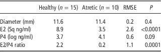
In the follicular fluid of 15 follicles, the steroid concentration were 79.21±9.6 ng/ml for E2 and 40.4±8.9 ng/ml for P4, with an E2/P4 ratio >1 (healthy antral follicles). A total of 10 follicles had an E2/P4 ratio of 0.2±0.3 and were considered atretic antral follicles (Table 1).
Diameter, 17 β-estradiol (E2) and progesterone (P4) concentrations, and E2/P4 ratio of bovine antral follicles

RMSE=root mean square error.
Variance heterogeneity of the E2 and P4 concentrations was corrected by logarithmic transformation for P4 and square root (Sq) for E2.
S1P and CER concentrations in cellular compartments of antral follicles
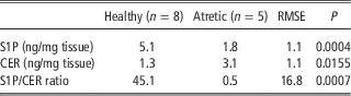
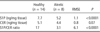
The S1P concentrations were significantly higher (P<0.05) in follicular compartments of healthy antral follicles than in atretic antral follicles (Table 2). In contrast, CER concentrations were only significantly higher in the granulosa cell of atretic antral follicles as compared with the healthy antral follicles (Table 2), but no differences were observed in the theca cell layer between healthy and atretic antral follicles (Table 3). In the granulosa cells, the S1P concentration was 45-fold higher than the CER concentration (Table 2). However, in the theca cell layer, S1P concentration was only 14-fold higher than CER (Table 3).
Sphingosine-1-phosphate (S1P), ceramide (CER) concentrations and S1P/CER ratio of granulosa cells from bovine antral follicles
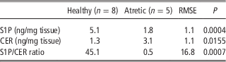
RMSE=root mean square error.
Variance heterogeneity of the sphingolipids concentration was corrected by logarithmic transformation.
Sphingosine-1-phosphate (S1P), and Ceramide (CER) concentrations, and the S1P/CER ratio in the theca cell layer of bovine antral follicles
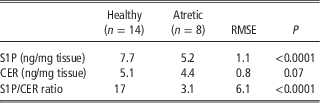
RMSE=root mean square error.
Variance heterogeneity of the sphingolipids concentration was corrected by logarithmic transformation.
Discussion
The development of follicles depends on trophic factors, such as gonadotropins and growth factors, which induce the proliferation and survival of cells (Rosales-Torres et al., Reference Rosales-Torres, Guzmán and Gutiérrez2012). It has been demonstrated that in bovine DFs there is an increase in LH receptor mRNA expression and anti-apoptotic genes, such as TIAF1, LASSY and LHCGR, which promote follicle development and E2 synthesis (Mihm et al., Reference Mihm, Baker, Ireland, Smith, Coussens, Evans and Ireland2006). In addition, it is well known that E2 promotes the granulosa cells proliferation and survival (Quirk et al., Reference Quirk, Cowan, Harman, Hu and Porter2004), and that angiogenesis is critical for the follicles development (Fraser, Reference Fraser2006), since the inhibition of vascular endothelial growth factor (VEGF), the main angiogenesis promoter, inhibited thecal blood vessels formation, as well as the development of antral follicles and ovulation in primates (Wulff et al., Reference Wulff, Wilson, Wiegand, Rudge and Fraser2002).
In the present study, we found that S1P concentrations in the granulosa and the theca cell layer were significantly higher in healthy than atretic antral follicles. Several studies have shown a role of S1P in the control of cell apoptosis and survival (Breslow and Weissman, Reference Breslow and Weissman2010). In culture, S1P reduced apoptosis in mesoangioblasts and in cardiac cells (Donati et al., Reference Donati, Cencetti, Nincheri, Bernacchioni, Brunelli, Clementi, Cossu and Bruni2007). Tao et al. (Reference Tao, Zhang, Vessey, Honbo and Karliner2007) found that decreased S1P synthesis reduced cell survival. Neutralisation of S1P with a monoclonal antibody blocked in vivo angiogenesis induced by fibroblast growth factor and VEGF (Argraves et al., Reference Argraves, Wilkerson and Argraves2010). It seems that the high S1P concentration found in healthy antral follicles, may play a critical role in protection, avoiding apoptosis and promoting angiogenesis.
Our study shows that CER was 138% higher in the granulosa cells of atretic antral follicles when compared with the healthy antral follicles. CER is an important molecule that induces cell cycle arrest and apoptosis (Schiffmann et al., Reference Schiffmann, Sandner, Birod, Wobst, Angioni, Ruckhäberle, Kaufmann, Ackermann, Lötsch, Schmidt, Geisslinger and Grösch2009). The addition of 100 µM CER to rat lung cells and prostate cancer cells increased BAX, cytochrome C and caspase expressions (Husari et al., Reference Husari, Dbaibo, Bitar, Khayat, Panjarian, Nasser, Bitar, El-Sabban, Zaatari and Mroueh2006). In addition, CER increased apoptosis, cytochrome C and capase-3 levels in cultures of rat granulosa cells (Chowdhury et al., Reference Chowdhury, Branch, Olatinwo, Thomas, Matthews and Thompson2011). Therefore, it is likely that CER could be involved in the granulosa cells apoptotic process that results in follicle atresia.
The relative levels of CER and S1P determine the fate of a cell (Sterin and Leocata, Reference Sterin and Leocata2007). For example, induction of apoptosis by imatinib in a human cell line (K562) increased the CER/S1P ratio, while sphingosine addition increased S1P and reduced this ratio (Baran et al., Reference Baran, Salas, Senkal, Gunduz, Bielawski, Obeid and Ogretmen2007). In a lung cell culture, induction of emphysema by angiogenesis inhibition, the CER/S1P ratio obtained was 1.1, while the addition of S1P reduced emphysema and the ratio CER/S1P to 0.7 (Diab et al., Reference Diab, Adamowicz, Kamocki, Rush, Garrison, Gu, Schweitzer, Skobeleva, Rajashekhar, Hubbard, Berdyshev and Petrache2010). Similarly, in the present study, the S1P/CER ratio was higher in healthy than atretic antral follicles.
In the theca cell layer, no differences were found in CER concentrations between healthy and atretic antral follicles. This may be because the granulosa cells that do not receive direct oxygenation are more sensitive to anoxia than theca cell layer (Rodgers and Irving-Rodgers, Reference Rodgers and Irving-Rodgers2010) and, therefore, the granulosa cells undergo apoptosis earlier (Rosales-Torres and Guzman, Reference Rosales-Torres and Guzmán2008). Several authors have shown that during atresia of bovine follicle, apoptosis occurs earlier in the granulosa cells then in the theca cell layer (Clark et al., Reference Clark, Irving-Rodgers, Dharmarajan and Rodgers2004). These studies could explain why differences in CER concentrations between healthy and atretic follicles were found in the granulosa cells, but not in the theca cell layer.
In different types of cells (i.e. cancer, lung, embryo and muscular cells), S1P production depends on SK1 phosphorylation by protein kinase C (PKC) (Kotelevets et al., Reference Kotelevets, Fabbro, Huwiler and Zangemeister-Wittke2012). In the granulosa cells of LH-dependent follicles, the binding of this hormone to its receptor promotes PKC activation (Gupta et al., Reference Gupta, Chapekar, Chhabra, Singh, Sinha and Luthra2012). Therefore, it is probable that SK1 phosphorylation in the granulosa cells occur as a consequence of LH-induced PKC activation, explaining the higher concentration of S1P found in healthy follicle in the present study.
In contrast, chelerythrine inhibition of PKC increases CER production in cell cultures (Chmura et al., Reference Chmura, Nodzenski, Crane, Virudachalam, Hallahan, Weichselbaum and Quintans1996). Therefore, the increase in CER concentrations found in the granulosa cells of antral atretic follicle (>8 mm) in our study, could be associated to the reduced LH stimulus reported in this type of follicle (Aerts and Bols, Reference Aerts and Bols2010). Furthermore, reduced LH stimulus in atretic follicle is linked to E2 production decrease and P4 production increase in the follicular fluid (Burke et al., Reference Burke, Cárdenas, Mussard and Day2005). Our results show that in the atretic follicles, in which P4 concentrations are dominant, the S1P/CER ratio diminishes drastically in the granulosa cells (S1P/CER 45.1 v. 0.5) and in the theca cells (S1P/CER 17 v. 3.1) compared with healthy follicles in which E2 concentrations are dominant.
The higher S1P and CER concentrations found in our study in the theca cell layer as compared with the granulosa cells, for both types of follicles, may be explained by the rich vasculature that exists in internal and external theca cells, and the resulting increase in endothelial cells (40%) (Martelli et al., Reference Martelli, Bernabó, Berardinelli, Russo, Rinaldi, Di Giacinto, Mauro and Barboni2009). It was shown that aortic bovine endothelial cells are rich in sphingomyelinases, which synthesise CER (Mulders et al., Reference Mulders, Peters and Michel2007). In addition, these cells are the main source of S1P since they express high levels of SKs and sphingosine phosphatases (Igarashi and Michel, Reference Igarashi and Michel2008). The data shown above support our results, which indicate that the theca cells layer compartment had the highest sphingolipid concentrations.
In conclusion, our results suggest that S1P and CER, as well as their ratio, may be associated with follicle status (healthy or atretic) in cattle. Although the granulosa cells produce lower amounts of sphingolipids than the theca cell layer, a significant difference in their concentration was evident when healthy and atretic antral follicles were compared, suggesting that the major changes in sphingolipid concentrations occur in this compartment, and that it is there where follicular atresia is triggered.
Acknowledgements
The authors thank CONACyT for a grant for project 50463-2 and Hernández-Coronado C thank CONACYT for a Master’s student grant.
Conflicts of Interest
The authors declare that there are no conflicts of interest that could be perceived as prejudicing the impartiality of the research reported.