Introduction
In this short review, we provide a summary of our recent findings from a series of related studies in sheep. Using a model of repeated maternal administration of alcohol (i.e. ethanol) during the third-trimester-equivalent of gestation, we assessed fetal and early postnatal functional and structural effects on several organs. We have focused on organs that are likely susceptible to fetal alcohol exposure and that could result in developmental programming for later onset disease. Organs studied were the brain, heart, kidney, lung and arteries. We address the potential role of fetal alcohol exposure in developmental programming of postnatal health and disease, and suggest new avenues of research in this field.
Incidence of fetal alcohol spectrum disorders (FASD)
Substantial numbers of infants are exposed to alcohol before birth. Recent surveys show that ∼30% of women in the United States of America (USA)Reference Ethen, Ramadhani and Scheuerle1 and ∼60% of women in AustraliaReference Colvin, Payne and Parsons2 drink alcohol at some point during their pregnancy. Alcohol consumption is typically highest during the first trimester with less consumed as the pregnancy progresses; however, 3% of women in the USA consume alcohol throughout pregnancy.Reference Ethen, Ramadhani and Scheuerle1 These statistics are of concern because maternal consumption of alcohol during pregnancy can result in a spectrum of developmental defects in the offspring referred to collectively as fetal alcohol spectrum disorders (FASD). The incidence of FASD is 20–50Reference May, Gossage and Kalberg3 and 94 per 1000 live births in the USA and Canada, respectively, but these rates may be under-estimated due to the reliance on mothers to self-report alcohol consumption. Detection of fatty acid ethyl esters in meconium may be a more objective measure of prenatal alcohol exposure.Reference Hutson, Rao and Fulga5 The most severe manifestation of FASD is the fetal alcohol syndrome (FAS), which is caused by repeated, heavy alcohol consumption during pregnancy. FAS is diagnosed by a triad of birth defects: growth restriction (pre- and postnatal), craniofacial dysmorphology and central nervous system (CNS) dysfunction.Reference Hoyme, May and Kalberg6 The incidence of FAS ranges from 2–7 per 1000 live births in the USA,Reference May, Gossage and Kalberg3 but is highest in South Africa where it has been reported to be 68–83 per 1000 live births.Reference May, Gossage and Marais7
Pre- and postnatal growth restriction
The developmental programming of adult-onset diseases, such as cardiovascular disease, due to low birth weight is now well-documented and accepted as a clinically important phenomenon.Reference de Boo and Harding8 Alcohol exposure during gestation can cause low birth weight,Reference Wright, Waterson and Barrison9 which can arise due to intrauterine growth restriction (IUGR)Reference Patra, Bakker and Irving10, Reference Jaddoe, Bakker and Hofman11 and/or preterm birth.Reference Parazzini, Chatenoud and Surace12, Reference O'Leary, Nassar and Kurinczuk13 Prolonged alcohol exposure (>10 g alcohol or 0.7 USA standard drinks per day) during early or late gestation may increase the risk of IUGR.Reference Patra, Bakker and Irving10, Reference Jaddoe, Bakker and Hofman11 Prenatal alcohol exposure also results in postnatal growth restriction in offspring,Reference Hannigan and Armant14, Reference Lumeng, Cabral and Gannon15 with reductions in body weight and height still evident in some individuals at 20 years of age.Reference Autti-Ramo, Fagerlund and Ervalahti16
Animal studies indicate that alcohol exposure can restrict fetal and/or postnatal growth depending on the degree of exposure. For example, in sheep, daily self-administered alcohol exposure throughout gestation can cause IUGR,Reference Potter, Belling and Mano17 whereas our own studies showed that prolonged, daily exposure in late gestation does not induce IUGR,Reference Kenna, De Matteo and Hanita18 although 3 days of alcohol exposure in late gestation apparently reduced fetal body weight.Reference Dalitz, Cock and Harding19 In rats, alcohol exposure (20% alcohol, 80% liquid diet) throughout pregnancy and lactation was associated with a reduction in postnatal growth, despite a normal birth weight.Reference Murillo-Fuentes, Artillo and Carreras20 Similarly, a mouse model of fetal alcohol exposure showed that mice born with a normal birth weight were growth restricted from 3 to 5 weeks postnatal age despite cross-fostering.Reference Kaminen-Ahola, Ahola and Flatscher-Bader21 In Japanese medaka fish (Oryzias latipes), alcohol exposure can reduce head width at any point during gestation; however, reductions in body length occur only following alcohol exposure in early or late gestation.Reference Oxendine, Cowden and Hinton22 Together these findings suggest that there are many variables, such as the duration, gestational timing and maximal dose of alcohol exposure, which can affect fetal and postnatal growth.
Fetal growth restriction caused by gestational exposure to alcohol is characterized by reduced body fat storesReference Church, Morbach and Subramanian23 and/or the number and type of skeletal muscle fibres;Reference David and Subramaniam24 these effects may be regulated by changes in insulin-like growth factors (IGFs). Increased or decreased concentrations of both IGF-1 and IGF-2 in offspring have been documented.Reference Aros, Mills and Iniguez25–Reference Aros, Mills and Iniguez30 The reason for this variability in IGF following prenatal alcohol exposure is likely due to species differences in animal models used (i.e. human, rat, sheep). Differences in maximal blood alcohol concentration or the gestational age at the time of exposure may also be contributing factors, as there is little consistency between studies in the procedures used to expose offspring to alcohol. Increased oxidative stress may also contribute to alcohol-induced fetal growth restriction, as administration of antioxidant peptides (NAPVSIPQ and SALLRSIPA) can alleviate fetal growth restriction in alcohol-exposed pregnant mice.Reference Spong, Abebe and Gozes31
Vascularization and growth of the placenta are important for maintaining fetal growth, and several studies suggest that the placenta may be highly susceptible to alcohol exposure. For example, during human pregnancy, consuming more than five USA standard drinks per week doubles the risk of placental-associated syndromes, defined as placental abruption, placental previa, preeclampsia, small for gestational age, preterm birth or stillbirth.Reference Salihu, Kornosky and Lynch32 Studies in rats show that daily alcohol exposure (1 g or 5 g/kg of maternal body weight) throughout pregnancy can cause irregular placental vascularizationReference Turan Akay and Arzu Kockaya33 and that alcohol-induced growth restriction is associated with increased placental weight.Reference Aufrere and Le Bourhis34 A combination of alcohol and tobacco smoke exposure in pregnant rats produced decreased placental weight, altered placental histology, including an increased number of hemorrhages and delayed fetal development.Reference Li, Yan and Wang35 The changes in fetal and placental growth were reversed by the concurrent administration of sodium ferulate, an antioxidant used to treat vascular disease.Reference Li, Yan and Wang35 Alcohol-induced IUGR may therefore be mediated, in part, by effects on placental growth and aberrant vascularization. The role of the placenta in alcohol-induced IUGR deserve further study.
Alcohol-related birth defects (ARBD)
Fetal structural malformations or ARBD can occur with prenatal alcohol exposure. Cardiac malformations, most commonly atrial or ventricular septal defects,Reference Hoyme, May and Kalberg6, Reference Nayak and Murthy36 occur in ∼20% of individuals exposed to alcohol in utero compared with 1.2% in control populations.Reference Tikkanen and Heinonen37, Reference Kvigne, Leonardson and Neff-Smith38 In FAS or FASD, a range of distinct facial anomalies is common: these include a smooth philtrum (the medial cleft between nose and upper lip), malformed noses (small, upturned, cleft or flat nasal bridge), “railtrack” ears, narrow ear canals, prominent/deformed pinna (external ear) and otosclerosis (bone overgrowth in the middle ear) and ocular defects such as microphthalmos (small eyes), epicanthus (skin fold over the upper eyelid) and strabismus (crossed eyes).Reference Stromland and Pinazo-Duran39, Reference Church, Eldis and Blakley40 Other malformations include kidney defects, such as renal hypoplasia, urethral obstructions and hydronephrosis.Reference Taylor, Jones and Jones41, Reference Assadi42
Alcohol-related neurodevelopmental disorder (ARND)
In children, prenatal alcohol exposure has been associated with decreased intelligent quotient, learning disabilities, memory impairment, visuo-spatial deficits, delayed language and motor development and increased risk of attention-deficit hyperactivity disorder.Reference Burd, Carlson and Kerbeshian43–Reference Streissguth, Barr and Sampson46 Prenatal alcohol exposure can affect multiple brain regions and functions, and can cause alterations in brain mass and neuronal organization during development.Reference Riley and McGee47, Reference Mattson, Crocker and Nguyen48 CNS dysfunction is a pre-requisite for the diagnosis of FAS and ARND, and is believed to be a result of CNS injury, although not all animal studies have detected clear evidence of brain damage in the fetus or neonate.Reference Kenna, De Matteo and Hanita18 Alcohol-induced brain injury can vary from none to extreme and may, in part, be due to genetic differences, which can influence the susceptibility of the brain to alcohol.Reference Chen, Ozturk and Ni49
Maternal alcohol exposure in late gestation using a sheep model
Experimental studies of fetal alcohol exposure vary widely in terms of the dose, the temporal pattern (e.g. binge or acute) and duration of exposure, the gestational age of the offspring and the animal model used. Although several species have been used for studying the developmental effects of gestational alcohol exposure,Reference Cudd50 the sheep has numerous advantages, especially for physiological studies, over other species including rodents. Sheep have a long gestation (∼147 days), give birth to one or two offspring that have a similar body weight and gestational ontogeny of major organs to humans, and have a non-reactant uterus that allows surgical implantation of probes and catheters that permit physiological monitoring of the intact fetus in utero. The pharmacokinetics, including distribution and biotransformation, of alcohol in the maternal-fetal unit are similar for ovine and human pregnancies.Reference Clarke, Smith and Patrick51 Maternal-fetal weight of sheep is also similar to that of humans.
Animal studies are necessarily limited in terms of recreating a realistic model of human alcohol consumption, primarily because human alcohol intake during pregnancy is highly variable among subjects. Furthermore, nutritional deficits or alcohol tolerance may be observed in humans following chronic alcohol consumption prior to pregnancy, which is difficult to recreate in animal models. Therefore, our model of alcohol exposure did not attempt to mimic a common pattern of human alcohol consumption, but instead investigated the effects that may occur following alcohol exposure during the latter stages of pregnancy. We chose to expose fetuses to alcohol during the third-trimester-equivalent, as this is when key maturational processes occur in the fetus, such as white matter development in the brain,Reference Rees, Nitsos and Rawson52 nephrogenesis in the kidney,Reference Gimonet, Bussieres and Medjebeur53 maturation of cardiomyocytes,Reference Burrell, Boyn and Kumarasamy54 and maturation of the gas-exchanging region and surfactant system in the lung.Reference Alcorn, Adamson and Maloney55, Reference Burri56
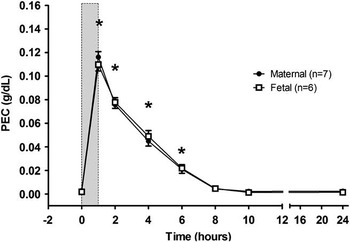
Alcohol was administered intravenously to pregnant ewes at either 1 g or 0.75 g/kg maternal body weight over 1 hour (h), which gave a maximal plasma alcohol (ethanol) concentration (PEC) of 0.11–0.12 g/dl in the ewe (range: 0.10–0.13 g/dl, coefficient of variation (CV): 11%) and fetus (range: 0.09–0.12 g/dl, CV: 12%; Fig. 1); this equates to ∼3–4 standard USA drinks ingested over 1 h. Alcohol was infused into the maternal jugular vein so that PEC could be reliably controlled. Maternal and fetal PEC reached similar peak values at the same time, and declined at approximately the same rate, indicating the rapid, bidirectional transfer of alcohol between the maternal and fetal circulations. Virtually all alcohol had been eliminated from maternal and fetal plasma by 8 h after the onset of each daily infusion. Maternal hepatic biotransformation of ethanol to acetaldehyde and acetate is the major mechanism of alcohol elimination from the maternal-fetal unit.Reference Clarke, Smith and Patrick51
Maternal and fetal plasma ethanol concentration over time in relation to infusion onset (time = 0). Shaded area indicates the time of the 1-hour daily infusion of ethanol (0.75 g/kg maternal body weight). *P < 0.05 between alcohol and control groups. From Kenna et al.Reference Kenna, De Matteo and Hanita18; reproduced and adapted with permission of the publisher. PEC, plasma ethanol concentration.

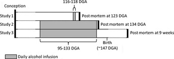
Our laboratory has conducted three different experiments on the fetal and postnatal effects of maternal alcohol administering during the third-trimester-equivalent (Fig. 2). The first of these studies examined the effects of administrating alcohol for 1 h per day to the pregnant ewe from 116 to 118 days of gestational age (DGA) on the fetal brain at 123 DGA (0.84 of term).Reference Dalitz, Cock and Harding19 The second study used a longer period of maternal alcohol administration with pregnant ewes receiving alcohol infusions for 1 h per day from 95 to 133 DGA. We examined effects on the fetal brain, lungs, kidneys, heart and small arteries at 134 DGA (0.9 of term);Reference Kenna, De Matteo and Hanita18, Reference Goh, Bensley and Kenna57–Reference Sozo, O'Day and Maritz59 alcohol administration was stopped at 133 DGA due to the risk of preterm birth.Reference Parazzini, Chatenoud and Surace12 The third study examined the effects of this long alcohol exposure regimen used in our second study on postnatal outcomes; that is, fetuses exposed to alcohol for 1 h per day from 95 to 133 DGA were allowed to be born naturally, and were then studied at 9 weeks of age.Reference Sozo, Vela and Stokes60 The gestational period of alcohol exposure and the PEC were similar among all three studies and the experiments were performed at the same facility, using the same equipment, and in sheep from the same supplier.
The experimental protocol of the three models of fetal alcohol exposure in late gestation used in our studies. Ethanol (1 g/kg maternal body weight in study 1; 0.75 g/kg maternal body weight in studies 2 and 3) was administered for 1 hour daily over the alcohol infusion period. DGA: days of gestational age. Term is equivalent to ∼147 DGA.

Effects of daily 1 h alcohol exposure for 3 days
In this study eight fetal sheep were exposed to a daily 1 h alcohol infusion (1 g/kg maternal body weight) for 3 days, from 116 to 118 DGA, via the maternal jugular vein; eight control fetuses received saline infusion.Reference Dalitz, Cock and Harding19, Reference Gatford, Dalitz and Cock27 The maximal fetal and maternal PEC of 0.11 ± 0.01 g/dl were reached at 1 h after the start of the infusion. Fetal tissue was collected at 123 DGA, 5 days after the last alcohol infusion. Daily alcohol infusion increased fetal arterial lactate concentrations at 6 h from the start of the daily alcohol infusion, but did not affect fetal arterial partial pressure of oxygen (PaO2) or carbon dioxide (PaCO2), arterial saturation of oxygen (SaO2), pH, blood glucose concentration or mean arterial pressure (MAP)Reference Dalitz, Cock and Harding19. Pro-inflammatory cytokines, interleukin (IL)-1β, IL-6 and tumor necrosis factor-α (TNF-α) were also measured in fetal plasma, but their concentrations were similar to controls.Reference Dalitz, Cock and Harding19 At necropsy, a mean decrease of 500 g (19%) in body weight was seen in alcohol-exposed fetuses compared with controls.Reference Gatford, Dalitz and Cock27 There was a transient decrease in plasma IGF-1 concentration in ewes at 30 h and decrease in IGF-2 concentration in fetuses from 24 to 54 h after the first alcohol infusion, which could account for the decrease in fetal body weight.Reference Gatford, Dalitz and Cock27
The fetal brain was examined with particular emphasis on white matter development that occurs in late gestation in sheep.Reference Rees, Nitsos and Rawson52 Three days of alcohol exposure during this period resulted in subcortical white matter injury, as defined by microglia/macrophage infiltration, axonal disruption, increased apoptosis, astrogliosis and altered glial cell morphology in 4 out of 8 (50%) fetuses examined.Reference Dalitz, Cock and Harding19 Three of the remaining four fetuses demonstrated astrogliosis and increased apoptosis in cerebral white matter.Reference Dalitz, Cock and Harding19 Thus, 7 out of 8 (88%) alcohol-exposed fetuses showed evidence of white matter vulnerability to alcohol, which could induce CNS dysfunction; fetal control brains did not show any sign of overt injury. Importantly, there was a positive correlation between the maximal fetal PEC and the degree of white matter injury among fetuses.Reference Dalitz, Cock and Harding19 Other studies have demonstrated correlations between peak PEC and effects in offspring.Reference Bonthius, Goodlett and West61–Reference Pierce and West64 The relationship between maximal PEC and effects on the fetal brain may explain the variability in brain injury that occurs in individuals with FASD.Reference Gladstone, Nulman and Koren65
Effects of a prolonged period of daily 1 h alcohol exposure
To determine the effects of a longer period of daily alcohol exposure on fetal development we infused alcohol from 95 to 133 days of gestation, with necropsy at 134 days (term ∼147 DGA). A protocol of daily maternal alcohol administration (0.75 g/kg maternal body weight) was used, as in the 3-day study, to achieve a maximal PEC of 0.11–0.12 g/dl (Fig. 1).
Physiological effects
When measured at 131–133 DGA, there were no significant effects of alcohol exposure on fetal heart rate (HR), MAP,Reference Kenna, De Matteo and Hanita18 electrocorticographic (ECoG) activity or sagittal sinus blood flow as an indicator of cerebral blood flow (unpublished observations). Prenatal alcohol exposure can initially impair fetal biophysical parameters, such as fetal breathing movements, electro-ocular activity (EOG) and/or ECoG, in humans and sheep.Reference McLeod, Brien and Loomis66, Reference Smith, Brien and Carmichael67 However, variables, such as the gestational maturity of the fetus and the duration of alcohol exposure, can impact biophysical parameters such as fetal breathing movements,Reference Smith, Brien and Carmichael67, Reference Bocking, Carmichael and Abdollah68 which may have occurred in our long-term alcohol exposure model.
In relation to the start of the daily alcohol infusion maternal pH was decreased at 2–4 h, blood glucose concentration was decreased at 1–4 h and blood lactate concentration was increased at 1–6 h before returning to baseline valuesReference Kenna, De Matteo and Hanita18. In the fetus, there was a similar increase in blood lactate concentration at 4–10 h from the start of the daily alcohol infusion, whereas fetal pH and blood glucose concentration were unaffected.Reference Kenna, De Matteo and Hanita18 We further observed that fetal PaO2 and SaO2 were decreased at 10 and 6–10 h, respectively, whereas maternal oxygenation was unaffected.Reference Kenna, De Matteo and Hanita18 All fetal arterial blood measurements returned to baseline values by 23 h after the end of the daily alcohol infusion.Reference Kenna, De Matteo and Hanita18
Fetal and maternal responses to daily alcohol infusion were more pronounced in this model of repeated alcohol exposure than in the 3-day exposure study. This may be due to either the duration of maternal alcohol exposure or the greater gestational age of the fetus (131–133 v. 123 DGA) at the time of measuring responses. Our study showed for the first time that maternal daily alcohol exposure could cause delayed and prolonged changes in maternal and fetal homeostasis, which could affect fetal development. The main physiological effects of this more prolonged daily exposure to alcohol were transient increases in maternal and fetal blood lactate concentration, decreases in maternal blood glucose concentration and pH and decreased fetal oxygenation, effects not seen with the shorter (3 days) treatment regimen.
Surprisingly, there was no evidence of fetal growth restriction after this longer (39 days) period of daily alcohol exposure; fetal body weight was not different between alcohol-exposed and control groups at 134 DGA,Reference Kenna, De Matteo and Hanita18 or at birth when animals proceeded to term.Reference Sozo, Vela and Stokes60 In this latter group, postnatal growth up to 9 weeks was also similar in the alcohol and control groups.Reference Sozo, Vela and Stokes60 In alcohol-exposed fetuses at 134 DGA, the ponderal index (body weight/crown-rump lengthReference May, Gossage and Kalberg3) was lower than in controls, indicating that these fetuses were thin relative to length.Reference Kenna, De Matteo and Hanita18 However, there was no decrease in ponderal index in the cohort of alcohol-exposed offspring that were born naturally at term, indicating that the prenatal reduction in body fat and/or muscle does not persist following the cessation of alcohol exposure at 133 DGA. Thus, fetal growth restriction was not a feature of this model of prolonged, daily alcohol exposure in late gestation.
Brain development
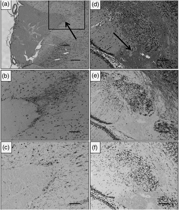
The fetal brain was examined at 134 DGA following 39 days of alcohol exposure.Reference Kenna, De Matteo and Hanita18 Based on our previous finding that 3 days of alcohol exposure causes white matter damage,Reference Dalitz, Cock and Harding19 we expected that 39 days of exposure would result in more severe white matter injury. We evaluated astrocyte and microglia/macrophage density, percentages of activated microglia, myelinating oligodendrocytes and apoptotic cells, and the number of blood vessels per unit area of brain tissue, but found no differences between alcohol-exposed and control fetuses,Reference Kenna, De Matteo and Hanita18 although the presence of more subtle brain injury may have been revealed by the use of more quantitative methods (e.g. stereology). In three out of eight (38%) alcohol-exposed fetuses, there were small subarachnoid hemorrhages in the cerebral cortex and/or cerebellum (Fig. 3);Reference Kenna, De Matteo and Hanita18 we did not detect any hemorrhages in control fetal brains. Blood vessel rupture could be due to changes in blood vessel wall composition and/or function, for which we have some evidence (see section “Small artery development”). Cerebral hemorrhage following prenatal alcohol exposure, especially during development, could put the neonate at increased risk of cerebral dysfunction, a key manifestation of the CNS injury of the FAS and ARND.Reference Stratton, Howe and Battaglia69 The differences seen between our 3- and 39-day alcohol models with respect to white matter injury and cerebral hemorrhage are worthy of further investigation, as they raise the possibility of white matter repair following initial alcohol exposure, but increasing risk of cerebrovascular injury with prolonged exposure.
Subarachnoid (a–c) and cerebellar hemorrhages (d–f) following 39 days of daily alcohol (0.75 g/kg maternal body weight) exposure in late gestation (95–134 days of gestational age). Hematoxylin and eosin (H&E) stained section of a cerebral hemisphere (a) showing a subarachnoid hemorrhage. The square in (a) is magnified to demonstrate (b) astroglial (glial fibrillary acidic protein (GFAP)) and (c) microglial (ionized calcium binding adaptor molecule 1 (IBA-1)) responses in tissue surrounding the hemorrhage. H&E section of the cerebellar hemisphere (d) shows a parenchymal hemorrhage and subsequent damage to the cerebellar cortex. The parenchymal hemorrhage is also associated with astroglial (GFAP; e) and microglial (IBA-1; f) responses. Arrows indicate the site of the hemorrhage. Scale bars: (a) 200 μm and (b–f) 100 μm. From Kenna et al.Reference Kenna, De Matteo and Hanita18; reproduced and adapted with permission of the publisher

Small artery development
Direct application of alcohol to isolated smooth muscle cells from mature pig coronary arteries or intact coronary arteries results in an increase of intracellular Ca2+ and smooth muscle contraction.Reference Kuroiwa, Aoki and Kobayashi70 These effects are modified by the presence of the endothelium, indicating complex interactions of alcohol with vascular tissue that include smooth muscle contraction via increases in intracellular free Ca2+ and endothelial release of vasodilators.
Little is known about the effects of alcohol exposure during pregnancy on the development of the vascular system. Although vascular development begins early, it is not until very late in gestation and early postnatal life that collagen and elastin (which provide strength and elasticity in vessel walls) reach maximal production.Reference Berry, Looker and Germain71, Reference Davis72 Alterations to blood vessel wall structure, such as changes in collagen and elastin content or in extracellular matrix cross-linking, can alter endothelial and smooth muscle function and arterial wall stiffness.Reference Humphrey and Na73 In pregnant sheep, a long (30–82 days) exposure to a relatively high concentration of alcohol (maximal PEC of ∼0.22 g/dl) once a day (5 days on, 2 days off per week) resulted in increased cerebrovascular relaxation responses when isolated vessels were tested in vitro.Reference Mayock, Ngai and Mondares74 In human umbilical arteries taken from newborn infants exposed to alcohol during gestation (17–20 standard drinks per trimester), endothelium-dependent relaxation was not affected, but the sensitivity of smooth muscle cells to vasoconstrictors was reduced.Reference Iveli, Morales and Rebolledo75 Aortas of adult rats exposed to binge alcohol exposure during gestation have reduced α1-adrenoceptor-mediated contraction and impaired endothelium-dependent relaxation.Reference Turcotte, Aberle and Norby76 Furthermore, 9-year-old children, exposed to alcohol in utero but not diagnosed with FASD, had increased aortic wall stiffness when compared with control children;Reference Morley, Dwyer and Hynes77 increased arterial wall stiffness is a clinical predictor of cardiovascular disease.Reference DeLoach and Townsend78 Together, these studies indicate that prenatal alcohol exposure affects arterial function and stiffness, which persist into childhood and may be the early expression of vascular dysfunction leading to cardiovascular disease in later life.
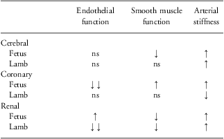
We have examined passive mechanical properties (arterial wall stiffness) and endothelial (nitric oxide, prostanoid and endothelium-dependent hyperpolarizing factor(s)) and smooth muscle function in ovine fetal and postnatal small arteries from five vascular beds (cerebral, coronary, renal, femoral and mesenteric) following daily alcohol exposure for 39 days during late gestation. At 134 DGA and 9 weeks after birth, we observed widespread changes in endothelial and smooth muscle function and passive mechanical properties, including arterial wall stiffness.Reference Parkington, Kenna and De Matteo79–Reference Tare, Kenna and Sozo82 Alcohol exposure resulted in region-dependent changes in vascular function and arterial wall mechanics which differed between the fetal and postnatal lamb. Changes observed in endothelial and smooth muscle function and arterial wall stiffness in arteries from the brain, heart and kidneys are summarized in Table 1. These changes, if they persist into later life, are likely to alter cardiovascular function and increase the risk of cardiovascular disease; for this reason they are the subject of on-going investigation. Changes in arterial mechanics and function could explain the cerebral hemorrhage that we observed in some fetuses after 39 days of alcohol exposure. Importantly, such changes could have serious consequences if they persist into postnatal life; the effects of challenges, such as exercise or stress, may further exacerbate the cardiovascular phenotype. Arterial dysfunction can restrict regional blood flow, indicating that key organs such as the brain, kidneys and heart may be at increased risk of damage following alcohol exposure in late gestation.
Effect of prenatal alcohol exposure on vascular function and wall stiffness in small cerebral, coronary and renal arteries from fetuses (134 days of gestation) and postnatal lambs (9 weeks of age)
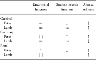
Comparison of vascular function and wall stiffness in cerebral, coronary and renal arteries between alcohol-treated fetuses (n = 4–9) and lambs (n = 6–10) and control fetuses (n = 4–9) and lambs (n = 5–8). Ethanol (0.75 g/kg maternal body weight) was infused via the maternal jugular vein for 1 hour daily from 95 to 133 days of gestation. ↑, significant increase in the ethanol treatment group compared with controls; ↓, significant decrease in the ethanol treatment group compared with controls; ns, not significantly different. (Unpublished observations: see text for details.)
Heart development
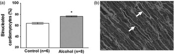
The fetal heart was the only organ in which we observed evidence of altered growth following 39 days of daily alcohol exposure. In this study, we observed that the fetal heart-to-body weight ratio was greater in alcohol-exposed fetuses than in controls.Reference Goh, Bensley and Kenna57 Specifically, there was a greater left ventricular and septal wall volume in alcohol-exposed fetuses.Reference Goh, Bensley and Kenna57 This left ventricular hypertrophy was attributed to an increase in cardiomyocyte size and a 12% increase in the proportion of mature, binucleated cardiomyocytes relative to immature, mononucleated cardiomyocytes (Fig. 4a).Reference Goh, Bensley and Kenna57 The increase in heart growth appears to be due to a direct trophic effect on the cardiomyocytes rather than a hemodynamic effect, as fetal and postnatal MAP were not affected. Other agents, including angiotensin II, have been found to induce cardiac hypertrophy in the absence of hemodynamic changes.Reference Kim and Iwao83 Toxicants including cocaine and acetaldehyde (the principal metabolite of ethanol) have also been shown to induce cardiac hypertrophy.Reference Chen, Tu and Purdon84 Although we do not know the fetal concentration of acetaldehyde in our study, the cardiac hypertrophy that we observed could be a result of elevated acetaldehyde levels.
Percentage of binucleated cardiomyocytes (a) within the left ventricle and cardiac septum in saline and alcohol-exposed fetal hearts at 134 days of gestation following 1 hour daily alcohol (0.75 g/kg maternal body weight) infusions from 95 to 133 days of gestational age. *P < 0.05 between alcohol and control groups. Representative left ventricular section (b) showing mono- (smaller) and bi-nucleated (larger) cardiomyocytes (arrows). Cardiomyocytes are stained with wheat germ agglutinin-Alexa Fluor 488 and TO-PRO-3. Scale bar = 8 μm. From Goh et al.Reference Goh, Bensley and Kenna57; reproduced and adapted with permission of the publisher.

In vitro studies show that alcohol exposure can increase the number of mature cardiomyocytes by increasing the number of cells in the GO/G1 phase of the cell cycle (indicative of mature cells that have exited the cell cycle),Reference Adickes, Mollner and Makoid85 which is consistent with the findings of our in vivo pregnant sheep study. As mature binucleated cardiomyocytes are larger than immature mononucleated cardiomyocytes (Fig. 4b), the increased proportion of binucleated cardiomyocytes likely contributes to the overall increase in left ventricular and septal size in the alcohol-exposed fetal lambs. In addition, mRNA expression of the cardiomyocyte growth factor IGF-1 was increased in alcohol-exposed fetal hearts, which may also contribute to the increased size of the cardiomyocytes.Reference Goh, Bensley and Kenna57
We observed an increase in the mRNA expression of the pro-apoptotic genes, caspase-3 and BAX, in the alcohol-exposed hearts.Reference Goh, Bensley and Kenna57 Alcohol exposure during pregnancy in rats can increase the expression of markers of cardiomyocyte apoptosis, decrease the heart to body weight ratio and increase cardiac contractile function in offspring.Reference Ren, Wold and Natavio86 However, in our model, the increase in caspase-3 and BAX mRNA expression was not associated with an increase in cardiac cell death. The functional consequences of these effects on cardiomyocyte growth and subsequent left ventricular hypertrophy remain to be investigated.
Kidney development
An examination of the fetal kidney at 134 DGA following prolonged (39 days) alcohol exposure during late gestation revealed no overt signs of injury or changes in kidney weight or glomerular volume.Reference Gray, Kenna and Bertram58 Furthermore, there were no differences in mRNA expression of several key genes responsible for kidney development and function such as the renin–angiotensin system, IGFs and sodium transporters.Reference Gray, Kenna and Bertram58 Fetal renal function was not assessed in this study, but amniotic fluid composition, which can be used as an indicator, showed no differences between control and alcohol-exposed groups with respect to concentrations of Na+, K+, Cl−, creatinine, urea, uric acid and total protein, suggesting that fetal renal function was not overtly compromised.
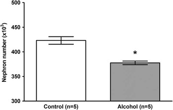
Nephrogenesis is normally complete by 120–130 DGA in sheep;Reference Gimonet, Bussieres and Medjebeur53 hence we were able to assess the effects of the prolonged alcohol exposure on final nephron number. We observed that total nephron number was decreased by 11% in the alcohol-exposed fetuses (Fig. 5).Reference Gray, Kenna and Bertram58 This decrease may have been due to a reduction in renal branching morphogenesis, which is actively occurring at ∼100 DGA in the fetal sheep,Reference Gimonet, Bussieres and Medjebeur53 as alcohol has been shown to reduce branching in organ-culture studies of the kidney.Reference Gray, Cullen-McEwen and Bertram87 As no new nephrons are produced following the completion of nephrogenesis, it is expected that the observed reduction in nephron number would persist into postnatal life. Previous studies have shown that a reduction of 20–30% in nephron number is associated with elevated blood pressure in later life in sheepReference Wintour, Moritz and Johnson88 and rodents,Reference Langley-Evans, Welham and Jackson89–Reference Wlodek, Mibus and Tan91 but it is unclear what effects an 11% reduction in nephron number would have on blood pressure in later life. As mentioned previously, there was no difference in MAP or HR in our alcohol-exposed 9-week-old lambs compared with controls.Reference Goh, Bensley and Kenna57 However, in pregnant rats, two doses of alcohol, resulting in similar maximal PEC and nephron deficit to those obtained in our study, produced elevated blood pressure in offspring at 6 months of age.Reference Gray, Denton and Cullen-McEwen92 Potentially, a small decrease in nephron number when combined with other cardiovascular risk factors, such as obesity and/or a poor diet, could increase the risk of hypertension in later life.
Total nephron number in saline and alcohol-exposed fetal kidneys at 134 days of gestation after 39 days of daily 1-hour ethanol infusions (0.75 g/kg maternal body weight). *P < 0.05 between alcohol and control cohorts. From Gray et al.Reference Gray, Kenna and Bertram58; reproduced and adapted with permission of the publisher.

Lung development
In sheep as in humans, lung development begins in utero and continues after birth. In both species, alveolarization begins during late gestation and continues into postnatal life.Reference Alcorn, Adamson and Maloney55, Reference Burri56 Repeated daily alcohol exposure did not alter lung morphology or lung growth in our fetuses at 134 DGA or in postnatal lambs at 9 weeks of age.Reference Sozo, O'Day and Maritz59, Reference Sozo, Vela and Stokes60 We observed an increase in collagen Iα1 mRNA and an increase in collagen protein deposition in fetal lungs, which could affect lung compliance.Reference Sozo, O'Day and Maritz59, Reference Sozo, Vela and Stokes60 Although this increase in collagen did not persist postnatally,Reference Sozo, O'Day and Maritz59, Reference Sozo, Vela and Stokes60 if present at term or preterm birth, it could adversely affect lung compliance at the onset of gaseous ventilation. We also measured mRNA expression of pro-inflammatory cytokines and observed that IL-1β and IL-8 mRNA expressions were reduced in the fetal lungs; however, there was no change in cytokine mRNA expression at 9 weeks after birth.Reference Sozo, O'Day and Maritz59, Reference Sozo, Vela and Stokes60 The alcohol-induced reductions in fetal IL-1β and IL-8 mRNA expression indicate impairment in the innate immune status of the lung during development, which could affect the ability of the lung to clear pathogens.
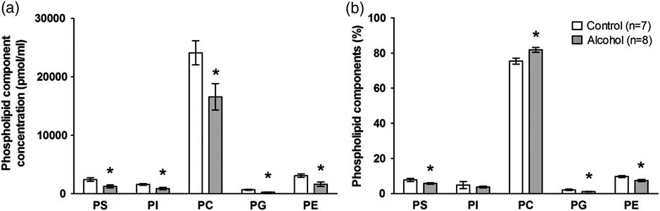
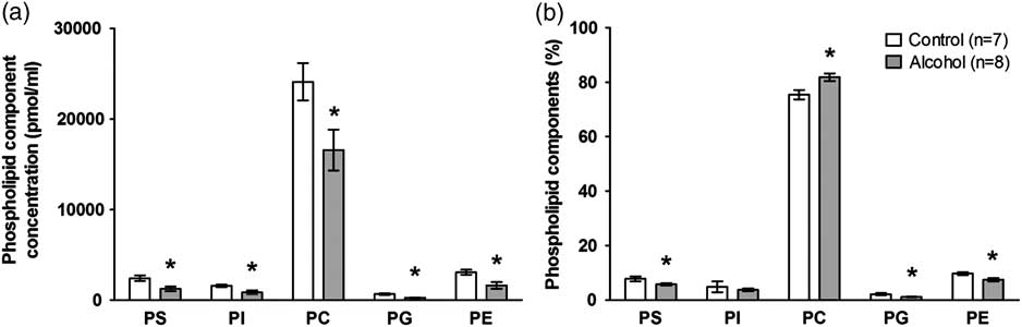
Lung surfactant comprises ∼90% lipids (90–95% of which are phospholipids) and ∼10% surfactant proteins, and acts to decrease surface tension and to assist in the immune response to respiratory pathogens. In amniotic fluid collected at 134 DGA, we observed that 39 days of alcohol exposure led to an overall reduction in phospholipid concentration, with a decrease in the proportion of the phospholipids, phosphatidylserine (PS), phosphatidylglycerol (PG) and phosphatidylethanolamine (PE) (Fig. 6).Reference Sozo, Vela and Stokes60 Amniotic fluid primarily comprises of fetal lung liquid and fetal urine. Thus, changes in lung liquid secretion rates, fetal swallowing and/or fetal urine production could account for the decrease in the proportion of phospholipids. This change in phospholipids could result in increased surface tension in the lung, which could contribute to respiratory distress at birth. However, alcohol-exposed lambs that were born at term (∼12 days after the final maternal alcohol infusion) showed no respiratory dysfunction at birth. The proportion of phosphatidylcholine (PC), which is the major phospholipid in surfactant and is involved in decreasing surface tension, was increased in amniotic fluid taken from alcohol-exposed fetuses; this may have compensated, in part, for the reduced phospholipid concentration.Reference Sozo, Vela and Stokes60 In bronchoalveolar lavage fluid from postnatal lambs, surfactant phospholipid composition was not different from that of controls,Reference Sozo, Vela and Stokes60 suggesting that recovery of surfactant production occurs after alcohol exposure is discontinued.
Surfactant phospholipid component concentration (a) and relative percentage of surfactant phospholipid classes (b) in amniotic fluid at 134 days of gestation following 39 days of daily alcohol (0.75 g/kg maternal body weight) infusions. Phospholipid classes: phosphatidylserine (PS), phosphatidylinositol (PI), phosphatidylcholine (PC), phosphatidylglycerol (PG) and phosphatidylethanolamine (PE). *P < 0.05 between alcohol and control cohorts. From Sozo et al.Reference Sozo, Vela and Stokes60; reproduced and adapted with permission of the publisher.
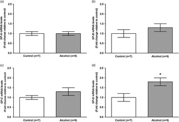
Surfactant proteins (SP)-A and SP-D have immunoregulatory roles, whilst SP-B and SP-C help to decrease surface tension in the lung. The gene expression of SP-A and SP-B was reduced in fetal lung tissueReference Sozo, Vela and Stokes60 indicating that the innate immunity of fetal lung may be compromised and lung compliance may be decreased. In 9-week-old alcohol-exposed lambs, the mRNA expression of SP-D was increased in lung tissue (Fig. 7).Reference Sozo, Vela and Stokes60 This increase in SP-D may be associated with an increased pro-inflammatory state, since previous studies have shown that SP-D not only controls pro-inflammatory responses,Reference Haczku93 but SP-D expression can be stimulated by an increase in pro-inflammatory cytokines.Reference Ikegami, Whitsett and Chroneos94 Furthermore, SP-D can control T-helper (Th)2-type inflammation in the airways and hence is involved in regulating allergic airway inflammation.Reference Haczku95 Given the known roles of SP-D in controlling pulmonary immunity, further studies are required to determine if prenatal ethanol exposure increases susceptibility to infection and allergic sensitization later in life. The alcohol-induced changes that we observed in the innate immune status of the fetal and postnatal lung may increase the risk of lung infection after birth, and thus warrant further investigation.
Relative surfactant protein mRNA expression levels (SP-A (a), SP-B (b), SP-C (c) and SP-D (d)) in lung tissue collected at 9 weeks after birth from control lambs or lambs exposed to daily 1-hour infusion of ethanol (0.75 g/kg maternal body weight) infusions for 39 days during late gestation (95–133 days of gestation). *P < 0.05 between alcohol and control cohorts. From Sozo et al.Reference Sozo, Vela and Stokes60; reproduced and adapted with permission of the publisher.

Relevance of the pregnant sheep models to FASD and FAS
In the 3-day alcohol exposure study, the alcohol-exposed fetuses were lighter than controls. However, neither fetal nor postnatal growth restriction occurred in our 39-day maternal alcohol administration model, with a reduction in ponderal index, but not body weight, observed at 134 DGA. The lack of persistent IUGR may indicate that placental function or maternal nutrition was not persistently affected by the alcohol protocol that we used. Structurally, many of the organs collected at necropsy were not different. Indeed, the only organ grossly affected was the fetal heart, which displayed signs of left ventricular hypertrophy that was attributed to an increase in cardiomyocyte size or maturity, rather than in the number of cardiomyocytes. We observed white matter injury in the 3-day alcohol model, but there was less evidence of overt cerebral injury in the 39-day model. Therefore, our exposure regimen of ∼3–4 standard USA drinks over 1 h for 39 days in late gestation appears to reflect the more subtle effects associated with FASD, rather than the more severe phenotype of FAS.
Repeated maternal alcohol administration decreased maternal blood glucose concentration and arterial pH, and increased blood lactate concentration.Reference Kenna, De Matteo and Hanita18 In the fetus, these alcohol treatments caused increased blood lactate concentration and decreased arterial oxygenation.Reference Kenna, De Matteo and Hanita18 Except for an increase in fetal arterial lactate concentrations, such changes in arterial blood composition were not seen in the 3-day alcohol exposure study, which may be the result of the shorter sampling protocol used in the 3-day study. It is likely that the changes observed in maternal blood lactate and pH were caused by the increased intracellular nicotinamide adenine dinucleotide hydride content during the hepatic metabolism of ethanol and acetaldehyde, and consequent reduction of pyruvate to lactate in glycolysis. Our finding that alcohol exposure resulted in a small decrease in fetal blood oxygenation beginning at 6 h from the onset of alcohol infusion is novel and suggests a modest impairment of placental function, perhaps as a result of acetaldehyde or other long-lasting ethanol metabolites, as the PEC in both the ewe and fetus was negligible at this time.Reference Kenna, De Matteo and Hanita18 The presence of only mild fetal hypoxemia following prolonged alcohol exposure is consistent with our failure to detect IUGR and more overt structural changes in the fetal brain, which might have been expected if more profound chronic fetal hypoxia had occurred.
Prenatal alcohol exposure in late gestation and cardiovascular risk
Prenatal alcohol exposure may affect heart and vascular development, thereby increasing the risk of cardiovascular disease in offspring. We observed that fetal alcohol exposure inhibited nephrogenesis, and together with left ventricular hypertrophy and altered small artery wall stiffness and function, these changes could impact adversely on systemic cardiovascular function in later life. Prolonged daily fetal alcohol exposure was also associated with cerebral hemorrhage, which suggests alterations in cerebral artery structure and/or function. Currently, there are no data on the prevalence of cardiovascular disease in older individuals affected by FASD. However, one study has suggested an association between FAS and mid-aortic syndrome, which is an uncommon syndrome characterized by the narrowing of the abdominal aortic wall and branches, resulting in hypertension.Reference Cura, Bugnone and Becker96 On the basis of our findings, the risk of cardiovascular disease in individuals exposed to alcohol prenatally in late gestation could be increased, and this should be a focus of future epidemiological studies.
Prenatal alcohol exposure in late gestation and risk of compromised pulmonary immunity
Our finding that prenatal alcohol exposure altered surfactant in the fetal lung is consistent with other studies in sheep.Reference Lazic, Wyatt and Matic97, Reference Lazic, Sow and Van Geelen98 In our model, we observed an 80% increase in SP-D mRNA expression in alcohol-exposed 9-week-old lambs.Reference Sozo, Vela and Stokes60 SP-D primarily acts to protect the lungs from airborne pathogens by maintaining innate immunity in the lung; such an increase in SP-D could enhance the innate immunity of the lung. It is possible that our observed increase in SP-D in postnatal lambs may be a response to an overall reduction in pulmonary immune function following prenatal alcohol exposure. This speculation is supported by our finding that SP-A mRNA expression in lung tissue of alcohol-exposed fetuses was approximately one-third of control levels at 134 DGA, indicating compromised fetal immune status.Reference Sozo, O'Day and Maritz59 The expression of SP-A, which also plays an immunoregulatory role, and IL-1β and IL-8 mRNA was decreased in the fetal lungsReference Sozo, O'Day and Maritz59 and TNF-α mRNA expression was increased in the placenta.Reference Kenna, De Matteo and Hanita18 It is unclear what effect these changes could have on overall immunity, but they may result in postnatal changes to immune function that could increase the risk of infection. Children with FAS are known to suffer more frequently from respiratory and other infections.Reference Johnson, Knight and Marmer99, Reference Church and Gerkin100
Alcohol dosage regimen and cerebral injury
The 3-day maternal alcohol administration protocol showed that seven out of eight exposed fetuses had some form of brain injury.Reference Dalitz, Cock and Harding19 Surprisingly however, following 39 days of alcohol exposure, we found little evidence of brain injury, although subarachnoid and cerebellar hemorrhages were observed.Reference Kenna, De Matteo and Hanita18 This finding is interesting, as it indicates that gray and white matter in the fetal brain may adapt to episodic, long-term exposure to alcohol, but cerebral blood vessels may become progressively more vulnerable to damage. The molecular basis of these putative repair, adaptation, and cerebrovascular changes is currently unknown, and further investigation is clearly needed.
Conclusions
On the basis of our findings, it appears that moderate, daily maternal alcohol intake during late gestation can cause a wide range of effects on the fetal brain, heart, small arteries, lungs and kidneys, some of which persist after birth. It appears that fetal adaptation to repeated alcohol exposure can occur. Our data, together with those of other studies, suggest that the cardiovascular and immune systems may be at higher risk of dysfunction in postnatal life. Additional research into the long-term cardiovascular and immunological effects of moderate prenatal alcohol exposure, particularly when challenged by lifestyle factors and infection, is warranted. It is important to recognize that many of the changes we have observed in fetal organ development will likely have long-term effects on health and disease during postnatal life. In order to fully understand the effects of alcohol exposure on the developing fetus, it is essential to examine multiple organs at the microscopic and molecular levels. Finally, in view of our findings, pregnant women should be advised to abstain from consuming alcohol.
Acknowledgments
The authors are grateful for the assistance of Natasha Blasch, Ray Alexander, Victoria Stokes and Alex Satragno. These projects were funded by the Canadian Institutes of Health Research (NET-54014), the National Health and Medical Research Council of Australia, ANZ trustees and the National Heart Foundation of Australia. We acknowledge funding from the NH&MRC and the Victorian Government's Operational Infrastructure Support Program to D. Walker in partial support of this work.