Introduction
Nature uses composite constructions to generate an amazing array of materials with tailored properties for specific applications. Although much can be learned from the intricate way nature combines components, which often in themselves have poor or limited mechanical properties, into materials with impressive properties, the development of biocomposites attempts to unravel nature’s complex system to obtain biofibers for use in conventional composites technologies. Biofibers can be used in short fiber applications such as thermoplastic injection moulding or can be made into blown or woven mats and cloth for use with thermosetting resins. This review will concentrate on lignocellulose fibers, fibers extracted from plant materials consisting largely of cellulose but with varying amounts of other substances such as lignin, pectin, waxes, and extractives. Lignocellulose fibers are the most commonly used natural fiber reinforcement in biocomposites. ChitinReference Paillet and Dufresne1 and cellulose produced from bacteriaReference Favier, Chanzy and Cavaille2 are also receiving some research attention, but this is largely as sources of nano-reinforcements. Nano-cellulose fiber composites are reviewed briefly in a subsequent section. The full potential for natural fibers in biocomposites is yet to be achieved, but they show potential because they are sustainable, have potentially high specific stiffness and strength, and produce less wear than glass fibers in the processing of thermoplastic composites.
It should be recognized that the term “biocomposite” is used for a variety of different composite materials in which either the matrix or reinforcement or both are a biomaterial or a biosourced material. Consequently, the term can cover a material containing bioresin + biofibers, bi-sourced resin + biofibers, synthetic resins + biofibers, bioresin or biosource resin with synthetic reinforcement, or more complex mixtures such as those found in nature. Although most synthesized biocomposites currently use lignocellulose fibers and a synthetic polymer matrix, there is growing interest in replacing the synthetic matrix with bioresins or biosourced resins.
Lignocellulose biofibers
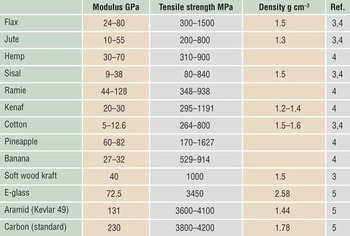
Modern materials engineers are attracted to the high stiffness of the primary cellulose crystal. The theoretical modulus of a cellulose crystal has been given as 250 GPa,Reference Bledzki and Gassan3 which compares favorably with carbon fiber and other high-technology reinforcements. However, in plant fibers, the high modulus cellulose crystals are combined with materials of much lower modulus. Moreover, the cellulose crystals are often aligned at an angle to the axis of the plant fiber. Both factors reduce the overall modulus of the plant fiber. Table I shows indicative values for the modulus of a number of natural lignocellulose fibers, and it can be seen that the modulus varies from 5 to 128 GPa. The upper end of this range is comparable to that of E-glass, the most commonly used glass fiber material. These upper end values of ∼70–100 GPa combined with their low density make natural fibers potentially attractive for use in composites.
Indicative properties of some natural and synthetic fibers.

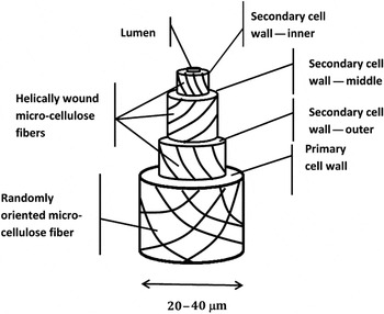
The reasons for the significant variation in properties of natural fibers are best understood through the structures from which they are derived. There are a large number of plant species from which cellulose-based fibers can be derived, but in each case, the cellulose fibers are part of a complex microstructure, consisting of crystalline microfibrils of cellulose that are in a matrix consisting of a hemicellulose, lignin, pectin, and extractives. These structures are well documented for wood pulp, and John and ThomasReference John and Thomas6 have reviewed them for natural fiber production. A typical arrangement of the microcellulose fibers is illustrated in Figure 1. During cell growth, a primary cell wall is first formed in which there is a somewhat random alignment of cellulose microfibrils, as shown in Figure 1. A secondary wall then forms within the primary wall, and this consists of three layers in which the cellulose microfibrils have much more precise orientations within each layer. The spiral angle and volume fraction of microfibrils in the thick middle layer of the secondary wall are considered to have the greatest influence on the fiber properties. Within the secondary cell wall is a hollow tube called the lumen. In a living plant, this lumen may contain water and nutrients, but after extraction for composite use, the lumen may collapse or remain as a tube within the fiber.
Typical arrangement of microcellulose fibers in the cell wall.

These fibers are themselves components of a higher level composite structure that makes up the stems, leaves, or trunk of the plant. Each plant varies in the details of its physical structure, and, moreover, the structure will change within regions of the plant and at different stages of its growth (see Reference Reference Cronier, Monties and Chabbert7 for a review of hemp plants).
This complex structure contains numerous variables that can affect the overall stiffness of the fiber. The stiffest phase, crystalline cellulose, is present in different amounts and with varying orientations to the axis of the fiber depending on factors such as the plant species, the position within the plant, the age of the plant, the growing conditions, and the extraction process for obtaining the fibers from the plant. Production of cellulose fibers from different sources requires the removal of plant material by the action of microorganisms or dew or water to release the wanted fiber (retting). This may be followed by scouring and bleaching that results in fibers with different surface energies and textures.Reference Ouajai and Shanks8–Reference Abdel-Halim, El-Rafie and Kohler10
Lignocellulose fiber composites
The discrepancy between the theoretical modulus of crystalline cellulose and cellulose fibers presents the materials engineer with essentially two choices. The plant fiber can be broken down either mechanically or chemically to provide fibers of a size small enough to eliminate the flaw as a potential property limiter. This yields a micro- or nanofiber that can approach the theoretical properties of the cellulose but creates problems of obtaining and maintaining good dispersion of the fibers in a composite resin. Alternately, the materials engineer can accept that the properties of the long fiber will not be optimal and design the composite material accordingly.
Long fiber composites
Although the properties of natural fibers are potentially attractive, many of the reported natural fiber composites do not achieve a level of stiffness and strength that are comparable to those obtained from synthetic fiber-reinforced composites. There are numerous reasons for this:
• The properties of natural fibers often vary depending on the source of the fiber and its treatment.
• The fibers contain numerous flaws.
• The surfaces of the fibers vary depending on the treatment of the fibers and may also vary along their length, making wetting of the fibers by the matrix potentially poor and variable.
• The fibers can absorb water, which degrades the properties of the composite.
Nature of the fiber surface
The interface region between the fiber and matrix in composites is critical to the ultimate properties of the composite. Good interfacial adhesion allows for transfer of stress to the reinforcing phase, which controls the modulus and strength of the composite, while debonding of the interface can introduce toughening mechanisms to the composite. It is therefore imperative that the interface is controlled.
Natural fibers contain a range of compounds. For example, raw hemp based fibers contain ∼65–80% cellulose, 5–20% hemicellulose, 2–7% lignin, 1–2.5% pectin, as well as extractives such as fats and waxes.Reference Cronier, Monties and Chabbert7, Reference Wang, Postle, Kessler and Kessler11, Reference Wang, Sain and Oksman12 Cronier et al.Reference Cronier, Monties and Chabbert7 also reported that the cellulose content increased continuously with the age of the plant, whereas with the onset of flowering, the proportion of hemicellulose started to decrease in contrast to that of lignin. Since lignin is more hydrophobic than hemicellulose, this has the potential to alter the wettability of the fiber by different resins. It should be noted that these are average compositions determined by bulk analysis of the fibers but do not necessarily give the surface composition, which is critical to the fiber-matrix interface.
One of the few techniques to give the composition of the surface layer of a material is x-ray photoelectron spectroscopy (XPS). This technique yields elemental analysis of the top few nanometers of the surface and has been used extensively for the analysis of wood pulp.Reference Dorris and Gray13–Reference Johansson and Campbell15 The theoretical oxygen to carbon (O/C) ratio for cellulose is 0.8, but surface analysis of many fiber typesReference Johansson, Campbell, Koljonen, Kleen and Buchert16, Reference Mitchell, Carr, Parfitt, Vickerman and Jones17 gives a value for O/C of ∼ 0.3, and this has been confirmed in our laboratory for hemp fibers. Doris and GrayReference Dorris and Gray13 have shown that lignin in wood pulp has an O/C ratio of ∼0.3, so it is highly likely that many natural composites present a surface that is more like lignin than cellulose. Even after specific treatments to remove lignin, such as washing with NaOH or employing more vigorous processes used to delignify wood pulp, lignin can be found on the surface, as it has a tendency to precipitate back onto the surface.Reference Xu, Li and Zhang18, Reference Maximova, Österberg, Koljonen and Stenius19 Alternatively, many extractives have very low O/C ratios, so these also may be on the surface.
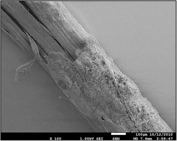
It is also possible that the surfaces of fibers are not uniform. Figure 2 shows a scanning electron microscopy (SEM) micrograph of a hemp fiber, where sections of the fibers have a crystalline appearance, while other sections are encrusted with amorphous-like material. This is consistent with other SEM images of fibers.Reference Hornsby, Hinrichsen and Tarverdi20 The XPS technique has a relatively large sampling area, so the O/C ratio obtained is an average value (usually several fibers). A low O/C ratio may then come from a region that contains surfaces that are cellulose with other regions that are lignin or extractives.
Scanning electron microscopy image of a fiber extracted from a dew retted hemp fiber mat showing variation in the surface condition along the fiber. Note the encrustation of the fiber with amorphous material in the bottom right and the more crystalline appearance of the fiber surface in the top left. Scale marker is 100 μm. (Image courtesy of Ron Rausch, Centre for Microscopy and Microanalysis, The University of Queensland.)

The nature of the surface will control the wetting of the fiber by the matrix, which in turn will affect the ability of the interface to transfer stress to the fiber. To quantify wetting, direct wetting studies can be conducted,Reference Pietak, Korte, Tan, Downard and Staiger9, Reference Liu, Wolcott, Gardner and Rials21 or the surface energy of the fiber surfaces can be studied using inverse gas chromatography.Reference Papirer, Brendle, Balard and Vergelati22, Reference Tze, Walinder and Gardiner23 Inverse gas chromatography provides measures of both the surface energy characteristics and the acid-base properties of a surface. Tze et al.Reference Tze, Walinder and Gardiner23 showed that for the case of cellulose/polystyrene composites, the fiber/matrix interactions, which play an important role in interface adhesion, could be predicted from the respective acid-base parameters of the fiber and matrix. Thus, inverse gas chromatography may provide a tool for designing surface treatments to control interfacial adhesion.
Surface modification
A logical response to the problems of incorporating a hydrophilic fiber into a hydrophobic resin is to modify the surface. There are numerous studies on the use of surface modifications or coupling agents to improve the interface between natural fibers and synthetic resins. In general, these have utilized ideas derived from processes used in conventional composites and adapted them to natural fibers. For example, since cellulose contains multiple hydroxyl groups, many researchers have used silane/titanate/zirconate coupling agents similar to those used for glass fibers. Xie et al. recently reviewed this area in detail.Reference Xie, Hill, Xiao, Militz and Mai24 It is thought that the silanes react with the hydroxyls on the natural fiber surface in a manner similar to the hydrolysis reaction known for glass fiber surfaces. However, the effect of silane treatments on the mechanical properties of natural fiber/polymer composites has been varied and inconsistent and probably requires careful matching of the type of silane and fiber treatment to the matrix resin used. An alternative to this is to use isocyanates, which are also known to react with hydroxyl groups.Reference Bledzki and Gassan3, Reference Maldas, Kokta and Daneaulf25, Reference Botaro and Gandini26 Many polyolefin thermoplastic composites containing various wood-derived reinforcements have used maleic anhydride grafted materials as coupling agents that draw on experience with glass fiber composites.Reference Mohanty, Dryal and Misra27, Reference Kazayawoko, Balatinecz and Matuana28 A third approach is to chemically modify the cellulose surface to make it more hydrophobic, such as by acetylation. This parallels work on other polysaccharides such as starch that have been modified in this way. Many of these approaches have had varying results, probably because too little attention has been paid to the real nature of the lignocellulose fiber surface.
Water sensitivity
Each repeat unit in the cellulose molecule has three hydroxyl groups with the ability for hydrogen bonding, which gives the crystalline cellulose its high melting point. Hydroxyl groups also have an affinity for water, and absorption and desorption of water can affect the properties of the fiberReference Dhakal, Zhang and Richardson29 and, consequently, the composite in different environments. The degradation of properties in the natural fiber composite could arise for a number of reasons. Cellulose molecules are held together with strong hydrogen bonding. Absorption of water can disrupt these hydrogen bonds and cause the cellulose to swell. This can reduce the properties of the fiber, but the swelling can also exert very high stresses on the matrix (as high as 165 MPaReference Mwaikambo and Ansell30), which may cause cracking of the relatively rigid matrix in thermosetting resins. Water can also reduce interfacial bonding, lessening stress transfer to the fiber while long-term exposure of some matrices such as polyesters to water may result in degradation of the resin.
Although coupling agents, such as silanes,Reference Xie, Hill, Xiao, Militz and Mai24 are known to give some improvement in the water sensitivity of the composites, this remains one of the largest challenges to be overcome before adoption of natural fiber composites in critical load-bearing applications. However, the natural hydrophobicity and low water permeability of most matrix resins provides a sufficient water barrier for natural fiber composites to be used in less critical applications such as panels in buses, trucks, and other land transport applications.
Nano-cellulose fiber composites
Cellulose crystals have a very high modulus, but their excellent properties are not fully utilized in the complex structure of plant tissue. Moreover, most plant fibers contain flaws due to variable growing conditions and random damage to plants from environmental conditions or from post-harvest handling and extraction of the fibers. These flaws limit both the stiffness and ultimate strength of the fibers. One way of better utilizing the stiffness of the cellulose crystal would be to break down the natural fiber to micro- or nanofibers and to disperse these as a reinforcing phase in resins. Favier et al.Reference Favier, Chanzy and Cavaille2 demonstrated the potential for this approach when they obtained a threefold increase in the dynamic storage modulus with a 3–6% addition of cellulose needles in a copolymer acrylate latex film.
Micro/nanofibrillation of natural fibers can be achieved by mechanical disintegration of wood fibers after enzyme pre-treatmentReference Ankerfors and Lindstrom31–Reference Revol, Bradford, Giasson, Marchessault and Gray33 or by acid hydrolysis of fibers with sulfuric acid.Reference Revol and Marchessault34, Reference Orts, Shey, Imam, Glenn, Guttman and Revol35 An alternative source of nano-cellulose is to use bacterial cellulose.Reference Favier, Chanzy and Cavaille2 Although the chemical structure and properties of the differently sourced nanofibers are similar (Orts et al.Reference Orts, Shey, Imam, Glenn, Guttman and Revol35 reported that acid hydrolyzed fibers have a modulus of 137 MPa, while bacterial fibers have been measured at 134 MPaReference Klemm, Schumann, Kramer, Hessler, Hornung, Schmauder and March36), their surface chemistries can be quite different. For example, sulfate ester groups may form on the surface of the acid hydrolyzed samples, which would alter the surface charge. Understanding and controlling these differences will be key to dispersion of the fibers in the matrix.
Summary
Natural fibers have the potential to be high specific modulus fibers that come from a sustainable resource. They are readily available and can be produced as byproducts of other agricultural activities. They are thus attractive as fibers for composites manufacture. Their potential has not yet been fully achieved and awaits better understanding and control of the fiber matrix interface and technologies to limit the effects of water uptake on the properties of the composites.