INTRODUCTION
The successful development of a recombinant vaccine against Taenia solium infection in pigs (Flisser et al. 2004; Gonzalez et al. 2005) represents the culmination of two decades of research on the application of recombinant oncosphere antigens as vaccines against infections with taeniid cestodes in their intermediate hosts. The foundations for this research were laid down in the 1930s when immunological investigations with Taenia species infecting laboratory animals indicated the clear potential for the development of effective vaccines (Miller, 1931a,b; Kan, 1934; Campbell, 1936). During the 1960s and 70s there was a resurgence in interest in the immunobiology of infection with taeniid cestodes in their intermediate hosts, underpinning the development of the 45W vaccine against Taenia ovis infection in sheep (Johnson et al. 1989), the first highly effective recombinant vaccine against a parasitic infection (Cox, 1993).
The T. ovis vaccine provided a model from which subsequent vaccines were developed (Table 1). Homologues of the host protective To45W and To18 antigens (Johnson et al. 1989; Harrison et al. 1996) were identified in the genomes of Taenia saginata (Lightowlers, Rolfe and Gauci, 1996b) and T. solium (Gauci, Flisser and Lightowlers, 1998; Gauci and Lightowlers, 2001) which allowed the relatively rapid development of recombinant vaccines against these species. Homologues of the host-protective antigens of Taenia spp. were not evident in the genome of Echinococcus granulosus, however a similar approach to that which had been successful for the identification of host protective oncosphere proteins of T. ovis was adopted for E. granulosus (Heath and Lawrence, 1996), leading to the successful development of a vaccine against hydatid infection in livestock animals (Lightowlers et al. 1996a).

While a wealth of data has been accumulated over the past decade or more on the effectiveness of Taenia and E. granulosus vaccines in both laboratory and field trials, none of these vaccines has been released for commercial use. This review highlights some seminal contributions to knowledge that underpinned the successful development of anti-cestode vaccines, considers the current status of these vaccines and examines the impediments to their commercial use and what steps might be taken to overcome these impediments.
SEMINAL CONTRIBUTIONS TO CESTODE IMMUNOLOGY
Demonstration of immunity to a metazoan parasite
In the introduction to his 1931 publication in the Journal of Preventive Medicine, Harry M. Miller (1931b) of Washington University, St Louis Missouri, states “Very few attempts have been made to immunize mammals against their metazoan parasites, and of the cases in the literature no one presents satisfactory evidence to show that it is possible.” He proceeded to detail the results of his experiments with Taenia taeniaeformis (formally known as Cysticercus fasciolaris) infections in rats in which he was able to induce a reliable, high level (>90%) immunity against a challenge infection. During his investigations, he serendipitously discovered that rats which harboured even a few metacestodes from an earlier exposure to the parasite were immune to a subsequent challenge infection (Miller, 1931a). He showed that this immunity persisted even after the surgical removal of the parasite from an infected animal (Miller and Massie, 1932), leading the way to his discovery that immunity could be passively transferred with either serum from infected rats (Miller and Gardiner, 1932) or via colostrum from dam to offspring (Miller, 1932).
Definition of concomitant immunity and active immunisation in a large animal species
A series of articles in the journal Nature in the 1960s heralded an extraordinary, lifelong contribution to the biology, control and especially the immunology of cestode infections by Michael Gemmell. Gemmell extended the observations made earlier by Froyd and Round (1959) who observed that intermediate hosts of taeniid cestodes could be infected with metacestode larvae after the injection of artificially hatched and activated oncospheres. Sheep or rabbits that had been exposed to an initial infection either orally or by injection of activated oncospheres were immune to a subsequent challenge infection and a considerable degree of cross-immunity was evident between different taeniid cestode species (Gemmell, 1962a,b; 1964a,b). Gemmell interpreted his findings to indicate that there were two phases in the elaboration of immunity – one acting at the level of the intestinal wall and the other acting on the early stages of the development of the metacestode. This distinction was made on the basis of the presence or absence of macroscopic non-viable lesions evident following the challenge infection (indicating immunity acting on the developing metacestode) or the absence of any macroscopic lesion (indicating immunity acting at the intestinal barrier). It remains unclear whether Gemmell was correct in his interpretation of his evidence in favour of immunity occurring at the level of the intestine. There is ample data which indicate that different immune responses act at the early stage of an infection and later during the development of the metacestode (Miller, 1931b; Bogh et al. 1988, 1990). However, Gemmell's implication that immunity was being expressed at the level of the gut may have been off the mark because the absence of a macroscopic lesion could be associated with the death of the parasite in the very early stages of its development rather than be evidence of a lack of penetration past the intestinal mucosa per se. Indeed, the effectiveness of immune serum in passively transferring protection to a naive recipient, without the generation of macroscopic lesions from the challenge parasites, even when the transfer of the serum was up to two days after an infection has been administered (Kan, 1934), argues in favour of the potential for post-gut immune responses to kill the early developing parasites without the generation of a macroscopic lesion. Gemmell later recognised the potential for protective immune responses to occur in the very early stages of the development of the parasite, possibly after penetration of the gut, and he referred to these as being “pre-encystment” immune responses (Gemmell and Soulsby, 1968).
Gemmell (1964a) found that he could induce a high level of immunity only by injection of living parasites and not by the injection of eggs that had been killed by freezing and thawing prior to injection. He concluded that the protective immune responses were “likely to be metabolic, possibly enzymatic, rather than purely somatic complexes”.
Restriction of host-protective antigens to the early developing metacestode
While working at the Australian National University, David Heath undertook experiments to determine the antigenically protective phase of larval development of Taenia pisiformis in rabbits (Heath, 1973a,b). He cultured oncospheres and developing metacestodes in vitro for varying periods of time before transplanting them into rabbits and subsequently determining whether exposure in vivo to parasites of differing ages led to the induction of immunity against a subsequent oral challenge infection with eggs. He found that injection of living oncospheres or developing larvae up to 18 days of age induced complete protection against development of any parasites from the challenge infection. Older developing parasites did not induce a similarly effective immune response and implantation of mature larvae failed to induce any immunity. These data indicate that the host-protective antigens are associated particularly with exposure to the early developing parasite, rather than the mature metacestode. Heath included one immunisation group in which the immunizing oncospheres were described as having been killed by treatment with 4% formaldehyde prior to their injection into rabbits. The animals immunized with this preparation were 100% immune from the subsequent oral challenge infection with T. pisiformis eggs. This finding appeared to contrast with Gemmell's earlier observations using Taenia hydatigena in sheep (Gemmell, 1964a), where injection of killed eggs (frozen at −70 °C for 24 h) had failed to induce a protective response whereas injection of the same number of viable eggs had induced near total protection. However, Heath (1973b) described the development of larvae associated with the injection site in animals injected with both live and formalin-treated oncospheres and hence the significance of his observations in relation to Gemmell's hypothesis, that protection was associated with exposure to living parasites, rather than to parasite components per se, remained unresolved.
Host-protective antigens are secreted products
Michael Rickard undertook experiments specifically to test Gemmell's hypothesis that immunity was only stimulated by exposure to the developing (living) parasite. At that time, diffusion chambers were being used in immunological and cancer research (Biggs and Eiselein, 1965; Nettesheim, Nakinodan and Chadwick, 1966) and they were beginning to be used in studies on immunity to parasites (Petithory and Rousset, 1965; Hillyer et al. 1970). Rickard applied diffusion chamber technology to determine whether the exposure of the hosts of T. ovis (sheep) or T. taeniaeformis (rats) to parasites implanted intraperitoneally in diffusion chambers (pore size 0·22 μm) would render them immune to a challenge infection with their respective parasite species (Rickard and Bell, 1971a,b,c). Hosts in which the diffusion chambers had been implanted were found to be immune (Table 2), confirming that direct contact with the parasites was not required but rather suggesting that the protective antigens were contained in products excreted or secreted by the parasites. Rickard proceeded to demonstrate that sheep and cattle could be successfully vaccinated against T. ovis and T. saginata, respectively, using antigens obtained from their oncospheres in in vitro culture (Rickard and Bell, 1971b; Rickard and Adolph, 1976).

A mechanism to account for variations in innate susceptibility to infection
The importance of the early stages in the development of taeniid cestodes to protective immune responses in intermediate hosts was highlighted in research by Graham Mitchell and his colleagues on the ontogeny of protective antibodies in different inbred strains of mice (Mitchell, Rajasekariah and Rickard, 1980). Their research revealed that different mouse strains differed greatly in their “innate” resistance to infection with T. taeniaeformis. Recognising that early research had shown protective serum antibodies were associated with the development of immunity in mice, they investigated the rate of development of protective antibodies in inbred strains of mice that differed substantially in their susceptibility to T. taeniaeformis infection. While some inbred strains were found to be relatively resistant to infection, the homozygous nude mutants of the same strain were highly susceptible to infection, but they could be completely protected by injection of a relatively small volume (0·25 ml) of serum from non-mutant mice, taken 4 weeks after infection with the parasite (Mitchell, Goding and Rickard, 1977). Mitchell and his colleagues used passive protection in nude mice as their assay for the ontogeny of host-protective antibodies in serum taken at various times after an infection in different strains of mice (Table 3). They found that mice that were relatively insusceptible to infection with T. taeniaeformis developed protective antibodies more rapidly than mice of a strain that was more susceptible to infection. In the data shown in Table 3, serum taken at 5 days post infection from C57Bl/6 (resistant) and C3H/He (susceptible) mice did not contain protective antibody. However by 9 days post- infection, the sera of C57Bl/6 mice contained highly effective levels of protective antibody whereas the serum from C3H/He mice was much less protective. At later times, sera from both resistant and susceptible strains of mice had high levels of protective antibody. Hence, variations in innate susceptibility between the different inbred strains of mice was associated with the rate of elaboration of protective antibodies prior to the parasite undergoing a transition from being susceptible to immune attack, to being insusceptible.

Taken together these data provided strong evidence indicating the susceptibility of the early stages in the development of taeniid cestodes to immune attack, suggesting that vaccines might be developed and based on antigens derived from the oncosphere. This led, following the advent of recombinant DNA technology in the early 1980s, to the successful development of vaccines against T. ovis (1989), T. saginata (1996), E. granulosus (1996) and T. solium (2004).
CURRENT STATUS OF CESTODE VACCINES
Taenia ovis
The metacestode of T. ovis infects the striated muscle tissues of sheep, causing the disease known as ovine cysticercosis, and dogs act as definitive hosts. Detection of cysticerci in sheep meat may lead to a carcase being downgraded or being condemned as being unfit for human consumption. A recombinant vaccine was developed as a commercial product to assist with the prevention of ovine cysticercosis (Johnson et al. 1989; Harrison et al. 1996; Lawrence et al. 1996).
It is more than a decade since Rickard and colleagues published their article entitled: Taenia ovis recombinant vaccine – “quo vadit” (Rickard et al. 1995). In this paper the authors state “ … research to develop efficacious vaccines for important parasitic diseases will not necessarily result in products being marketed for control of these diseases”. The 45W-based vaccine against T. ovis infection in sheep, first described by Johnson et al. (1989), was supported by a multinational animal vaccine manufacturing company. The vaccine progressed through all stages of development, including production scale-up (Dempster, Robinson and Harrison, 1996), safety, efficacy and field trails (Lawrence et al. 1996), achieving registration for commercial use (in New Zealand). Regrettably the vaccine has never been marketed. One of the factors that led to commercial support for research into a vaccine for T. ovis in sheep was the cost to the sheep meat industries of New Zealand and Australia in particular, of the presence of lesions or foreign objects in sheep meat exported to the United States of America (US). Intensive inspections of meat landed in the US were occurring at a time when there was political lobbying occurring there by farmers who were concerned about the commercial impact of meat importation on the domestic meat production industry. One of the types of lesion being detected was T. ovis. This provided an extra impetus to the commercial potential for a T. ovis vaccine. However, by the time the vaccine became a practical reality, the political situation in the US had altered, rejection of whole containers of imported sheep meat was no longer occurring and hence an important commercial driver for T. ovis control had changed. Other political and industry changes had occurred over this period also in New Zealand and these too lessened the prospects for success of a commercial vaccine against T. ovis in sheep. When the time came for the commercial sponsor's marketing group to consider all of the marketing issues surrounding the release of a new product for T. ovis control, the likelihood of commercial returns was judged to be insufficient to support the marketing of the vaccine. Despite the publication which described the T. ovis vaccine being acknowledged as the first description of an effective recombinant vaccine against any parasite, and the discovery being recognised as a milestone in the history of parasitology (Cox, 1993), the vaccine was not a commercial success. Nevertheless, the technology remains available. Should the economic situation regarding T. ovis change in the future to be more favourable towards commercial release of the vaccine, it would be a relatively simple exercise to bring this about.
One of the potential spin-offs from the T. ovis project was the possibility of utilizing T. ovis as a model for the development of practical vaccines against other taeniid cestode parasites. These included parasites with commercial potential (e.g. T. saginata) and/or of health significance (E. granulosus and T. solium). Despite this, the commercial backers of the T. ovis vaccine project chose to withdraw from further work in the field. Nevertheless, work did continue at the University of Melbourne first on the development of a vaccine against cysticercosis in cattle, and later in pigs, as well as on the development of a vaccine against hydatid disease in collaboration with Dr David Heath of the Wallaceville Animal Research Centre in New Zealand.
Taenia saginata
Taenia saginata causes taeniasis in humans, although this does not typically lead to serious medical consequences. The cysticercus occurs in the striated muscles of cattle. Carcases and meat detected as infected with the parasite are down-graded or condemned as being unfit for human consumption. The parasite is common throughout the developing world where poorly cooked or raw beef meat is eaten and where unsanitary disposal of sewage occurs. It also occurs commonly in some first world countries where cultural practices favour the eating of raw or poorly cooked beef meat (Murrell et al. 2005).
Knowledge about the host protective recombinant antigens of T. ovis was used to clone proteins expressed by homologous genes in T. saginata. This led to the relatively rapid development of a highly effective vaccine against cysticercosis in cattle (Lightowlers et al. 1996b). However, despite the extraordinary effectiveness of the vaccine in repeated experimental protection trials in cattle (typically >95% protection), and recognition of the extent and cost of cysticercosis in cattle around the world (Geerts, Kumar and Abbeele, 1980; Nadzhafov, 1987; Mikityuk and Kuznetsova, 1991; Mobius, 1993; Kanev et al. 1995; Murrell et al. 2005), commercial animal vaccine companies have not shown interest in exploitation of this vaccine. There is an increasing awareness of food quality and safety issues which may lead to a change in regulatory attitudes to T. saginata infection in cattle. This could change the prospects for commercial application of a T. saginata vaccine, particularly in Europe where people persist in eating raw beef meat, potentially exposing themselves to taeniasis. For the time being, the TSA9/TSA18 vaccine for T. saginata, described by Lightowlers et al. (1996b), remains in commercial limbo, with the T. ovis vaccine.
Taenia solium
Along with T. saginata, T. solium also causes taeniasis in humans. The normal intermediate host for T. solium is the pig in which cysticerci encyst in the muscles as well as other tissues. However, unlike T. saginata, the eggs of the T. solium tapeworm are infective also for humans. Ingestion of T. solium eggs by humans leads to the development of cysticerci in body tissues, with the parasite commonly having the propensity to encyst in neural tissue and causing a disease known as neurocysticercosis.
Due to the medical importance of T. solium, there has been considerable interest in the development of a vaccine to assist with control of transmission of the parasite. These efforts have focused on development of a vaccine for pigs in the belief that by preventing cysticercosis in pigs, the parasite's life cycle would be broken and the source of taeniasis and cysticercosis in humans would be decreased or eliminated. Much of this research has been undertaken by groups in Mexico using either parasite extracts (Molinari et al. 1997) or defined antigens (Huerta et al. 2001). These studies have made significant progress, however they have not been successful in inducing the levels of protection that had been observed for T. ovis or T. saginata using oncosphere antigens.
One of the longer-term goals of the research undertaken in the 1980s on vaccination against T. ovis was the development of a vaccine to assist in the control of transmission of T. solium. The strategy that was adopted was to use T. saginata as a model to determine whether information about the protective antigens of T. ovis could be used to facilitate the development of an effective vaccine against a closely related taeniid species. The reason for taking this approach was because, although T. saginata was a relatively difficult (involves an obligatory human host) and expensive (experiments with cattle) parasite on which to undertake vaccine research, T. solium represented a substantially more challenging target. T. solium taeniasis is restricted to the developing world, people with T. solium taeniasis are commonly unaware they are infected, and patients with T. solium taeniasis represent only a small fraction of the population even in highly endemic regions (Garcia et al. 2003). In addition, handling the parasite eggs requires special care and facilities because of their potential for infecting humans. The successful development of the vaccine against T. saginata, based on cloning and expression of mRNA encoding the homologues of the host-protective antigens of T. ovis, provided confidence that a similar strategy could be effective for T. solium also.
Genes were identified in T. solium with close DNA sequence homology to those encoding the host protective antigens of T. ovis (Gauci et al. 1998; Gauci and Lightowlers, 2001) and the associated proteins were expressed in E. coli. To date, data from four vaccine trials have been published in which these antigens were used to vaccinate pigs against an experimental challenge infection with T. solium eggs (Flisser et al. 2004; Gonzalez et al. 2005). Additional data are now available from a fifth trial. These trials were undertaken in four different countries (Mexico, Cameroon, Peru, Honduras) by four different groups of researchers. Two protective antigens have been defined – TSOL18 and TSOL45 -1A. One of these antigens, TSOL18, has induced more than 99% protection in each of the five vaccine trials undertaken to date (Table 4).

Much further research is required to be carried out before the TSOL18 vaccine could be recommended for use as a new control measure for T. solium. Among the issues that need to be determined and optimized are: (1) Duration of protection; (2) Antigen dose, immunisation schedule, adjuvant; (3) Potential for co-delivery with other porcine vaccines; (4) Protection of neonates; (5) Scaling up of antigen production; (6) Safety and (7) Efficacy against field-derived infection Research has been initiated in these areas and it is anticipated that small-scale field trials will be carried out in a number of endemic countries with the vaccine in 2008/9.
Echinococcus granulosus
Cystic hydatid disease caused by infection with E. granulosus is responsible for substantial human morbidity and mortality worldwide (Eckert, Gemmell and Soulsby, 1981). The parasite is carried by dogs and other canids as definitive hosts with a wide range of mainly herbivorous mammals acting as intermediate hosts. Humans become infected by accidentally ingesting tapeworm eggs derived from the faeces of an infected dog. Although there is sylvatic transmission of the parasite, many human infections arise because of infections occurring with the adult tapeworm in domestic dogs which are kept as working and/or pet dogs. Sheep play a major role in hydatid disease transmission leading to human infections. Application of an effective vaccine to reduce hydatid infection in livestock would be likely to have a substantial impact on the rate of transmission of the disease to humans.
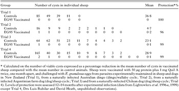
E. granulosus belongs to the same family of cestode parasite as the Taenia species and many aspects of its immunological relationship with its intermediate host are similar to that occurring with Taenia species. For example, infected animals show a significant degree of resistance to re-infection (Gemmell, 1966; Heath et al. 1979a), there is cross-immunity between infection with Taenia species and E. granulosus (Gemmell, 1966; Heath et al. 1979a) and sheep can be immunized against an experimental infection with E. granulosus using oncospheres or their secreted products (Heath et al. 1981; Osborn and Heath, 1982). For these reasons, it was considered likely that the vaccine development approach which had been used successfully for the Taenia species could also be successful for E. granulosus. Initial investigations in which genomic DNA from E. granulosus was probed with labeled cDNA encoding the host protective To16, To18 and To45W antigens of T. ovis failed to identify any homologues of these genes in E. granulosus. However, the strategy by which the native host-protective antigens of T. ovis had been initially identified was likely to be applicable also to E. granulosus. Heath and Lawrence (1996) undertook antigen fractionation studies which identified host protective native oncosphere proteins. One of these antigens was successfully cloned, expressed and shown to be capable of inducing a high level of protection against experimental challenge infection with E. granulosus eggs in sheep (Lightowlers et al. 1996a, 1999; Table 5).

The EG95 vaccine has been licensed by the University of Melbourne and AgResearch New Zealand to a commercial group which has established vaccine production facilities in Beijing, China with a view towards manufacture of GMP-quality vaccine for sale in China and elsewhere. Production-scale vaccine batches have been prepared and safety and efficacy data obtained. The original schedule for licensing and sale of the vaccine has blown out and it remains to be seen whether this source of commercialization will be successful. Extensive field testing of the vaccine has been undertaken by Dr David Heath and his Chinese colleagues (Heath, Jensen and Lightowlers, 2003) and schedules have been recommended for effective application of the vaccine in some endemic situations (Heath et al. 2002; Torgerson and Heath, 2003).
IMMUNE MECHANISMS INDUCED BY ONCOSPHERE ANTIGEN VACCINES
The majority of investigations of immune mechanisms against an egg-induced infection with taeniid cestode parasites have been undertaken on animals that have developed immunity following infection or following immunisation with native oncosphere antigens, rather than following immunisation with recombinant antigens. Immunity induced following an infection can be passively transferred to naive recipients using serum or colostrum from infected donors. This has been demonstrated to be true for T. taeniaeformis infections in mice and rats (Miller and Gardiner, 1932; Kan, 1934; Musoke and Williams, 1975; Mitchell et al. 1977, 1980), infections with T. pisiformis in rabbits (Kerr, 1935; Nemeth, 1970) as well as Taenia hydatigena (Gemmell, Blundell Hasell and MacNamara, 1969; Gemmell et al. 1990; Jacobs et al. 1994) and T. ovis (Heath et al. 1979b; Gemmell et al. 1990) in sheep. Evidence to support the occurrence of protective antibodies following infection with E. granulosus is not so clear cut (Heath, Lawrence and Yong, 1992; Dempster, Harrison and Berridge, 1995) although transfer of a significant level of protection with colostral antibodies has been achieved in sheep (Dempster et al. 1995) as well as in a mouse model of infection with E. granulosus (Dempster et al. 1991). Immunization with oncosphere antigens, rather than infection, also leads to protective antibodies that can be transferred via either serum or colostrum in the case of T. taeniaeformis in mice (Lightowlers, Rickard and Mitchell, 1986), T. ovis (Rickard and Arundel, 1974; Heath et al. 1979b) and T. hydatigena (Jacobs et al. 1994) in sheep and T. saginata in cattle (Rickard, Adolph and Arundel, 1977).
Direct evidence for antibodies playing a crucial role in vaccine-induced immunity to taeniid cestodes induced by recombinant oncosphere antigens comes from in vitro oncosphere killing assays. Heath and his colleagues pioneered the use of this technology for assaying protective serum antibodies against infection with E. granulosus (Heath and Smyth, 1970; Heath and Lawrence, 1981, 1996; Heath et al. 1992). Subsequently, the assay was applied to investigations of immune responses in sheep induced by the EG95 vaccine (Woollard et al. 2000a,b, 2001) where it was found that the level of protection in vivo correlated well with the efficacy of oncosphere killing in vitro. The potential value of the technique has been extended greatly by the work of Kyngdon and his colleagues (Kyngdon et al. 2006) who have established new in vitro oncosphere killing assays for T. pisiformis, T. ovis, T. saginata and T. solium. Kyngdon et al. (2006) showed that immunity induced using the TSA9/TSA18 recombinant vaccine against T. saginata in cattle and the TSOL18 and TSOL45 vaccines against T. solium infection in pigs are associated with specific serum antibodies that kill homologous oncospheres in vitro (Figs. 1, 2). The assays require a source of complement (Heath and Lawrence, 1981; Kyngdon et al. 2006), supporting earlier evidence that host-protective antibodies require complement in order to exert their protective effects in vivo (Mitchell et al. 1977). These data provide strong evidence to support the role played by complement-fixing antibodies as an immune effector mechanism induced by the recombinant cestode vaccines. While the potential contribution of other immune effector mechanisms cannot be excluded, serum antibodies alone are sufficient to account for the high levels of protection induced by the vaccines.

Fig. 1. Appearance of activated Taenia saginata oncospheres after 10 days in vitro culture. (A) A viable T. saginata larva after 10 days of in vitro culture in the presence of normal bovine serum. At this time, T. saginata larvae were approximately 60–100 μm in diameter, contained a defined cystic cavity, and were similar in appearance to T. ovis and T. solium larvae after 10 days of in vitro culture in the presence of normal serum. (B) A non-viable T. saginata larva after 10 days of in vitro culture in the presence of immune bovine serum. The margin of the larva is no longer visible, and the hooks (arrow) appear disordered within an enlarged covering of serum precipitate. Splayed hooks indicate a lack of development past post-oncospheral reorganisation. At this time, the appearance of T. saginata larvae was similar to the appearance of T. ovis and T. solium larvae after 10 days of in vitro culture in the presence of immune serum. (C) A live T. saginata larva stained with methylene blue after 10 days of in vitro culture in the presence of normal serum. Live larvae stained intensely blue, and the projections from the tegument stained less intensely. Live T. solium larvae had a similar appearance when stained with methylene blue. (D) A non-viable T. saginata larva stained with methylene blue after 10 days of in vitro culture in the presence of immune serum. The larva stained only very lightly; hooks are visible (arrow). Non-viable T. solium larvae had a similar appearance when stained with methylene blue. Scale bars=40 μm. Reproduced from Kyngdon et al. (2006) with permission.

Fig. 2. Percentage of non-viable Taenia spp. larvae after 10 days in vitro culture in the presence of either normal or immune serum from the homologous host species. (A) Activated Taenia pisiformis oncospheres cultured in vitro in the presence of pre-immune sera (Group 1) or immune sera (Group 2) from rabbits immunized with T. pisiformis oncospheres; and pre-immune sera (Group 3) or immune sera (Group 4) from rabbits infected with T. pisiformis eggs. Specific killing of T. pisiformis larvae was demonstrated in Groups 2 and 4. (B) Activated T. ovis oncospheres were cultured in the presence of pre-immune sera (Group 1), or immune sera (Group 2) from sheep immunized with T. ovis oncospheres; and pre-immune sera (Group 3) or immune sera (Group 4) from sheep infected with T. ovis eggs. Specific killing of T. ovis larvae was demonstrated in Groups 2 and 4. (C) Activated T. saginata oncospheres were cultured in the presence of sera from cattle immunized with: GST (Group 1); TSA18-GST+TSA9-GST (Group 2); MBP (Group 3); or TSA18-MBP+TSA9-MBP (Group 4). Specific killing of T. saginata larvae was demonstrated in Groups 2 and 4. (D) Activated T. solium oncospheres were cultured in the presence of sera from pigs immunized with: GST (Group 1); TSOL18-GST (Group 2); GST and MBP (Group 3); or TSOL45-GST and TSOL45-MBP (Group 4). Specific killing of T. solium larvae was demonstrated in Groups 2 and 4. Each column=mean percentage of non-viable Taenia spp. larvae in a replicate culture of one serum within the group. Error bars=standard deviation. Reproduced from Kyngdon et al. (2006) with permission.
COMMERCIAL VERSUS ORPHAN VACCINES
The recombinant cestode vaccines have been remarkable successful; none however have been adopted for practical use at this time. The principal reason why there has been a failure to date to adopt vaccination as a control strategy for taeniid cestode infections relates to the assessment of cost versus benefit. For T. ovis and T. saginata, the principal drivers for vaccine development are commercial – these parasites do cause economic losses to the sheep and cattle meat industries. In the case of T. saginata, the parasite does infect humans as the obligatory definitive host but the medical consequences of the taeniasis are usually minor. The drivers for control of hydatid disease and T. solium cysticercosis relate almost entirely to the human health although there are also direct economic losses due to hydatid infection in livestock and due to porcine cysticercosis.
Hydatid disease is widespread around the world (Eckert et al. 2001); many of the locations of high endemicity occur in developing countries. However hydatid disease remains endemic in many first world countries despite the existence of specific disease control campaigns over the past 50 years (Eckert et al. 2001), a number of which continue to this day. There are two major problems with establishing and sustaining hydatid disease control efforts. Firstly, the significance of the parasite relates to human health whereas transmission control measures must be carried out in animals. Thus, implementation of control requires activity is to be carried out in the veterinary/agriculture sector but with demand coming from the health sector. The second major problem with hydatid control is the poor efficacy of currently available control measures as they are applied in practice, and the consequent long duration of control programmes before they have substantial and sustainable effects on transmission (Eckert et al. 1981). The EG95 vaccine has the potential to solve this second problem. Mathematical modeling of hydatid disease transmission predicts that control would be more efficient and would occur in a shorter timeframe if it were to incorporate vaccination of livestock animals (Torgerson, 2003; Torgerson and Heath, 2003). However, unless governments were to mandate hydatid vaccination, there is little economic incentive for livestock owners to invest resources in hydatid disease vaccination and hence there is a lack of commercial interest in manufacture and marketing of an hydatid vaccine. Nevertheless, those governments which are currently investing in hydatid control through traditional methods (largely dog control and anthelmintic treatment) could consider reprioritizing their allocation of funding to include vaccination, thereby creating a demand and commercial opportunity.
The situation with regard to the commercial potential of any control measure for T. solium cysticercosis control is even more dire than it is for hydatid disease because transmission of T. solium is restricted entirely to the poorer parts of the developing world. Implementation of any control measure specifically for T. solium would only occur through the efforts of governments or philanthropic agencies. Here again, a new and highly effective vaccine attracts no commercial interest in manufacture or distribution of the product.
Investment in control measures for human and agricultural diseases is governed almost entirely by the economic impacts of the diseases in the developed world. The research and development cost for new treatments is expensive and carries a high risk of commercial failure. Batson (2002) suggests a figure in excess of US$100 M for the costs of developing a new human vaccine. Development of a new vaccine for animals may not be as expensive however most of the steps and risks involved would be common. In Fig. 3 Batson (2002) summarizes the risks involved in taking a new human vaccine from research to commercial application and the risks involved in developing a new animal vaccine would be similar. For those diseases where there is a clear commercial opportunity, corporations will compete to secure intellectual property and fund the entire cost of the programme. At the other extreme, control measures for diseases like neurocysticercosis require the entire development costs to be borne by the public sector. Many vaccine development projects fall somewhere between these extremes. In such cases research costs may be borne by the public sector alone or in partnership with the private sector. The extent to which further development costs would be met by commercial interests (e.g. field trials, scale-up costs, safety trials) would differ depending on the potential commercial value of the vaccine in each case. Commercial companies understandably seek disease interventions where the intellectual property can be protected and where a sufficient financial return could be guaranteed, if the project were successful, sufficient to recoup the investment costs and provide a profit for the shareholders.

Fig. 3. Risk at every stage of the vaccine life cycle. Reproduced from Batson (2002) with permission.
Over the past two decades there has been an increasing awareness of instances where human disease control or treatment measures have been available, where there is an obvious humanitarian demand, and yet there is a lack of commercial incentive for manufacture and distribution of these measures. These have become known as Orphan Products (Haffner, 1991) and, where the control measure involves vaccination, Orphan Vaccines (Lang and Wood, 1999). Attention has been focused on these products through initiatives such as the World Health Assembly's Expanded Program on Immunization, the US government's Orphan Drug Act, the Global Alliance for Vaccines and Immunization and the Malaria Vaccine Initiative, nevertheless there remain many more opportunities for support of Orphan Products, including the hydatid and T. solium cysticercosis vaccines.
ACHIEVING PRACTICAL USE OF THE HYDATID AND CYSTICERCOSIS VACCINES – AN ACTION PLAN
Government and philanthropic agency resources exist that could be allocated to support control measures for diseases like neurocysticercosis and hydatid disease. In order to secure financial support for the application of a new control strategy such as vaccination, the administrators of these resources need first to be made aware of the significance of the new development as well as the social and economic importance of the related diseases. How do we provide this information most effectively?
Collating the burden of disease
Perhaps the most critical issue in making a case for prioritizing valuable health funds for the control of hydatid disease or T. solium cysticercosis is providing accurate data on the social and economic burden of these diseases. Until recently, very little attention has been placed on collating these data so as to allow an estimation of the burden of hydatid disease and neurocysticercosis in recognized terms, such as Disability-Adjusted Life Years. Work has begun in this area (Budke et al. 2004; Carabin et al. 2005) but there is an urgent need for expansion of this effort so that the global burden of these diseases can be presented accurately.
Regional alliances
Those parts of the world with the highest prevalence of cystic hydatid disease and neurocysticercosis tend to occur within broad regions. Hydatid disease is a particular problem in Africa, South America, central and east Asia and the countries bordering the Mediterranean, while neurocysticercosis is most prevalent in Africa and central and south America. Countries which recognise these as important problems could make a stronger case for gaining international support for action towards controlling the diseases if they were to communicate and collaborate on this issue with other countries in their region. One such grouping has been formed involving countries of east and southern Africa with a shared interest in T. solium cysticercosis and taeniasis (Boa et al. 2003; Mukaratirwa et al. 2003; Phiri et al. 2003). This is a welcome development and could provide a model for the establishment of other groups representing regional areas for the purpose of voicing their concerns to international donors and agencies about cysticercosis or cystic hydatid disease. Groups such as these would have the potential to promote the value of a new disease control measure such as vaccination and assist in gaining support for the scale-up and distribution of the vaccines.
CONCLUDING REMARKS
Most if not all of the practical issues surrounding large-scale production and application of the EG95 hydatid vaccine have been solved and successful vaccine field trials have been completed (Heath et al. 2003). The vaccine has also been effective in experimental trials carried out in New Zealand, Australia, Argentina, Chile, China, and Romania (Lightowlers et al. 1996a, 1999; Heath et al. 2003; Torgerson and Heath, 2003; Morariu et al. 2005). While there has been interest expressed in using the vaccine from hydatid control authorities in a number of countries, the lack of availability of the product in sufficient quantities for practical use remains a stumbling block. The TSOL18 vaccine against T. solium, on the other hand, has only recently been proven to be effective in initial experimental trials. Research is progressing towards defining the operational characteristics of this vaccine. The vaccine has been successful in experimental trials completed by different groups of researchers in four different countries (Flisser et al. 2004; Gonzalez et al. 2005) and the target antigen appears to be identical in T. solium isolates from around the world (Gauci, Ito and Lightowlers, 2006). Here again the signs are encouraging and suggest that this vaccine could have substantial potential for assisting with the control of transmission of this important zoonotic disease. Neither of these vaccines is attractive to multinational vaccine manufacturers and there is no certain path to achieving commercial-scale production which would provide product for practical use. One option may be to seek to identify appropriate production capacity in the regions where the vaccines have application; an approach that is being investigated in relation to other orphan vaccines (Milstien, Batson and Meaney, 1997). In beginning to tackle these problems, the cestode vaccines appear to be the first recombinant helminth vaccines to make the transition beyond the proof-of-principle phase into tackling the issues of production and application.
ACKNOWLEDGEMENTS
Funding is acknowledged from the National Health and Medical Research Council of Australia and the Wellcome Trust.